MB 251 Exam 4
1/132
There's no tags or description
Looks like no tags are added yet.
Name | Mastery | Learn | Test | Matching | Spaced | Call with Kai |
|---|
No analytics yet
Send a link to your students to track their progress
133 Terms
Pathogen
Microbial parasite that causes disease/damage to host. Organisms causing disease in a host
Opportunistic Pathogen
Causes disease only in absence of normal host resistance
Colonization
When a microorganism is established and growing in a host, but not harming the host. Described non-infectious growth in host
Pathogen is present but does not cause disease
Infection
When a microorganisms penetrates host’s defenses to actively grow in a host and cause damage or impair host cell processes and functions
Process by which pathogen causes disease. Disease is present
Disease
Damage or injury to the host that impairs host function or kills them. Also used to provide a common name associated with the infection
Describes a scenario where a host is damaged. Disruption, lyced, being made to do something.
Ex. Typhoid Fever
Symptom
Subjective experiences felt by patient that cannot be directly observed by others. Vary from person to person. Something that the host is telling you that they are feeling
Ex. Stomach pain, headache, nausea, fatigue
Clinical Sign of disease
Indicators of disease that can be observed or measured and not influenced by the patient’s perception or interpretation. Things you can see on the outside
Ex. Fever, rash, blood cell counts
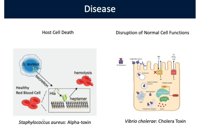
Cell Lysis and Death
Some bacteria have toxins that destroy cell membranes resulting in lysis/death
Ex. Staphylococcus aureus: Alpha-Toxin

Disruption of Cellular Function
Some bacteria produce toxins that don’t kill host cells but cause them not to function properly resulting in clinical signs of disease
Ex. Vibrio cholera: Cholera Toxin
Virulence'/Pathogenicity
Ability of a microorganisms to inflict damage on host
Ex. Highly virulent, Non-pathogenic
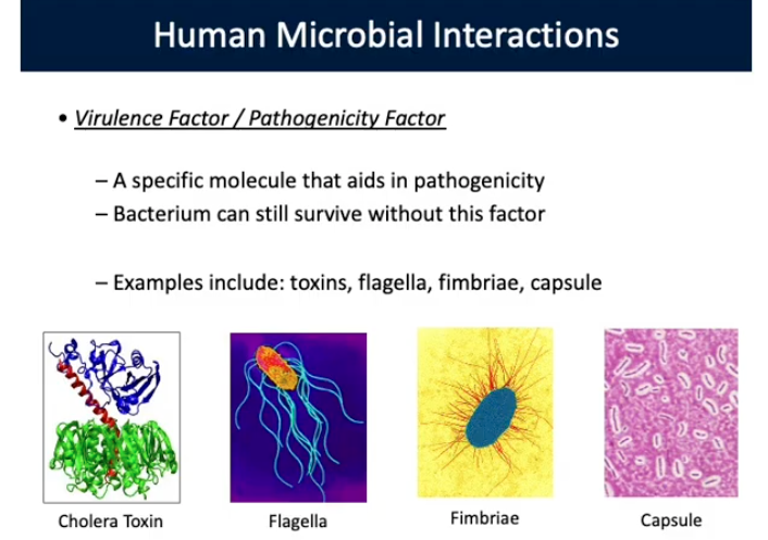
Virulence Factor/Pathogenicity Factor
Specific molecule that aids in virulence/pathogenicity
Bacterium can still survive without this factor
Not essential for survival in non-infection situation
EX. RNA Polymerase is not a virulence Factor
EX. Toxins, flagella, fimbriae, capsule

Virulence Factors (Specific)
Molecules produced by pathogens aid in ability of organisms to:
Colonization of a niche in the host (attachment to cells)
Invade/exit a host
Evade/inhibit host’s immune response
Get nutrition from host/environment
1st Mechanisms of Pathogenesis
Portals of Entry: Mucous membranes, Respiratory (inhalation)/Gastrointestinal(food/water)/Genitourinary(Sexual intercourse) tract, Conjunctiva, Skin (cut, bug bites), Patenteral Route
Site/Mechanisms by which a pathogen enters a host. Provides site where pathogen grows/multiplies.
Not always where infection happens. Some pathogens spread and disseminate in a host
Do not show site of infection
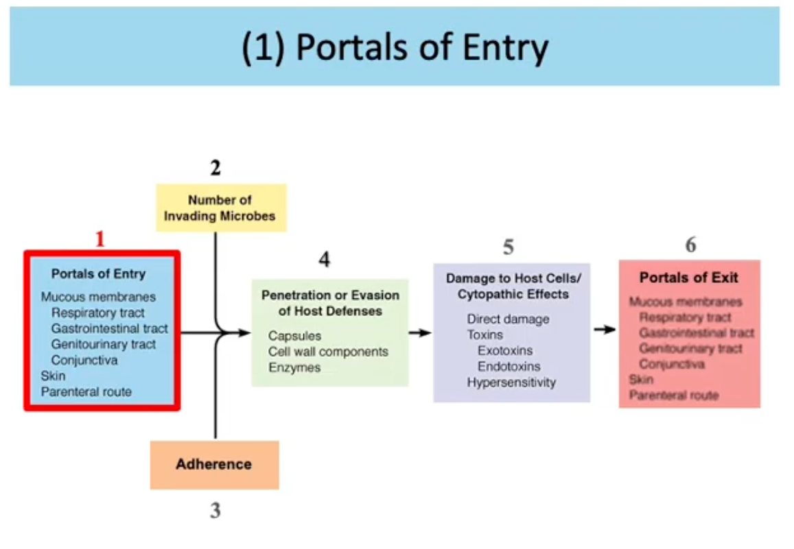
2nd Mechanisms of Pathogenesis
Number of invading Microbes. Virulence of a pathogen can be described by:
Experimental studies assessing the infectious dose
LD50: Lethal dose of toxin/agent/cells that kills 50% of hosts in population. Measured by amount of cells/amount of protein/compound
3rd Mechanisms of Pathogenesis
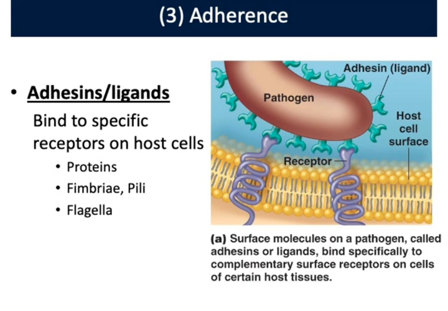
Adherence:
Adhesions/ligands: Bind to receptors of host cells (proteins, fimbriae, pili, flagella)
4th Mechanisms of Pathogenesis
Penetration/Evasion of Host Defenses: Capsules, Cell wall components, Enzymes
5th Mechanisms of Pathogenesis
Damage to Host Cells/Cytopathic Effects: Direct damage, Toxins, Exotoxins, Endotoxins, Hypersensitivity
Portals of Exit
Mucous membranes, Respiratory/Gastrointestinal/Gentourinary tract, Conjunctive, Skin, Parenteral Route
Dissemination
Occurs through bloodstream/lymphatic System. Spread/split of pathogens within host
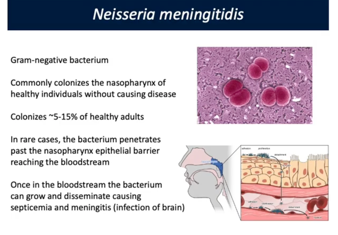
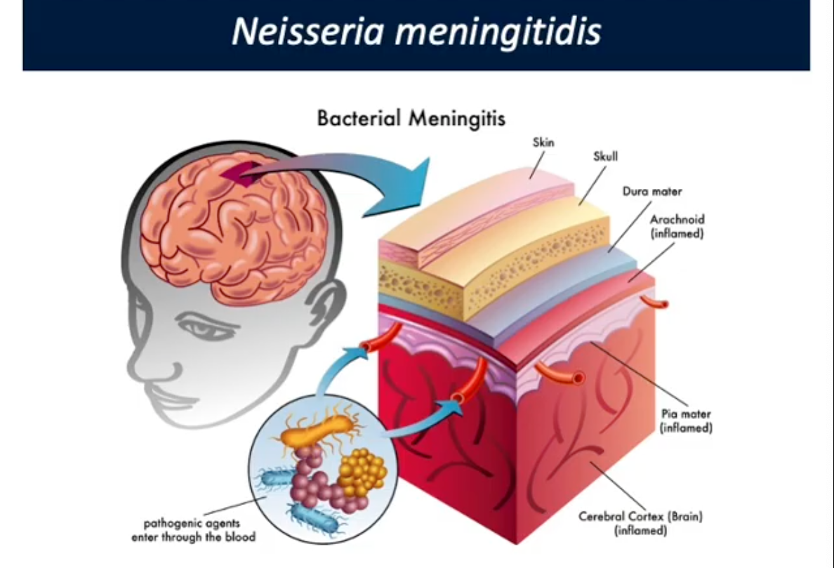
Neisseria meningitis
Gram negative. Colonizes nasopharynx of individuals without causing disease. 5-15% of health adults. Rarely penetrates nasopharynx epithelial barrier reaching bloodstream
Once in bloodstream, grows and disseminates causing septicemia and meningitis (infection of brain)
Immune system does not have access to the brain because process is very toxic/deadly
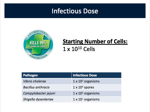
Infectious Dose (2nd Mechanisms of Pathogenesis)
Number of organisms needed to establish infection
LD50
Lethal dose of toxin/agent/cells that kills 50% of hosts in population. Measured by amount of cells/amount of protein/compound
Site of Infection
Portal of entry and receptor mediated adherence are 2 important factors affecting ability to cause infection. POE does not show site of infection
Some pathogens disseminate to different sites in host
Host receptors may be present on multiple cell types at different sites in host
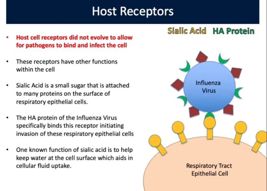
Host Receptors (3rd Mechanism of Pathogenesis)
did not evolve to allow for pathogens to bind and infect cell. Receptors have other functions in cell. Sialic Acid is a sugar attached to many proteins on surface of respiratory epithelial cells
The HA protein of Influenza Virus binds this receptor starting invasion of respiratory epithelial cells
Helps keep water at cell surface which aids in fluid uptake
Evasion of Host Defenses (4th Mechanisms of Pathogenesis)
Prevent phagocytosis by macrophages. Invisibility cloak
Ex. Streptococcus pneumonia
Enzymes:
Leukocidins: : lyse white Blood cells
IgA proteases: Destroy IgA antibodies;Haemophilus influenza
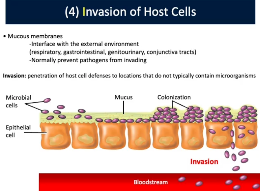
Invasion of Host Cells (4th Mechanisms of Pathogenesis)
Mucous Membranes: Interface with external environment (respiratory, gastrointestinal, genitourinary, conjunctiva tracts)
Normally prevent pathogens from invading
Invasion (4th Mechanisms of Pathogenesis)
Penetration of host cell defenses to locations that do not contain microorganisms
Ex. Listeria monocytogenes:
Gram positive. Facultative anaerobe, Psychotolerant Mesophile
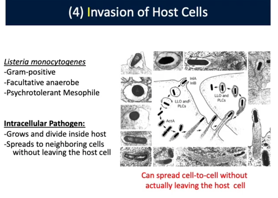
Intracellular Pathogen (Invasion/4th Mechanisms of Pathogenesis)
Grows and divide inside host. Spreads to neighboring cells without leaving the host cell. Can spread cell to cell without leaving the host cell
Toxin (5th Mechanisms of Pathogenesis)
Biologically produced poison that aids in pathogenicity
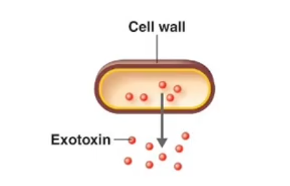
Exotoxin (5th Mechanisms of Pathogenesis)
A toxin that is secreted from the cell
Ex. Lipases: Hydrolyze phospholipids
Hemolysins: Lyse red blood cells. a = Incomplete Lysis (H2O2), b = Complete Lysis, y = No lysis
Leukocidins: Lyse white blood cells
Enterotoxin (5th Mechanisms of Pathogenesis)
Subset of exotoxin that specifically affects the small intestine
Ex. Cholera Toxin
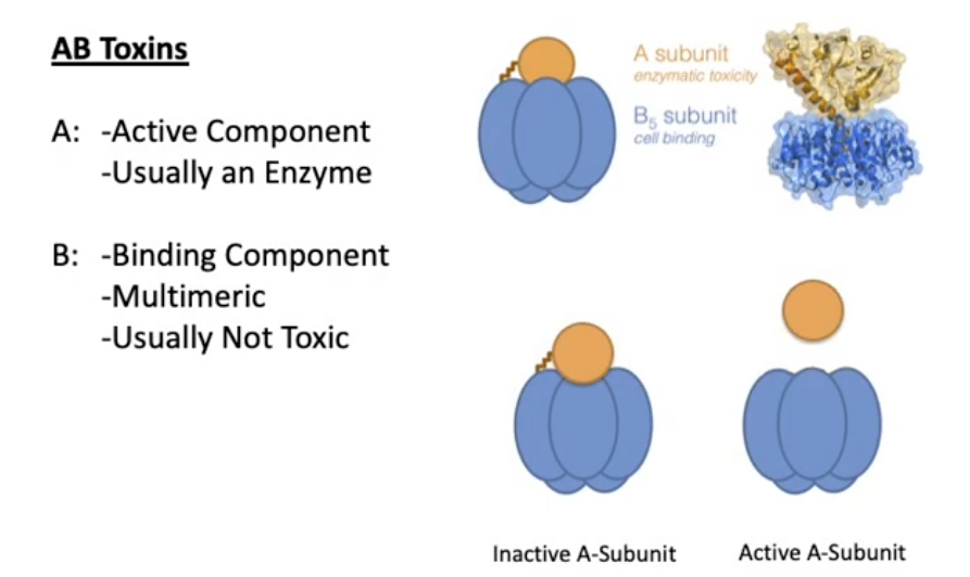
AB-Type Toxins (Exotoxin/5th Mechanisms of Pathogenesis)
AB Toxins:
A: Active Component; Usually Enzyme
B: Binding Component; Multimeric, Usually not toxic
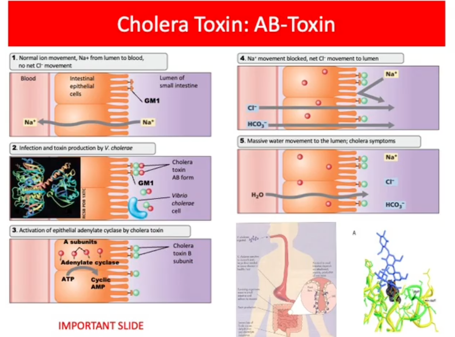
Cholera Toxin (5th Mechanisms of Pathogenesis)
Vibrio cholera: Gram-negative, calculative anaerobe, Aquatic Bacteria
Death due to dehydration from profuse diarrhea
Treatment is rehydration
Disease is mediated by enterotoxin: Target cells of the small intestine. Cholerae Toxin is an AB-type toxin
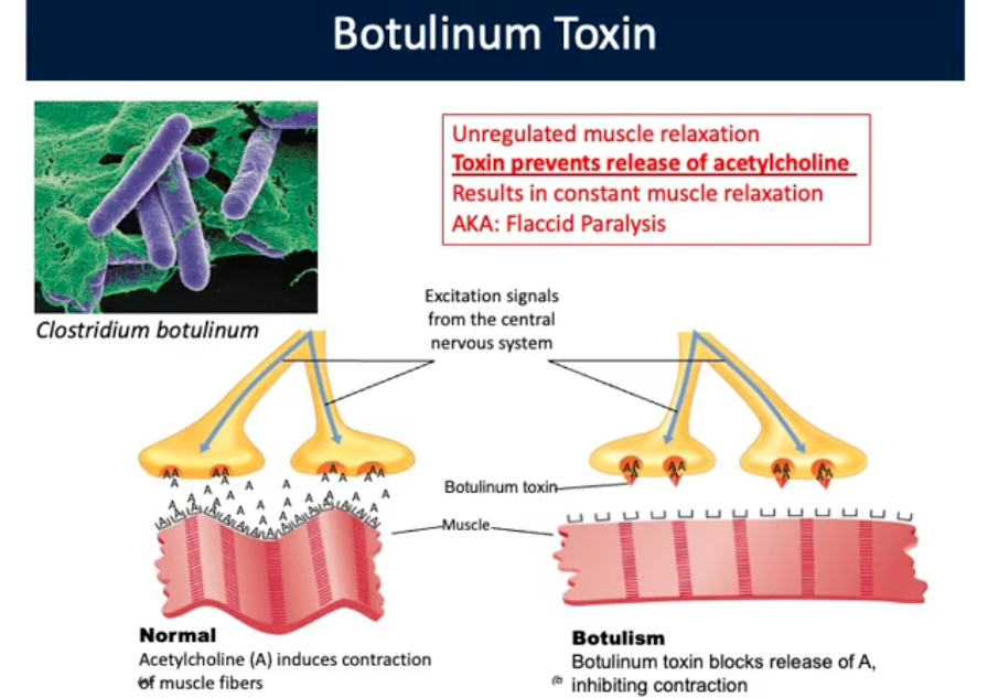
Clostridium botulinum (Toxigenic Clostridial Pathogens)
Gram Positive, Obligate Anaerobe, spore forming bacterium, found in soil.
Canned foods = good environment. Anaerobic atmosphere with nutrients for growth via fermentation —> CO2 —> Bloating can. Toxin is heat sensitive if not cooked can become intoxicated by eating toxin
Unregulated muscle relaxation. Toxin prevents release of acetylcholine —> Constant muscle Relaxation/Flaccid Paralysis
Present in Botox
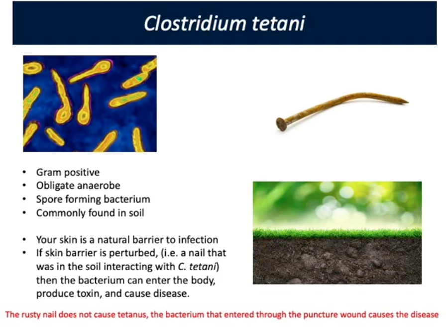
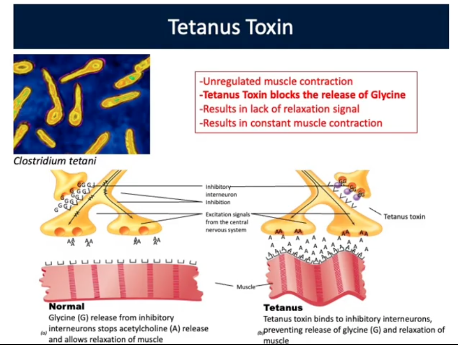
Clostridium tetani (Toxigenic Clostridial Pathogens)
Gram Positive, Obligate anaerobe, spore forming, found in soil
Skin is barrier to infection. If barrier perturbed bacterium enters body, produces toxin, causes disease
Rusty nail does NOT cause tetanus, bacterium entering body through puncture causes disease
Tetanus Toxin blocks release of Glycine
Results in lack of relaxation signal and constant muscle contraction
Portals of Exit (6th Mechanisms of Pathogenesis)
Mucous membranes, Respiratory (inhalation)/Gastrointestinal(food/water)/Genitourinary(Sexual intercourse) tract, Conjunctiva, Skin (cut, bug bites), Patenteral Route
John Snow
Considered the father of epidemiology, where he found that Vibrio cholera was the cause of cholera outbreaks associated with contaminated water
Which of the following virulence factors would allow a bacterial pathogen to evade a host immune response>
Capsule: protective layers surrounding some bacteria that help them avoid phagocytosis by immune cells such as macrophages and neutrophils. This allows the bacteria to evade the host immune response
Leukocidin Enzyme: Toxins that kill white blood cells (leukocytes). By destroying immune cells, they help the pathogen escape immune defenses
Virology
Study of Viruses
Virus
Genetic element that cannot replicate independently of a living host cell. Infect cells from all life
Classified by hosts they infect: Bacterial (Bacteriophages), Archael, Animal Viruses etc
Virus Particle (virion)
Extracellular form of virus. Exists outside hosts and starts transmission from 1 cell to another
Has Nucleic acid genome surrounded by protein coat and other layers of material
Obligate Intracellular: Rely on hosts for survival
Extremely small Genomes
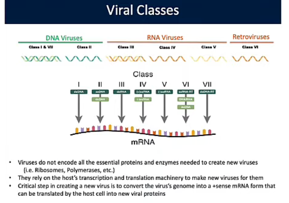
Viral Classes
Do not encode all essential proteins/enzymes needed to reproduce
Rely on host’s transcription/translation to reproduce
Convert virus’s genome into +sense mRNA form that is translated by host cell into new viral proteins
Encoded specialized enzymes host cell won’t have to convert genome to right mRNA form to be translated by host
Nucleocapsid (Viral Structure)
Complete complex of nucleic acid and protein packaged in the virion
Nucleic Acid (Viral Structure)
Genetic Material; Different tips from DNA to RNA
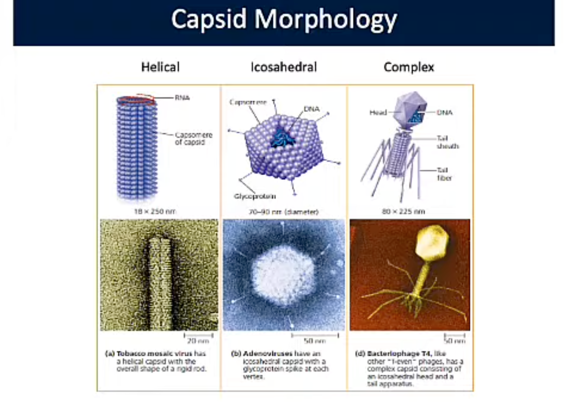
Capsid (Viral Structure)
Protein shell surrounding genome of virus particle.
3 Types: Helical, Icosahedral, and Complex
Capsomere (Viral Structure)
Individual protein subunit of capsid
Enveloped virus (Viral Structure)
Virus that contains additional layers around nucleocapsid
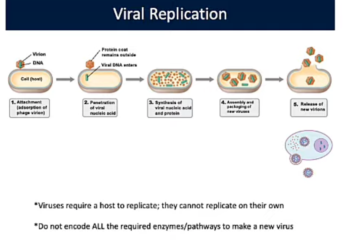
Viral Replication
Require a host to replicate; do not replicate on their own. Do not encode ALL required enzymes/pathways to make a new virus
Attachment; absorption of phage virion
Penetration of viral nucleic acid
Synthesis of viral nucleic acid/protein
Assembly and packaging of new virus
Release of new virions
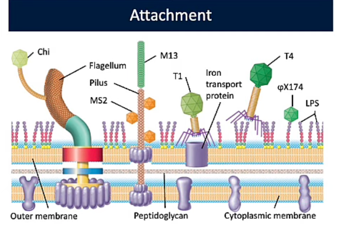
Attachment (Viral Replication PT1)
Attachment of virion (virus outside cell) to host cell. Receptor needed on surface of host and infecting virus (lock and key)
Receptors on host cell carry out normal function;Not made for Viral Attachment. Receptors = Proteins, carbs, glycoproteins, lipids, lipoproteins, complexes
T1 Bacteriophage: Iron uptake protein
T4 Bacteriophage: Polysaccharides on Outer Membrane
upside down y Bacteriophage: Maltose transporter
Influenza A Virus: Sialic Acid Residue
HIV: CCR5, CCR2, CXCR4 (T-cells)
No cell receptor = no binding = no infection
Mutation leads to loss of binding and infection
Reciprocal mutation enables binding of new mutated host receptor
Multicellular organisms: Infect tissues/cells where receptor Is present; not whole body
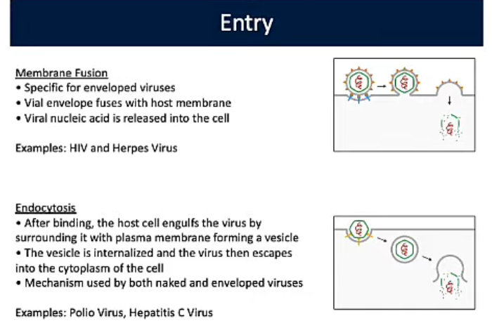
Membrane Fusion (Viral Replication PT2)
Specific for enveloped viruses, Vial envelope fuses with host membrane, Viral nucleic acid is released into the cell
Ex. HIV and Herpes

Endocytosis (Viral Replication PT2)
After binding, the host cell engulfs the virus by surrounding it with plasma membrane forming a vesicle
Vesicle internalized and virus escapes into cytoplasm of cell
Used by naked and enveloped virus
Ex. Polio Virus, Hep C Virus
Replication, Transcription, Translation (Viral Replication PT3)
Using the host to make all individual parts of a new virus
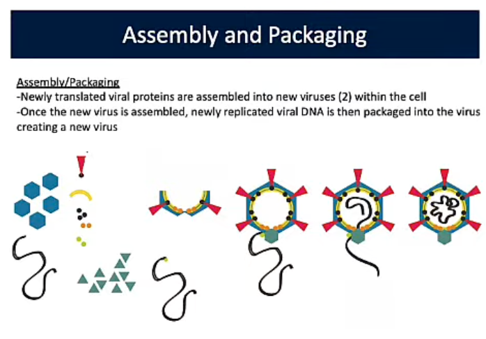
Assembly and Packaging (Viral Replication PT4)
Newly transcribed viral proteins assembled into new viruses (2) in cell. Newly replicated viral DNA is packaged into the virus creating a new one
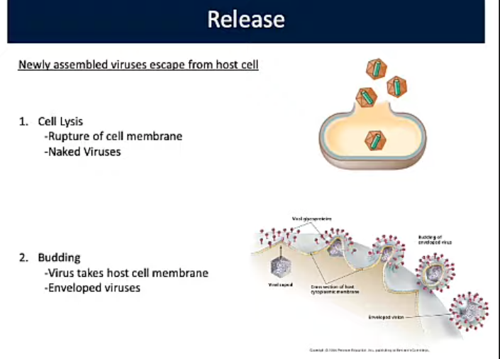
Release (Viral Replication PT5)
Newly assembled viruses escaped from host cell through:
Cell Lysis: Rupture of cell membrane/Naked Viruses
Budding: Virus takes host cell membrane/Enveloped Virus
Cell does not immediately die but soon does
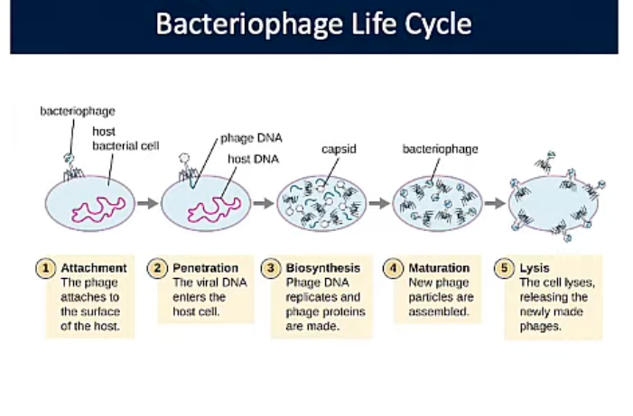
Bacteriophages Life Cycle
Viruses that infect bacteria
Attachment: Phage attaches to surface of host
Penetration: Viral DNA enters cell
Biosynthesis: Phage DNA replicates and phage proteins are made
Maturation: New phage particles are made
Lysis: Cell lyses, releasing newly made phages
Entry/Attachment (Bacteriophages Life Cycle)
Virus binds to surface of host cell and injects genetic material. Virus does not enter cell. Only viral nucleic acid enters cell
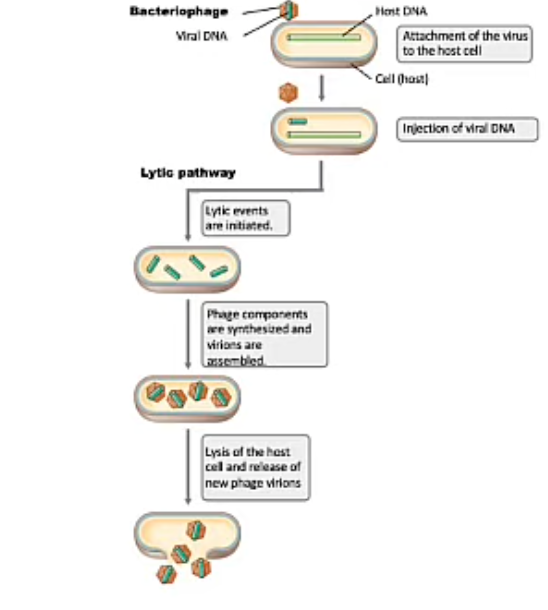
Lytic Bacteriophages (Bacteriophages Life Cycle)
Lytic Cycle: Viral DNA not integrated into host chromosome. DNA transfer by new virus infecting other host cells. Results in to lysis
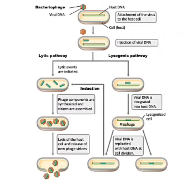
Lysogenic Bacteriophages (Bacteriophages Life Cycle)
DNA integrated into host chromosome (prophage). Transfer DNA by replication of host chromosome. No new phages made. Does not result in cell death.
Viruses become activated, start replicating and make new phages leading to lysis
Prophage: Bacteriophage (genome) that has recombined into bacterial host’s chromosome
Prophage (Lysogenic Bacteriophages Bacteriophages Life Cycle)
Bacteriophage (genome) that has recombined into bacterial host’s chromosome
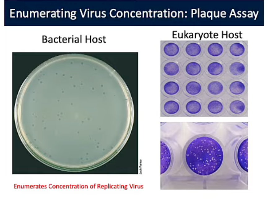
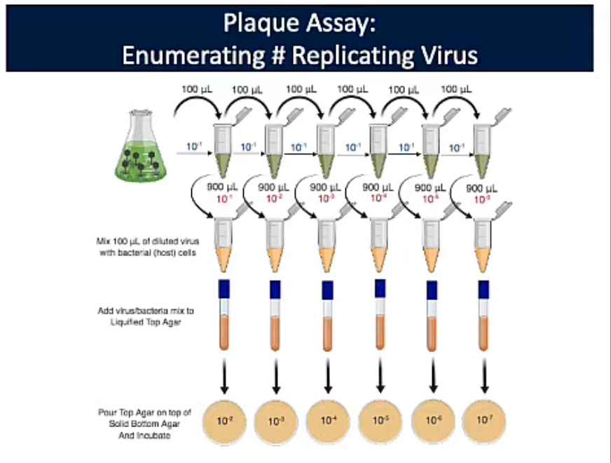
Plaque Assay
Enumerates Concentration of Replicating Viruses
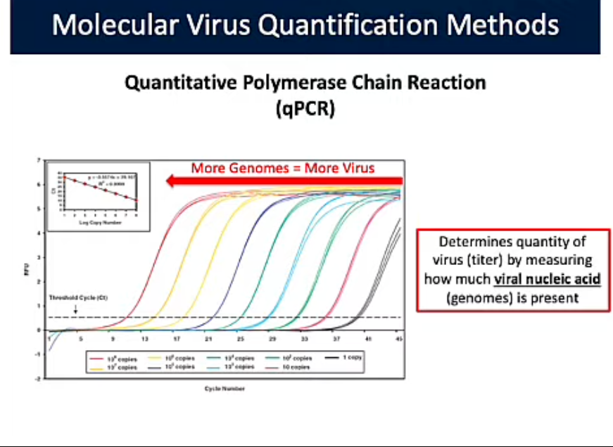
Quantitive Polymearse Chain Reaction (qPRC) (Molecular Quantification Methods)
Determines quantity of virus (titer) by measuring how much viral nucleic acid (genomes) are present. More phage you start with, the faster you can get to the threshold.
Threshold = # needed to show the signal is real; making that you can statistically/accurately quantify the # of virus genes present.
More Genomes = more virus
Red met/pases threshold at 10 cycles
Grey met/passed threshold 39 cycles
Modern molecular method for detecting a virus in a sample via viral DNA
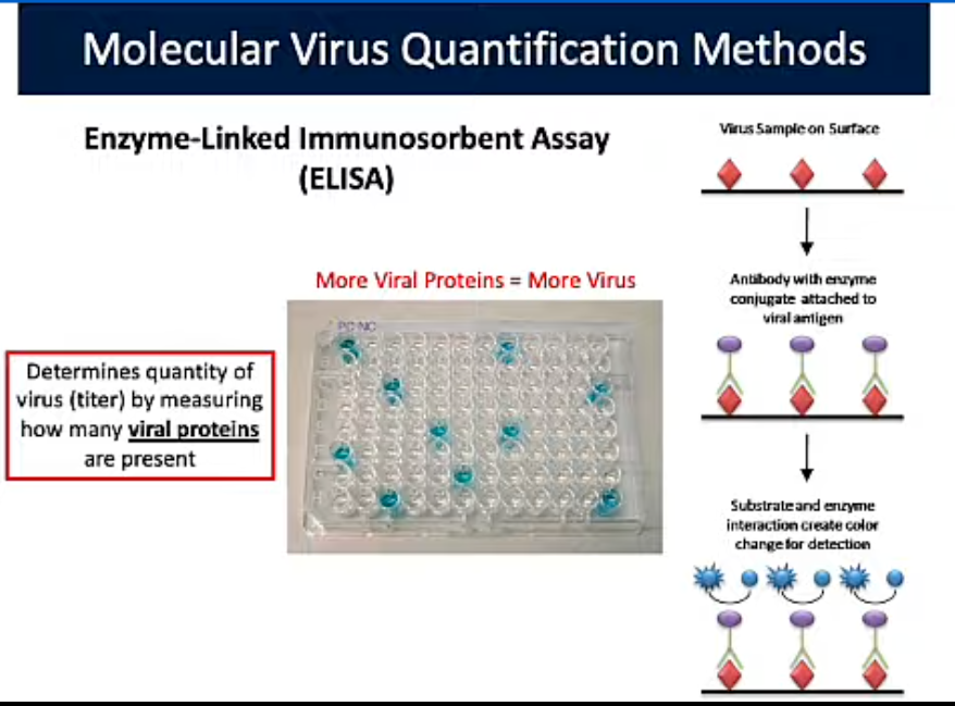
Enzyme-Linked Immunosorbent Assay (ELISA) (Molecular Quantification Methods)
Determines quantity of virus (titer by measuring how many viral proteins are present
More Viral Proteins = More Virus
Nucleic Acid PCR Test (COVID-19 Testing)
Test for active infection or those who are current asymptomatic carriers.
qRT-PCR based test
Detects SARS-CoV-2 RNA
Antigen Test (COVID-19 Testing)
Tests for current infection. detects presences of SARS-CoV-2 proteins. Not as sensitive as PCR test. Could result in false negative. Rapid/At home tests
Antibody Test
Detects previous exposure
HIV Infection
Infects CD4 T-Cells
Binds to CD4 and CCR5 receptors on host cell by glycoproteins on the virus
T-Cells are important cell of immune system allowing for adaptive (memory immunity)
Infection with HIV leads to death of CD4 T-cells
Loss of CD4 T-Cells results in loss of immune function (AIDS) leading to increased risk of infection or development of certain cancers
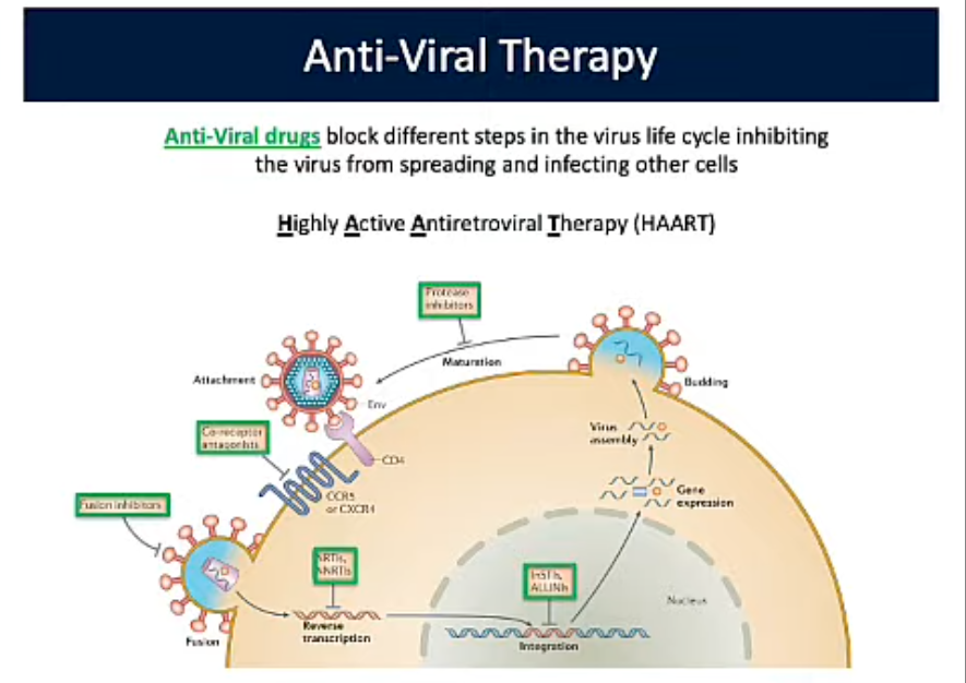
Anti-viral Therapy
Block different steps in the virus life cycle inhibiting the virus from spreading and infecting other cells
Highly Active Antiretroviral Therapy (HAART)
Combination Therapies
3-5 different antiviral drugs that help lower spread of drug resistant mutant virus. HIV has high mutation rate. If only using 1 odds of resistance are high. Using multiple resistance becomes very low
PrEP: Pre-Exposure Prophylaxis
Combination Anti-Viral drugs that work to inhibit parts of the HIV life cycle
Influenza Virus
Influenzavirus A, B, and C infect humans. Segmented Viral Genome. Composed of 8 pieces of linear RNA. 11 Protein encoding genes.
Neuraminidase
Hemagglutinin (HA)
Found on surface of virus.
Binds to cells; recognizes silica acid on cells. Cells of upper respiratory trace
Aids in entry of viral genome into cell. Fusion of host membrane with viral membrane
18 different subtypes (influenza A).
Neuramindase (NA)
Enzyme found on surface of virus.
Facilitates virus release from host cell. Cleaves silica acid; glycoprotein complexes
Cleaves neuraminic acid from respiratory tract muffins. Allows movement of virus to the target
Has 9 different subtypes (Influenza A)
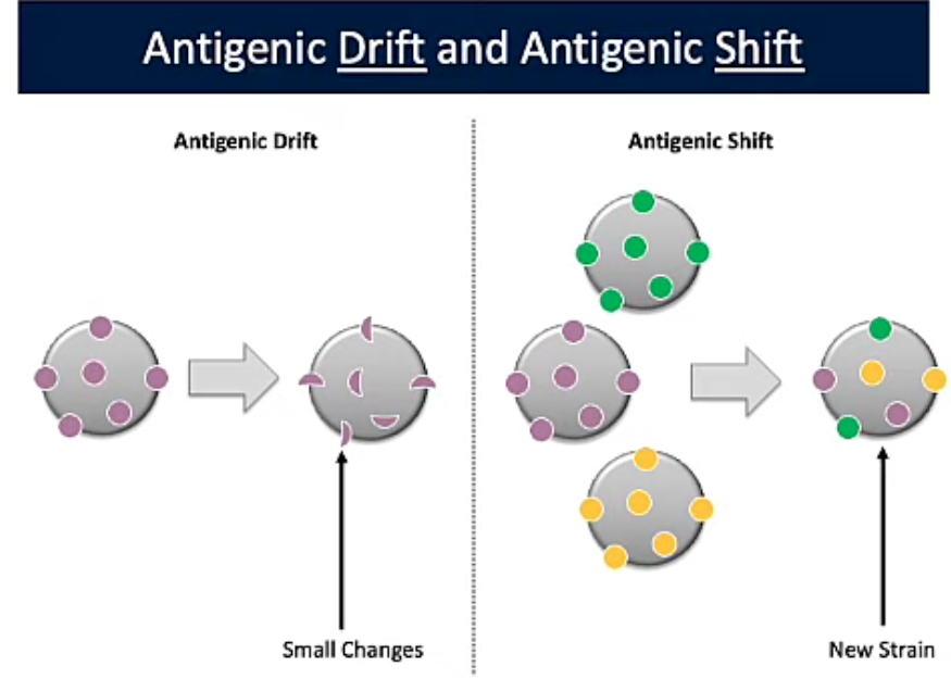
Reassortment (Antigenic Shift)
When 2 or more segmented RNA viruses infect a single cell, parts of each genome can be packaged into a single capsid, creating a new virus.
Can result in pandemics
Strain Variation and Vaccine Efficacy (Antigenic Drift)
If there is a mutation circulating strains, the immunity generated from the vaccine may not be as protective
Immunology
Study of physiological mechanisms that humans and other animals use to defend against invaders:
Bacteria, Viruses, Fungi, Parasites, Toxins
Immunity
Ability to ward off disease
Susceptibility
Lack of resistance to a disease
Physical Barriers
Skin: Dermis: Inner portion made of connective tisue
Epidermis: Outer portion made of tightly packed epithelial cells containing keratin, a protective protein
Shedding and dryness of skin works to inhibit microbial growth
Mucous Membranes: Epithelial layer that lines the gastrointestinal respiratory, and genitourinary tracts
Mucous: Viscous glycoproteins that trap microbes and prevent tracts from drying out
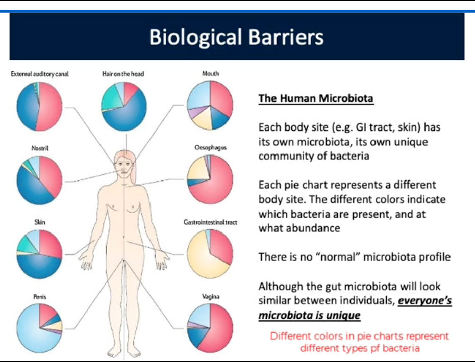
Biological Barriers
Human Microbiota: Microbes that live in and on our body that help prevent the entablement and growth of pathogenic microorganisms
Each body site has own microbiota/community of bacteria
Everyones’s microbiota is unique; but gut looks similar
Different colors in pie charts show different types of bacteria
Protects against invading pathogens
Antibiotics (Colonization Resistance)
Alter microbial populations allowing for pathogens to colonize/cause disease
Innate Immunity (Cellular Barriers)
Defenses against any pathogen; rapid, present at birth
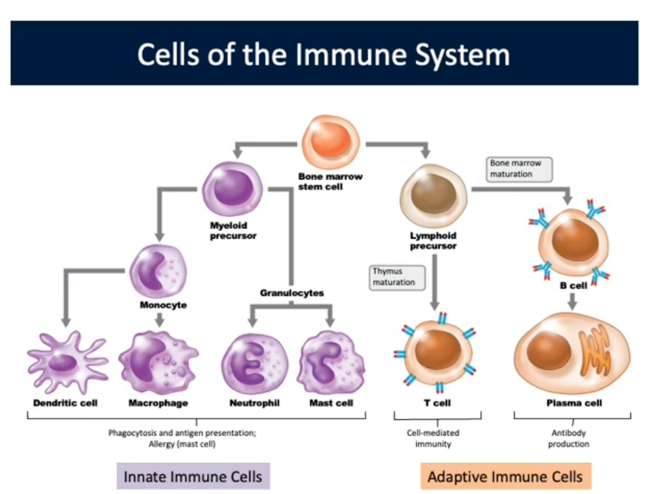
Adaptive Immunity (Cellular Barriers)
Immunity or resistance to a specific pathogen; slower to respond, has memory component
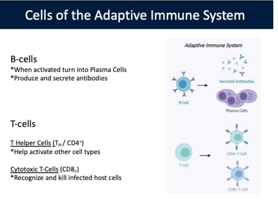
Specialized lymphocytes: T and B cells. Antibodies
B cells: When activated turn into Plasma Cells. Produce/secrete antibodies. In Lymph nodes, antigens activate B-cells into Plasma cells —> antibody producing factories. B-Cells secrete antibodies that disseminate through the body, including to the original site of infection to help control the infection
T cells: In Lymph nodes, Dendritic cells active T-Cells. Helper T-cells play crucial role in activating other immune cells. Activated helper T-cells migrate back to site of infection to recruit more Cytotoxic T-Cells and macrophages
T Helper Cells: Help Activate other cell types
Cytotoxic T-Cells: Recognize and kill infected hosts
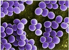
Staphylococcus aureus Skin Infection
Caused by Damage to skin barrier leading to Extracellular infection. Do not invade host cells. Are outside the host cell but the bacteria are still causing damage and disrupting host cell funct

Macrophages (First Reaction at Site of Infection)
First responders, circulating through the body looking for invaders. Recognize and phagocytose S. aureus to kill it. Submit chemical signals to recruit Neutrophiles to site of infection

Neutrophils (First Reaction at Site of Infection)
Phagocytose (ingest) S. aureus to kill. Release compounds to prevent pathogens from growing. Release compounds to recruit more neutrophils to site of infection
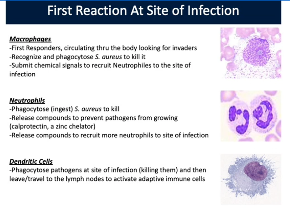
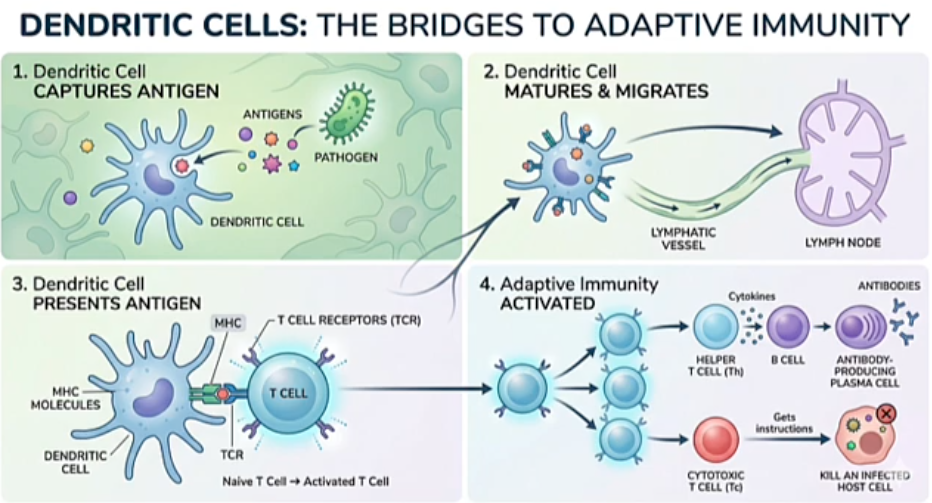
Dendritic Cells (First Reaction at Site of Infection)
Phagocytose pathogens at site of infection (killing them) and then travel to lymph nodes to activate adaptive immune cells
Su
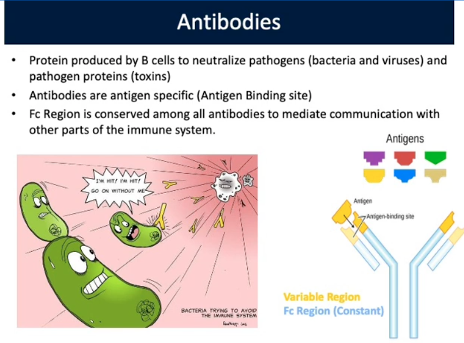
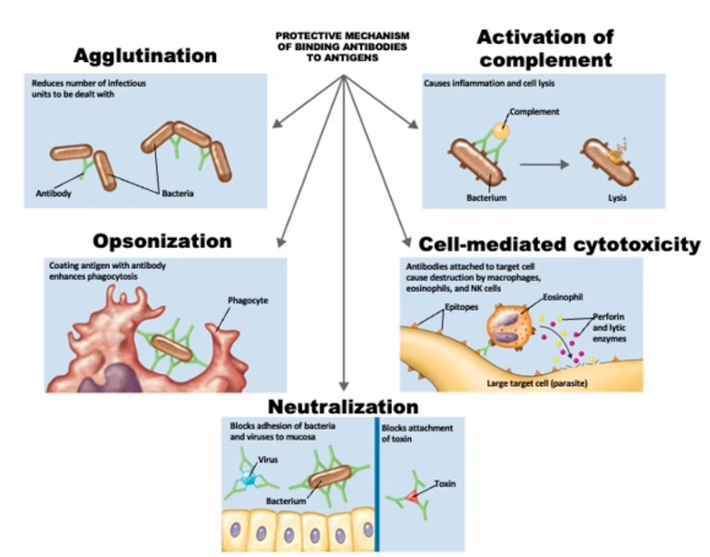
Antibodies
Protein produced by B-cells to neutralize pathogens (bacteria and viruses) and pathogen proteins (toxins). Neutralize visions before they bind and infect new cells
Antigen specific (Antigen Binding site)
Fc Region is conserved among all antibodies to mediate communication with other parts of immune system
Influenza Lung Infection
Influenza virus needs a host to replicate and survive. Intracellular (inside host cell)
Antibodies are not able to penetrate into host cell
Cytotoxic T-cells are deployed to kill the infected cell so that it does not create more new viruses that spread and infect more neighboring cells
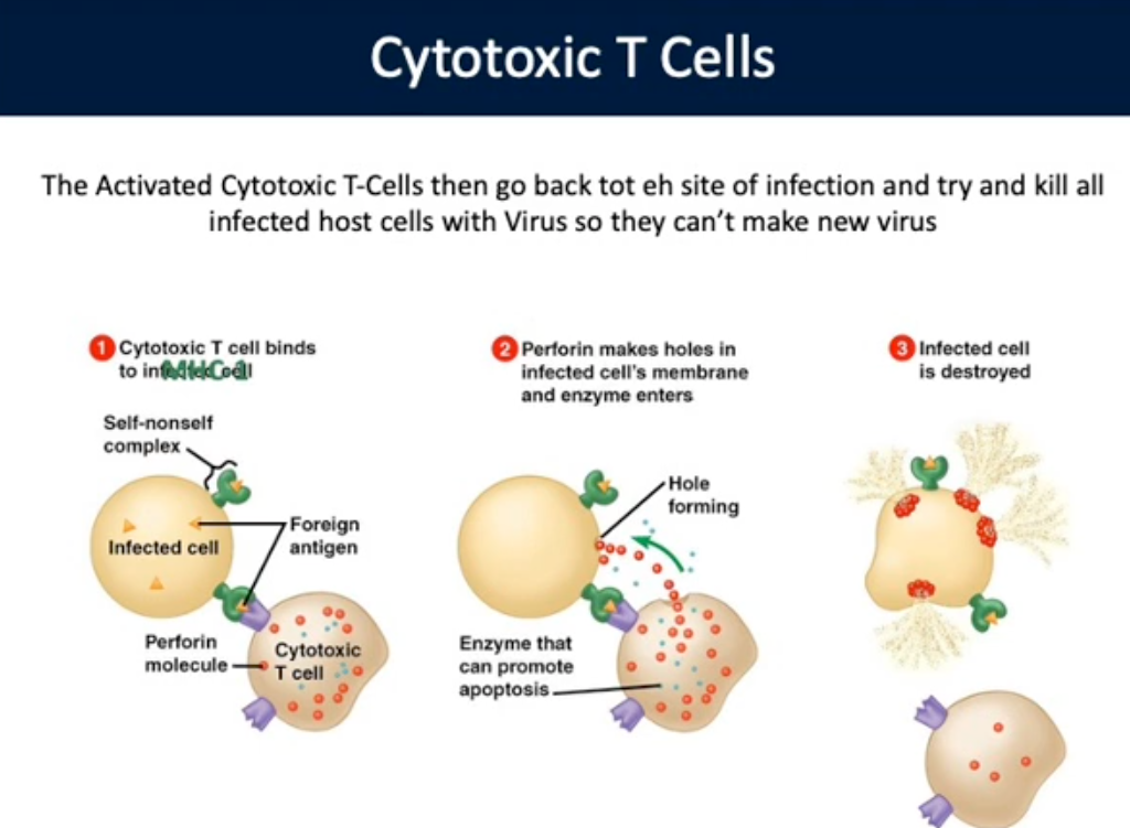
Cytotoxic T-Cells
Activated Cytotoxic T-Cells go back to site of infection and try and kill all infected host cells with virus so they can’t make new virus. Important for killing infected cells.
Kill cancerous cells similar to viral infected cell
Kills cancerous cell before it proliferates
Cancer and The Immune System
Main goal is to maintain homeostasis. An infection and cancer perturbs homeostasis. When a cell acquires a mutation that causes it to be cancerous it will present antigens on its surface stating that it’s sick/damaged and needs to be destroyed before proliferating out of control, causing cancer
Immunotherapy
CAR: Chimeric Antigen Receptors. Patient blood drawn to isolate own T-cells. T-cells are engineered to express antigens that mimic cancer cells. Engineered T-cells are transplanted back into patient to fight cancer
Antigen
Molecule capable of inducing an immune response
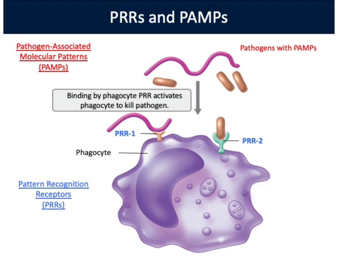
Recognize Foreign Pathogens
Host Immune Cell: Pattern Recognition Receptors (PRRs)
Membrane bound proteins
Pathogen: Pathogen associated molecular patterns (PAMPs)
Molecules associated with pathogen
Epitope
Specific molecule structure on an antigen that an immune cell receptor binds and responds to. Can be multiple epitopes per antigen
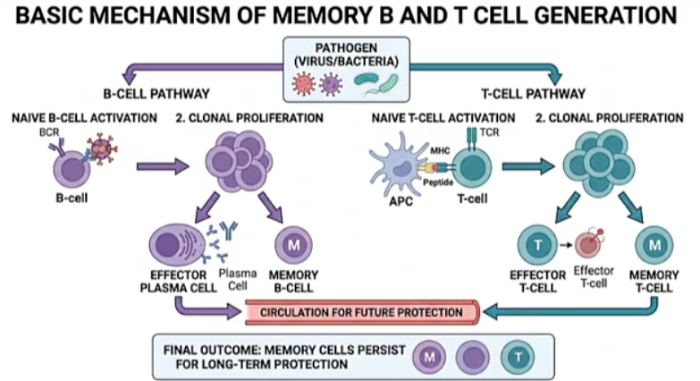
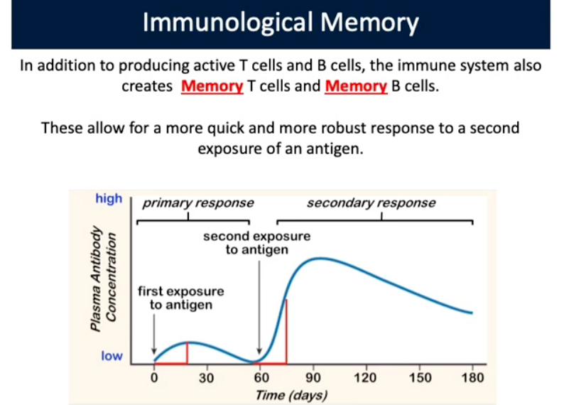
Immunological Memory
Produce active T-cells and B-cells, the immune system also creates memory T and B cells. Allow for quick/robust response to a second exposure of an antigen
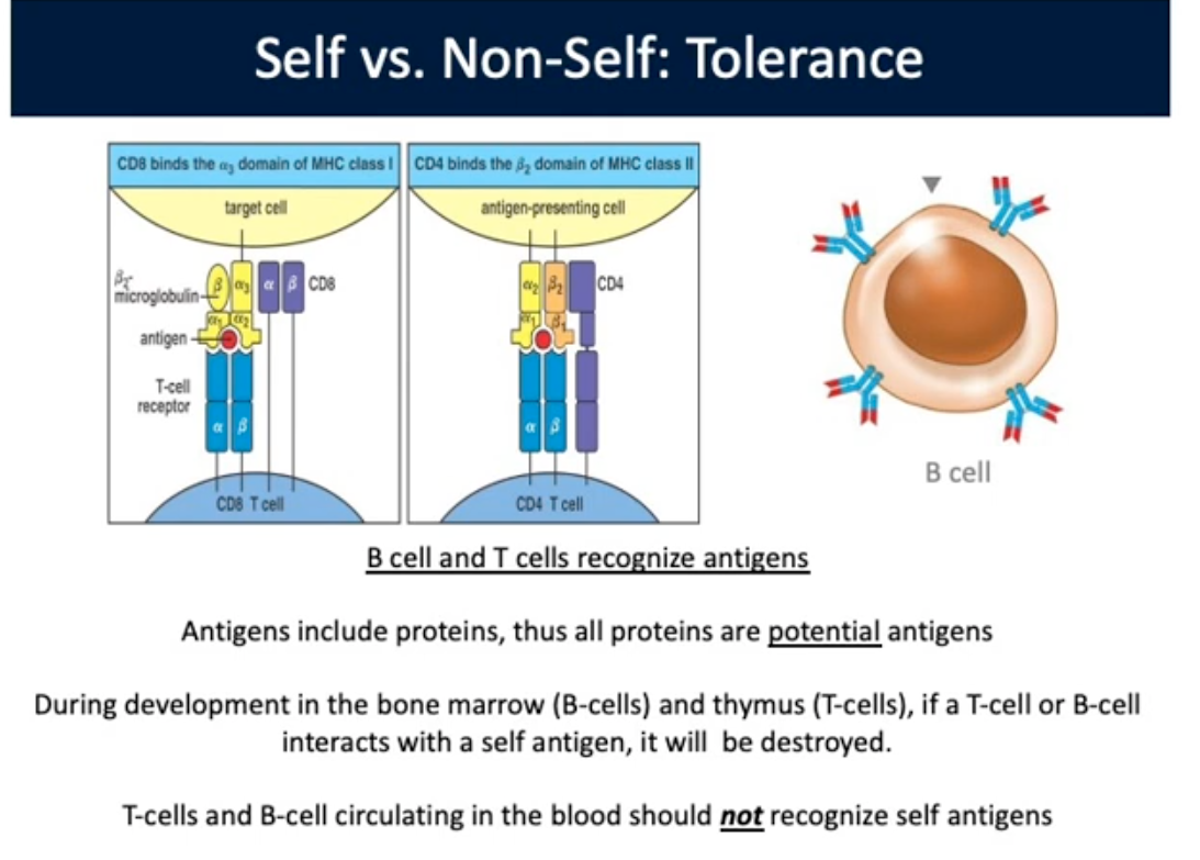
Self vs. Non-Self: Tolerance
Antigens include proteins, thus all proteins are potential antigens. During bone marrow developments, B-cells and thymus (T-cells), if a T-cell or B-cell interacts with self antigen, it will be destroyed. T and B cells circulate in blood should not recognize self antigens
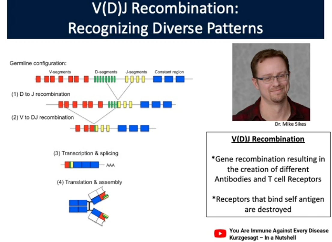
V(D)J Recombination
Gene recombination resulting in the creation of different Antibodies and T-cells Receptors. Receptors that bind self antigen are destroyed

Natural Immunity
Natural Active Immunity: Acquiring/clearing an infection that starts an adaptive immune response. Creation of memory B-cells and T-cells
Natural Passive Immunity: Maternal antibody transfer across the placenta, or in breast milk
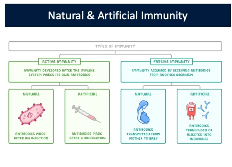
Artificial Immunity:
Artificial Active Immunity: Exposure to controlled dose of an antigen to induce activation of immune system (vaccination). Does not result in an active infection, but memory cells are generated
Active Passive Immunity: Administration of antiserum (antibodies) derived from an immune individual to treat infection. No memory cells produced
Antigen
Substance that provokes an adaptive immune response