MCB 2210 L23: Actin Binding Proteins + Microtubule Associated Proteins
 Call Kai
 Learn
Learn Practice Test
 Spaced Repetition
 Match
Match Flashcards
 Knowt Play
1/19

There's no tags or description
Looks like no tags are added yet.
Name | Mastery | Learn | Test | Matching | Spaced | Call with Kai |
|---|
No analytics yet
Send a link to your students to track their progress
20 Terms
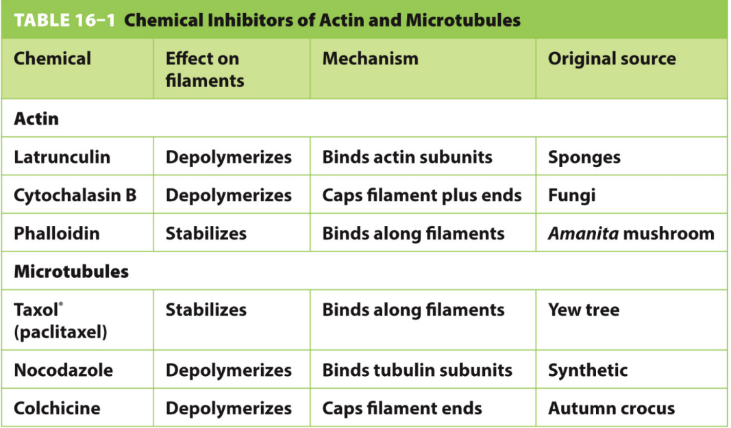
Define the 3 actin drugs and 3 microtubule drugs used to study polymerization
Actin
Phalloidin → stabilizes actin
Cytochalasin B → depolymerizes (capping)
Latrunculin → depolymerizes (sequestering)
Microtubules
Taxol → stabilizes MTs
Colchicine → depolymerizes MTs (capping)
Nocodazole → depolymerizes MTs (binding)
What are 4 factors that actin binding proteins (ABPs) control?
Actin assembly
Actin disassembly
Actin organization
Actin movement
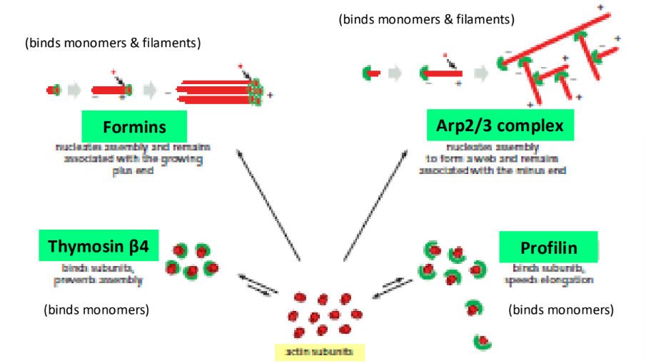
What are 2 types of actin binding proteins that bind to both monomers & filaments and nucleate actin?
What are 2 types of actin binding proteins that bind to monomers and control actin polymerization?
Formins = nucleate unbranched actin
Arp2/3 complex = nucleates branched actin w/ WASP protein
Profilin = actin polymerization (Exchange factor, ADP-actin → ATP-actin)
Thymosin β4 = sequesters protein → decrease actin polymerization → depolymerization
What are 2 types of actin severing proteins?
What 2 types of actin capping protein?
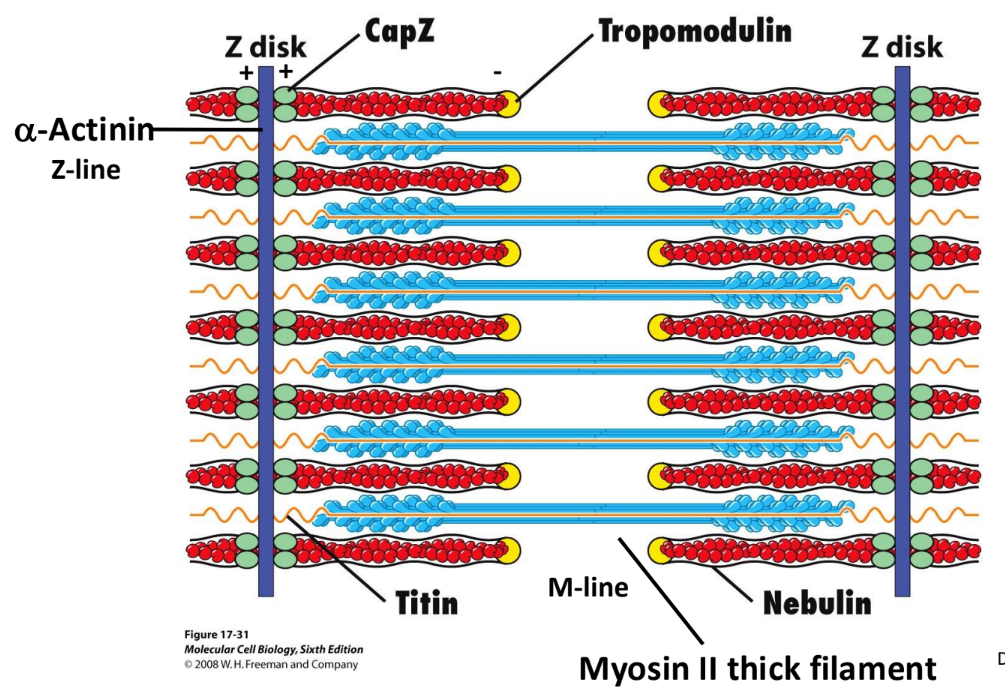
What is a common actin stabilizing protein in muscle cells?
Cofilin = sever (-) ends of actin
Gelsolin = sever actin filaments → cap (+) ends actin
Increase [Ca2+] activates gelsolin
CapZ = caps (+) ends of actin
Tropomodulin = caps (-) ends of actin
Tropomyosin = stabilizes actin filament in muscle cells (actin = contractile muscle)
Binds to sides → less likely G-actin monomer depolymerizes
![<ol><li><p><strong>Cofilin</strong> = sever (-) ends of actin</p></li><li><p><strong>Gelsolin</strong> = sever actin filaments → cap (+) ends actin</p><ol><li><p>Increase [Ca<sup>2+</sup>] activates gelsolin</p></li></ol></li></ol><p></p><ol><li><p><strong>CapZ</strong> = caps (+) ends of actin</p></li><li><p><strong>Tropomodulin</strong> = caps (-) ends of actin</p></li></ol><p></p><ol><li><p><strong>Tropomyosin</strong> = stabilizes actin filament in muscle cells (actin = contractile muscle)</p><ol><li><p>Binds to sides → less likely G-actin monomer depolymerizes</p></li></ol></li></ol><p></p>](https://assets.knowt.com/user-attachments/c453fba9-edef-408a-8e0f-51e4f5f88cdd.png)
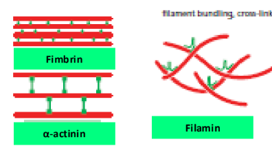
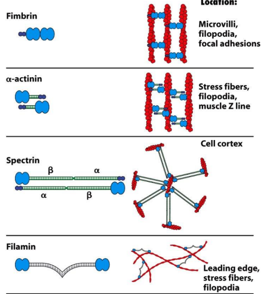
What are 3 types of actin cross-linking proteins?
Fimbrin = rigid, parallel, tightly bundled actin (protein = short)
α-actinin = rigid, parallel, not as tightly bundled actin (protein = longer)
Filamin = flexible, gel-like actin networks
What are the 2 types of actin membrane-linker proteins?
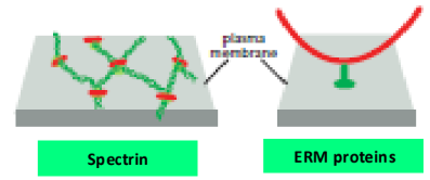
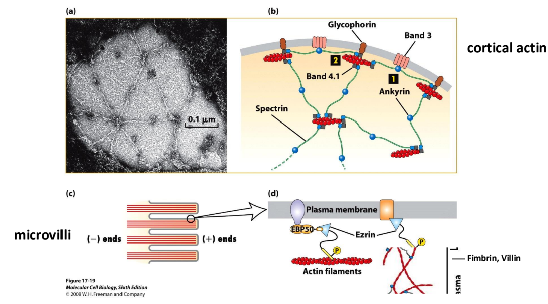
Spectrin = link actin to membrane
ERM Family proteins = link actin to membrane
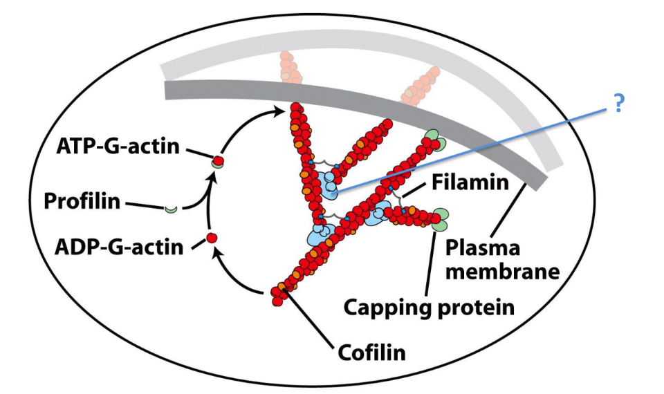
Describe how ABPs control assembly, disassembly, & organization of actin filaments
Help push membrane outwards
Arp2/3 Complex + WASP protein → branched actin
Filamin = stabilizes actin branchesx
CapZ = actin capping protein = caps (+) end of actin
Cofilin = actin severing protein → ADP-G-actin
Profilin = actin binding protein → ADP-G-actin → ATP-G-actin
What are cross-linking proteins + 2 types of longer, more flexible spacers, and 3 types of shorter, more rigid spacers?
Cross-linking proteins = 2 actin-binding domains separated by spacer domain
Longer, more flexible spacers → gel-like networks
Filamin
Spectrin
Shorter, more rigid spacers → tight bundles
Fimbrin
Villin
Fascin
α-actinin = crosslink filaments → parallel/antiparallel bundles → contraction
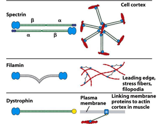
What are membrane-actin linker proteins and what are 3 types?
Membrane-actin linker proteins = attach cytoskeleton → plasma membrane
ERM proteins → link b/w F-actin + integral membrane proteins
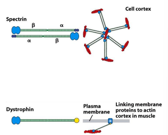
Spectrin → connect actin + membrane proteins
Dystrophin → connect actin + muscle cell membrane
Describe arrays of actin bundles that α-actinin can form
Form antiparallel arrays in contractile structures (stress fibers or sarcomeres)
Define cell cortex and primary actin binding protein associated with it.
What actin binding protein links actin to membrane of microvilli?
Cell cortex = actin-rich layer beneath plasma membrane
Spectrin = closely associated w/ cell cortex
Ezrin = links actin to microvilli membrane
What genetic mutation can lead to easily ruptured RBCs or hereditary spherocytic anemias?
What genetic mutations can lead to muscular dystrophy?
Spectrin-based skeleton of cell cortex = critical for RBC shape
Mutations in spectrin → easily ruptured RBCs
Dystrophin = actin cross-linking protein = link actin in muscle → membrane proteins
Mutations in dystrophin → muscle cell membrane weakens → ruptures
Define functions of 3 microtubule associated proteins. γ-TURC, +TIPS, Stathmin
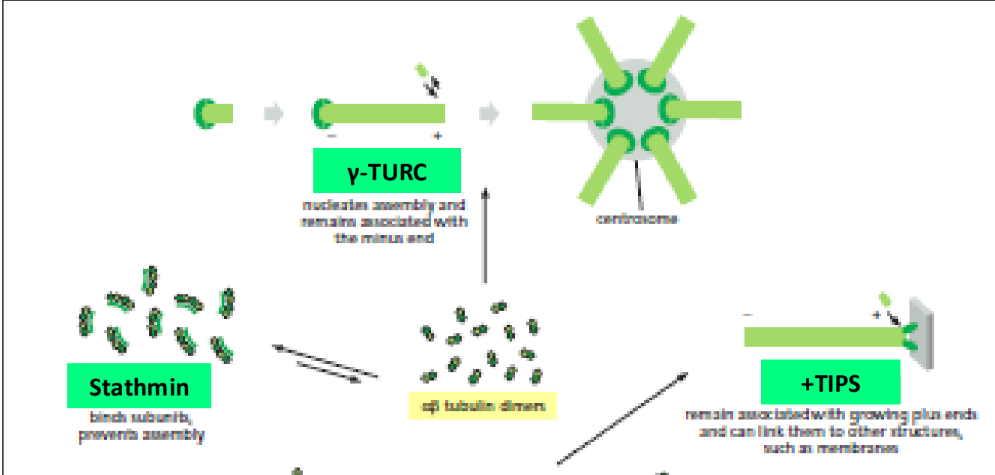
γ-TURC = nucleating protein @ centrosome, associated w/ (-) end
+TIPS = bind to GTP cap of MTs @ (+) end (Ex. EB1)
Can link to membrane
Fluorescently tagged to demonstrate MT (+) end dynamics
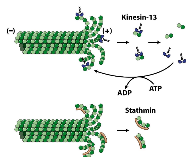
Stathmin = fragments of MT dimers
Bind subunits → prevent MT assembly
What are 2 types of microtubule associated proteins that depolymerize microtubules?
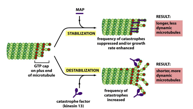
Kinesin-13 = MT motor protein that depolymerizes @ MT end
Force tubulin dimers → Δ conformation → catastrophe
Katanin = severing protein → depolymerizes @ MT middle
CHOPS → catastrophe @ segment ends
What are 3 types of microtubule associated proteins that stabilize MTs?
MAPs = stabilize MTs by binding along sides
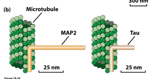
Tau = stabilize MTs by linking MTs → tightly bundled MTs
Taiwan = country on a MAP
MAP-2 = stabilize MTs by linking MTs → wider bundled MTs
What microtubule associated protein links microtubules to intermediate filaments?
Plectin
Pleakley = alien = IF MT (martians)
Describe mechanism of Kinesin-13 and Stathmin
Kinesin-13 = depolymerization @ MT end “13 = unlucky”
Strain protofilaments → curls
Peels away
DOES NOT INVOLVE GTP CAP HYDROLYSIS
Stathmin = disassembly from (+) end “Stalling”
GTP cap = GONE (rate of polymerization < rate of hydrolysis)
Bind when protofilaments = exposed
Take tubulin heterodimers out of circulation
Define polymer-binding MAPs + structure + 2 examples
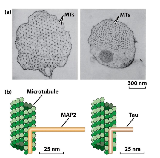
Polymer binding MAPs = stabilize MTs by binding to sides, enhance assembly (stabilize nuclei), organize MTs → bundles, mediate MT interactions w/ other proteins (Ex. Intermediate filaments + actin)
Polymer-binding MAPs = 2 major domains
MT binding domain = binds tubulin dimers together → stabilize MT
Projection domain = interact w/ MTs + other structures (Ex. intermediate filaments)
Tau + MAP2 = organize MTs in neuronal axons + dendrites
Spacing b/w MTs = greater for MAP2 than Tau
MAP2 (longer projection domain)
Note MAP2 + Tau = expressed in cells that do not normally form axons → long axon like structures are still induced
What do MAPs help do to microtubules?
Stabilize microtubules + enhance growth by suppressing frequency of catastrophes → enhance growth rate
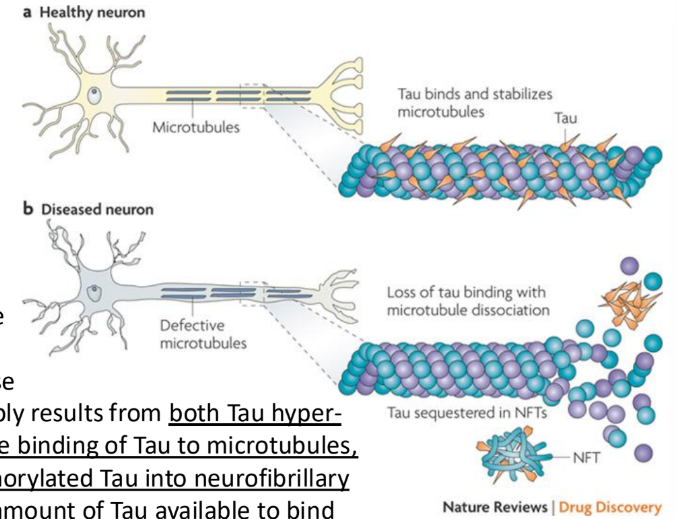
Which MAP is associated with Alzheimer’s disease? What are the is the two-step process that can explain this?
Tau = abundant in neurons + stabilizes MT
Tau hyperphosphorylation → reduces binding of Tau to MTs
Tau becomes “sticky” → accumulates + clumps
Sequestration of hyper-phosphorylated Tau into Neurofibrillary Tangles (NFTs) = reduces amount of Tau available to bind to MTs
Reduced Tau binding to MT → MT instability + reduced axonal support
Could contribute to Alzheimer’s disease