3.3.4.1 Mass Transport in animals
1/38
There's no tags or description
Looks like no tags are added yet.
Name | Mastery | Learn | Test | Matching | Spaced | Call with Kai |
|---|
No analytics yet
Send a link to your students to track their progress
39 Terms
Explain the role of red blood cells and haemoglobin in oxygen transport. (4 marks)
- Red blood cells contain many haemoglobin molecules and lack a nucleus, giving more space for haemoglobin and allowing a high surface area to volume ratio with a short diffusion pathway.
- At gas exchange surfaces, where the partial pressure of oxygen is high, haemoglobin binds to oxygen to form oxyhaemoglobin.
- Each haemoglobin molecule can carry four oxygen molecules, one per haem group.
- Near body cells where the partial pressure of oxygen is low, oxyhaemoglobin releases oxygen.
Draw a labelled diagram of the structure of haemglobin. (3 marks)
NOTE: You do not need to learn the alpha and beta chains - simply there to help you understand the structure.
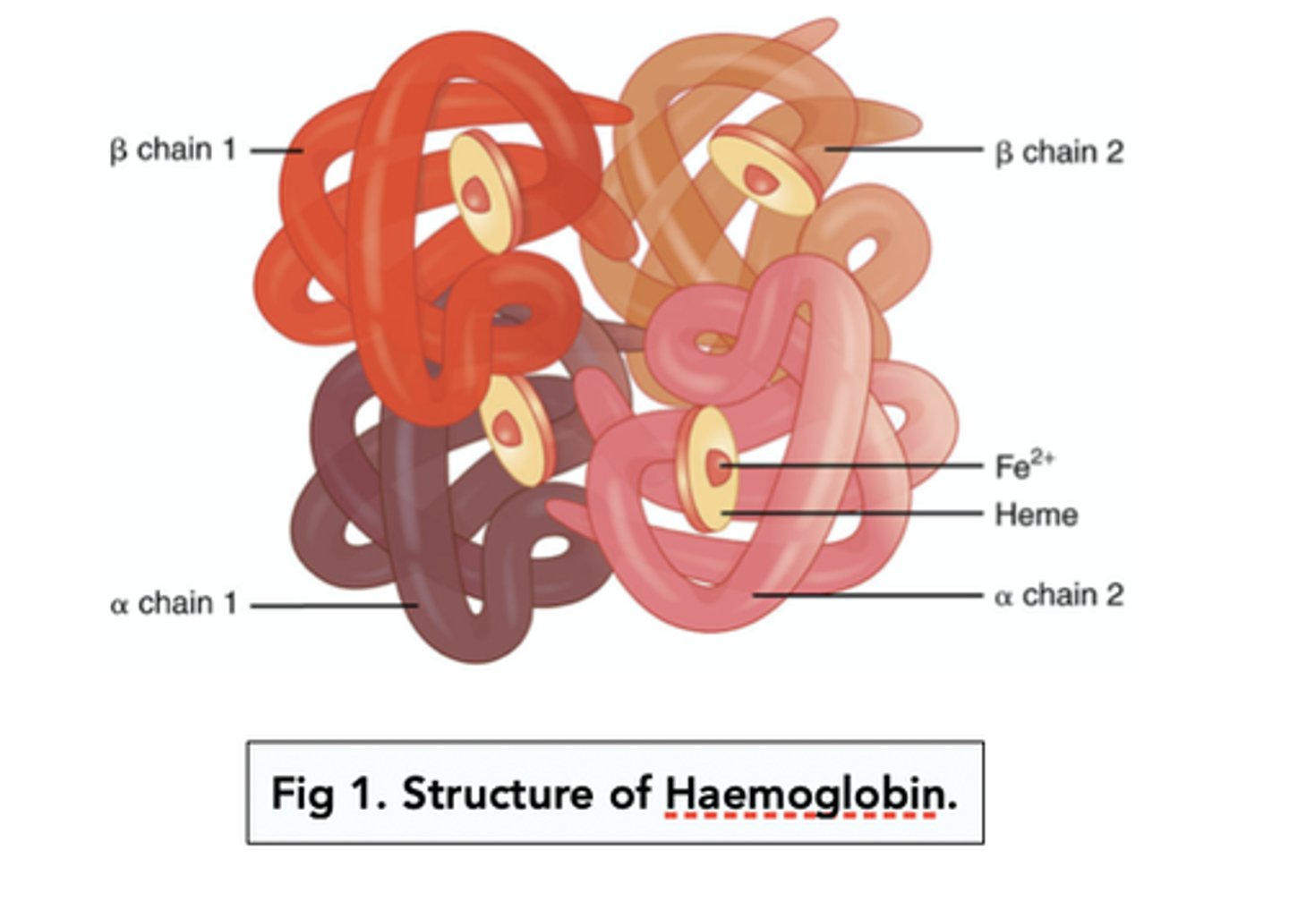
Describe the structure of haemoglobin. (3 marks)
- Haemoglobin is a protein with a quaternary structure.
- It is made from four polypeptide chains.
- Each chain contains a haem group with an iron ion (Fe²⁺).
Draw a labelled diagram of a general oxyhaemoglobin dissociation curve. (4 marks)
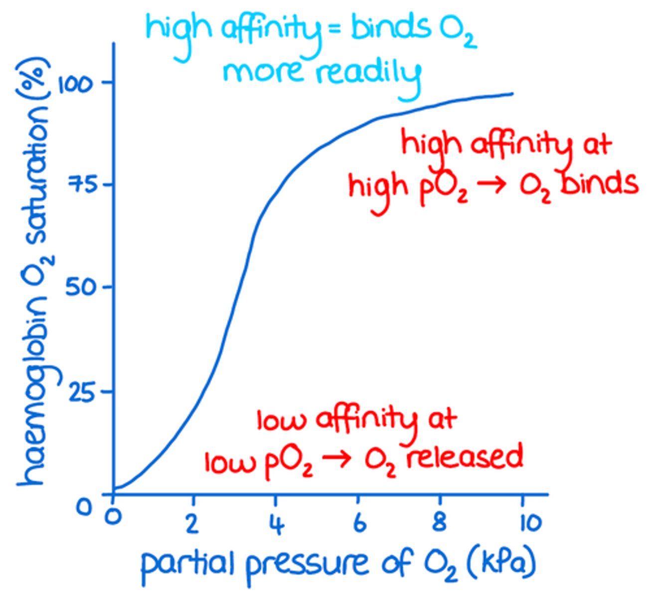
Describe the loading, transport, and unloading of oxygen in relation to the oxyhaemoglobin dissociation curve. (2 marks)
- In areas with low partial pressure of oxygen, such as respiring tissues, haemoglobin has a low affinity for oxygen, so it readily releases it and percentage saturation is low.
- In areas with high partial pressure of oxygen, such as at gas exchange surfaces, haemoglobin has a high affinity for oxygen, so it readily binds to it and percentage saturation is high.
Explain how the cooperative nature of oxygen binding produces an S-shaped oxyhaemoglobin dissociation curve. (2 marks)
- The first oxygen molecule binds to haemoglobin, altering its tertiary or quaternary structure.
- This exposes more haem group binding sites, making it easier for additional oxygen molecules to bind.
Describe evidence for the cooperative nature of oxygen binding. (2 marks)
- At low partial pressures of oxygen, the percentage saturation of haemoglobin increases only slightly as the first oxygen binds.
- At higher partial pressures of oxygen, the percentage saturation increases rapidly, showing that oxygen binds more easily after the first molecule has bound.
Define the Bohr effect. (2 marks)
- It is the influence of carbon dioxide concentration on the release of oxygen from oxyhaemoglobin.
- An increase in CO₂ shifts the oxyhaemoglobin dissociation curve to the right.
Draw and explain the effect of CO₂ concentration on the dissociation of oxyhaemoglobin. (6 marks)
- Increased CO₂ levels, for example during high respiration rates, lower blood pH, making it more acidic.
- This reduces haemoglobin's affinity for oxygen as its shape changes slightly.
- As a result, oxygen is unloaded more readily and more quickly to respiring tissues at a given partial pressure of oxygen.
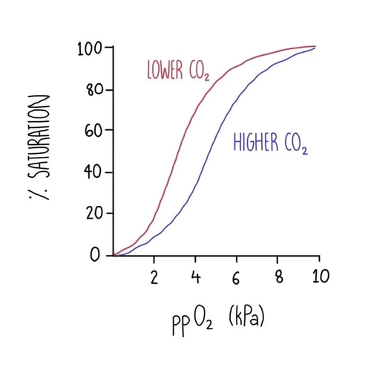
Describe evidence for the Bohr effect. (2 marks)
At the same partial pressure of oxygen, haemoglobin's percentage saturation is lower when CO₂ concentration is higher.
Explain the advantage of the Bohr effect during exercise. (2 marks)
- Increased oxygen unloading allows faster aerobic respiration.
- Reducing anaerobic respiration and increasing ATP production.
Explain why different types of haemoglobin have different oxygen transport properties. (3 marks)
- Different haemoglobin types have variations in their amino acid sequence.
- These changes alter the protein's tertiary or quaternary structure.
- This affects the protein's affinity for oxygen.
Explain how organisms can be adapted to their environments by having different types of haemoglobin. (4 marks)
- If the oxyhaemoglobin dissociation curve shifts to the left, haemoglobin has a higher oxygen affinity.
- Allowing more oxygen to bind at lower partial pressures, which is useful in low oxygen habitats (e.g. high altitudes, underground, or in foetuses).
- If the curve shifts to the right, haemoglobin has a lower oxygen affinity.
- Causing oxygen to unload more readily at respiring tissues, which is useful in animals with high metabolic rates or activity levels.
Describe the general pattern of blood circulation in a mammal. (2 marks)
- In the right side of the heart, deoxygenated blood is pumped to the lungs and returns oxygenated to the left side.
- In the left side of the heart, oxygenated blood is pumped to the rest of the body and returns deoxygenated to the right side.
State the importance of a double circulatory system. (3 marks)
- Prevents mixing of oxygenated and deoxygenated blood, ensuring blood reaching the body is fully oxygenated for aerobic respiration.
- Allows blood to be pumped to the body at a higher pressure after leaving the lungs.
- This enables quicker and more efficient delivery and removal of substances to and from body cells.
Draw a labelled diagram of the pattern of the double blood circulation in a mammals. (8 marks)
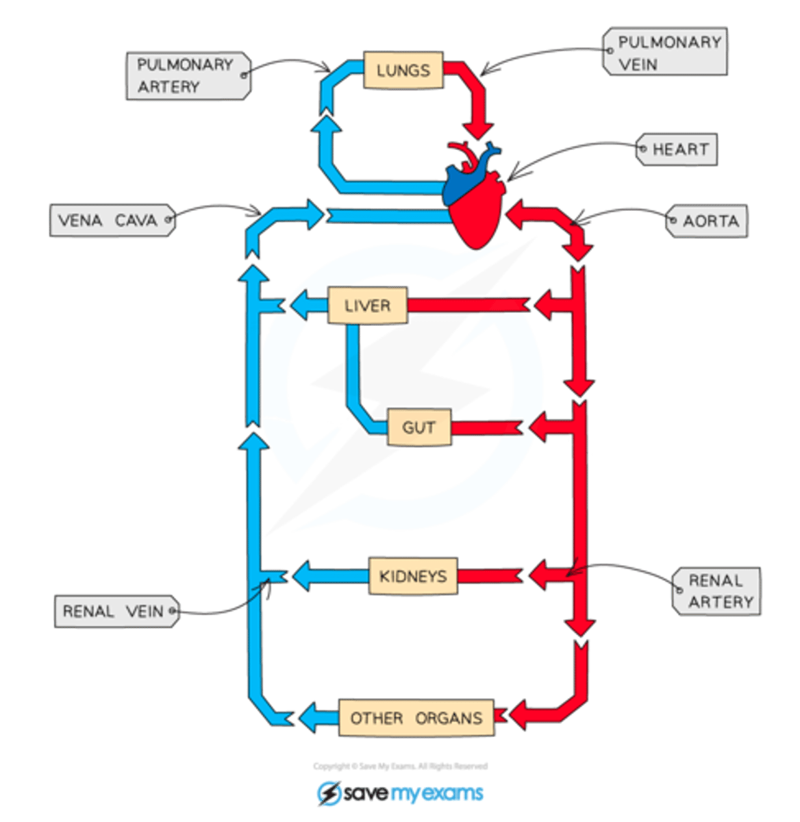
State the blood vessels entering and leaving the heart and lungs. (4 marks)
- Vena cava which carries deoxygenated blood from body tissues to the heart.
- Pulmonary artery which carries deoxygenated blood from the heart to the lungs.
- Pulmonary vein which carries oxygenated blood from the lungs to the heart.
- Aorta which carries oxygenated blood from the heart to body tissues.
State the blood vessels entering and leaving the kidneys. (2 marks)
- Renal arteries which deliver oxygenated blood to the kidneys.
- Renal veins which return deoxygenated blood from the kidneys to the vena cava.
State the blood vessels that carry oxygenated blood to the heart muscle. (2 marks)
- Coronary arteries which branch from the aorta and run over the surface of the heart.
- To supply oxygenated blood to the heart muscle.
Draw a labelled diagram to show the gross structure of the human heart. (6 marks)
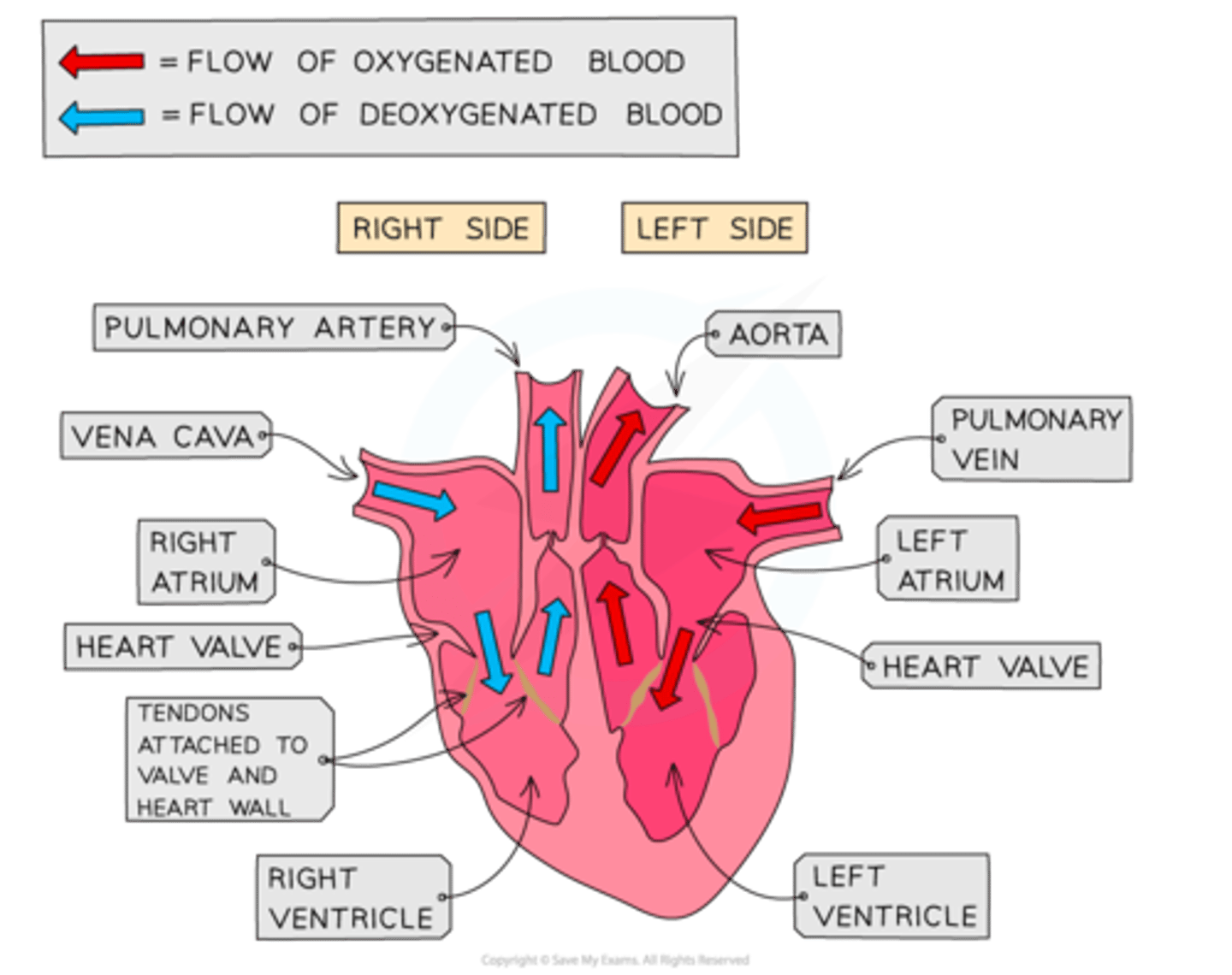
Explain why the wall of the left ventricle is thicker than the right. (2 marks)
- The left ventricle has more muscle to contract with greater strength.
- This allows it to generate higher pressure to pump blood throughout the whole body.
Define the cardiac cycle and state how heart rate can be calculated from cardiac cycle data. (2 marks)
- The series of events that occur during a single heart beat.
- Heart rate (beats per minute) = 60 ÷ length of one cardiac cycle in seconds.
Draw a labelled diagram of the cardiac cycle. (6 marks)
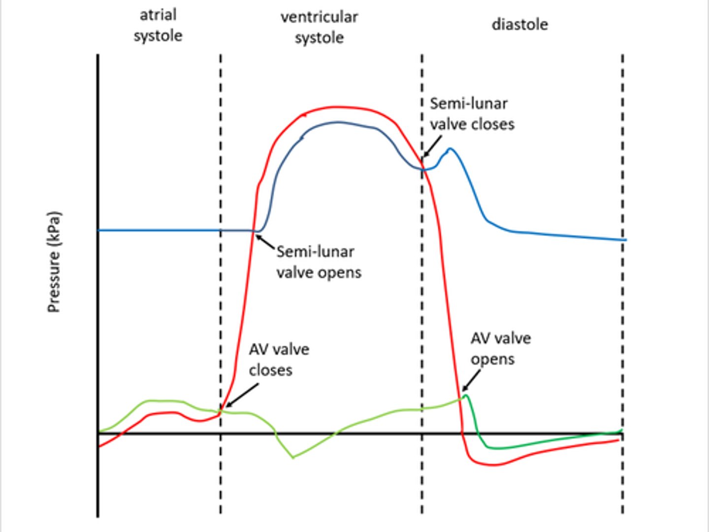
Explain the changes in pressure, volume, and valve positions during atrial systole. (4 marks)
- The atria contract, which decreases their volume and increases their pressure.
- Atrioventricular valves open when atrial pressure is greater than ventricular pressure.
- Semilunar valves remain shut because pressure in the arteries is higher than in the ventricles.
- Blood flows from the atria into the ventricles.
Explain the changes in pressure, volume, and valve positions during ventricular systole. (4 marks)
- The ventricles contract, causing their volume to decrease and pressure to rise.
- Atrioventricular valves close when ventricular pressure exceeds atrial pressure.
- Semilunar valves open when ventricular pressure exceeds arterial pressure.
- Blood is forced out of the heart into the arteries.
Explain the changes in pressure, volume, and valve positions during diastole. (2 marks)
- The atria and ventricles relax, increasing volume and reducing pressure.
- Semilunar valves close when arterial pressure is greater than ventricular pressure.
- Atrioventricular valves open when atrial pressure exceeds ventricular pressure.
- Blood enters the atria from veins and passes passively into the ventricles.
Explain how valve positions can be determined from pressure or volume graphs. (4 marks)
- Semilunar valves are closed when arterial pressure is higher than ventricular pressure, preventing backflow from arteries to ventricles.
- Semilunar valves open when ventricular pressure is greater than arterial pressure, allowing blood into arteries.
- Atrioventricular valves are closed when ventricular pressure exceeds atrial pressure, preventing backflow into the atria.
- Atrioventricular valves open when atrial pressure is greater than ventricular pressure, allowing blood to move into ventricles.
Describe the equation for cardiac output. (2 marks)
- Cardiac output = stroke volume × heart rate.
- Stroke volume is the volume of blood pumped per beat, and heart rate is the number of beats per minute.
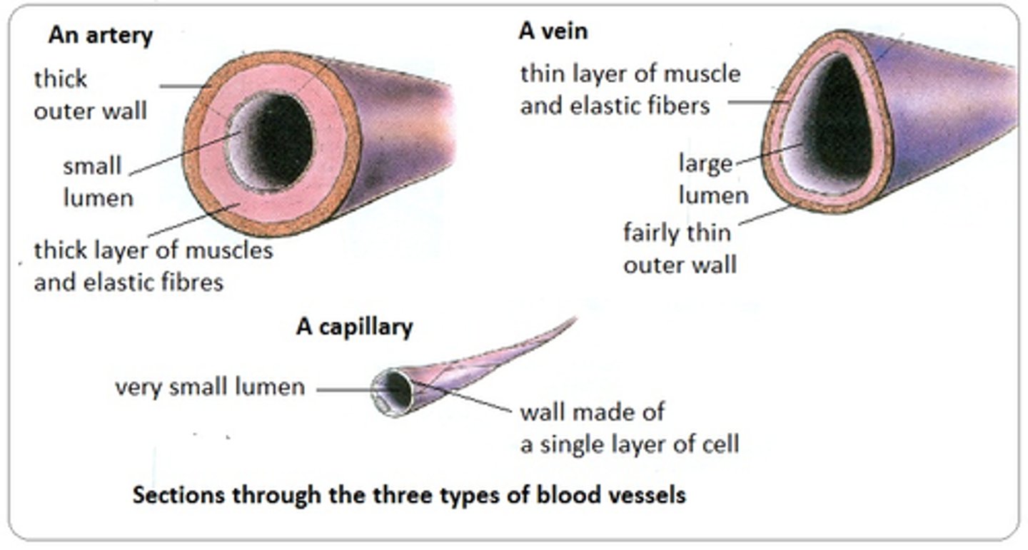
Explain how the structure of arteries relates to their function. (6 marks)
- Arteries carry blood away from the heart at a high pressure.
- Thick smooth muscle can contract to control and withstand blood pressure and flow.
- Thick elastic tissue stretches during ventricular contraction and recoils as ventricles relax, smoothing pressure surges and maintaining high pressure.
- A thick wall withstands high pressure and reduces the risk of bursting.
- A smooth, folded endothelium reduces friction and allows stretching.
- A narrow lumen helps maintain high blood pressure.
Explain how the structure of arterioles relates to their function. (3 marks)
- Arterioles have more smooth muscle than arteries, allowing lumen narrowing (vasoconstriction) to reduce blood flow to capillaries.
- Relaxation of the smooth muscle widens the lumen (vasodilation), increasing blood flow to capillaries.
- They have a thinner elastic layer than arteries, as pressure surges are lower further from the heart.
Explain how the structure of capillaries relates to their function. (4 marks)
- The wall is a single layer of endothelial cells, reducing diffusion distance.
- A dense network of capillaries increases surface area for diffusion.
- Their small diameter reduces blood flow speed, allowing more time for diffusion.
- Pores between endothelial cells allow larger substances to pass through.
Explain how the structure of veins relates to their function. (4 marks)
- Veins carry blood towards the heart at a low pressure.
- Veins have a wider lumen than arteries, reducing resistance to blood flow.
- They contain very little elastic and muscle tissue, leading to lower blood pressure.
- Valves prevent the backflow of blood.
Draw a labelled diagram of an artery, vein and capillary. (4 marks)
NOTE: Arterioles are a thinner extension of the arteries
Explain the formation of tissue fluid at the arteriole end of capillaries. (3 marks)
- High hydrostatic pressure inside the capillaries (caused by ventricular contraction) is greater than in the tissue fluid, producing a net outward force.
- This forces water and dissolved substances out of the capillaries.
- Large plasma proteins remain in the capillaries.
Explain the return of tissue fluid to the circulatory system at the venule end of capillaries. (4 marks)
- Hydrostatic pressure in the capillaries decreases as fluid leaves and due to friction.
- The concentration of plasma proteins increases, lowering the water potential in the capillaries below that of the tissue fluid.
- Water re-enters the capillaries by osmosis down the water potential gradient.
- Any excess fluid is drained into lymphatic capillaries and returned to the bloodstream via the veins.
State and explain causes of excess tissue fluid accumulation. (3 marks)
- A low protein concentration in plasma reduces the water potential gradient, causing more fluid to leave at the arteriole end and less to be reabsorbed at the venule end as the lymph system may not drain fluid quickly enough.
- High blood pressure, e.g. from excess salt, increases outward pressure at the arteriole end and decreases inward pressure at the venule end, causing greater fluid loss and reduced reabsorption.
- Blockage in lymph vessels prevents excess fluid from being drained and reabsorbed.
Define a risk factor and state examples of risk factors of cardiovascular disease. (3 marks)
- A lifestyle factor or substance in the body/environment linked to a higher chance of developing a disease.
- Shown to be associated with an increased incidence of disease.
- Examples include age, a diet high in salt or saturated fat, smoking, lack of physical activity, and genetic factors.
Describe how a heartbeat is initiated and coordinated (5 marks)
SAN sends wave of electrical impulses across atria causing atrial contraction
non-conducting tissue prevents immediate contraction of ventricles
AVN delays impulse whilst blood leaves atria and ventricles fill
AVN sends wave of electrical activity down bundle of his
causing ventricles to contract from base up
Explain how the heart muscle and the heart valves maintain a one way flow of blood from the left atrium to aorta (5 marks)
atrium has higher pressure than ventricle causing atrioventricular valves to open
ventricle has higher pressure than atrium causing atrioventricular valves to close
ventricle has higher pressure than aorta causing semilunar valves to open