ECG Interpretation
1/98
There's no tags or description
Looks like no tags are added yet.
Name | Mastery | Learn | Test | Matching | Spaced | Call with Kai |
|---|
No analytics yet
Send a link to your students to track their progress
99 Terms
List the Five Step Method in order:
Regularity
Rate
P Waves
PR Interval
QRS Complex
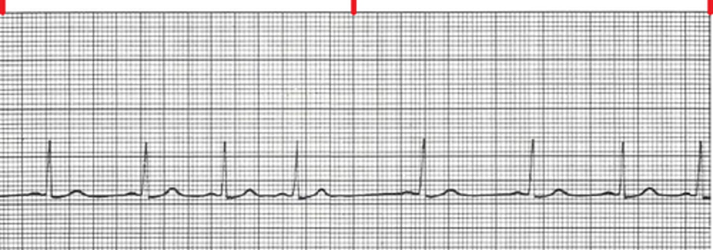
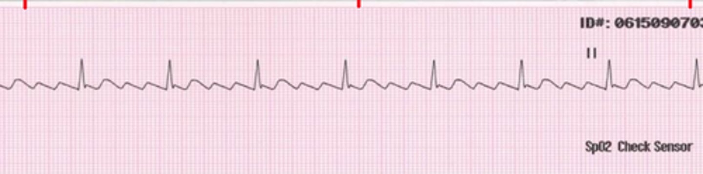
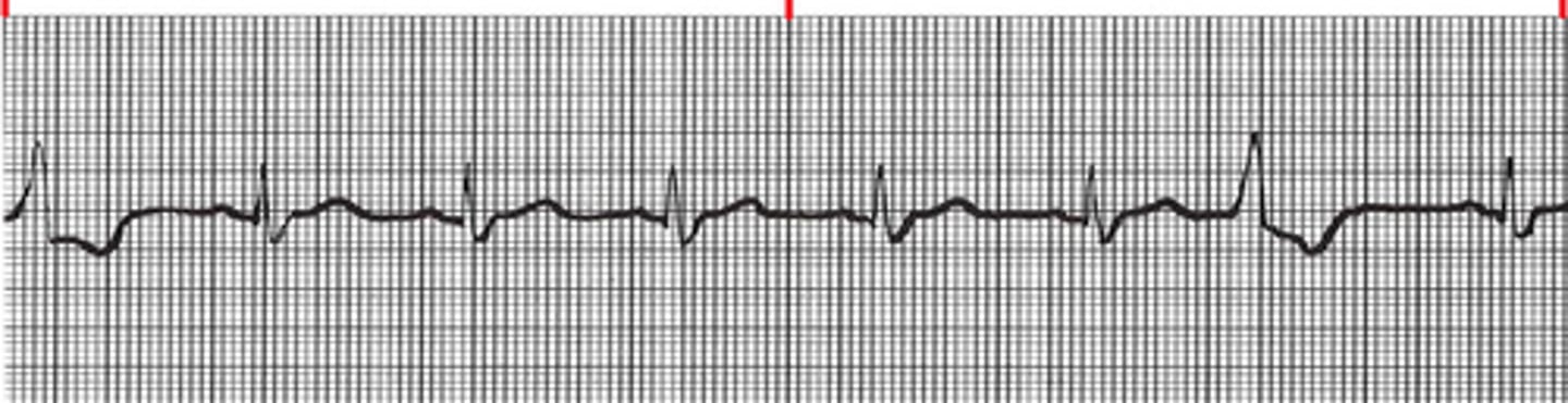
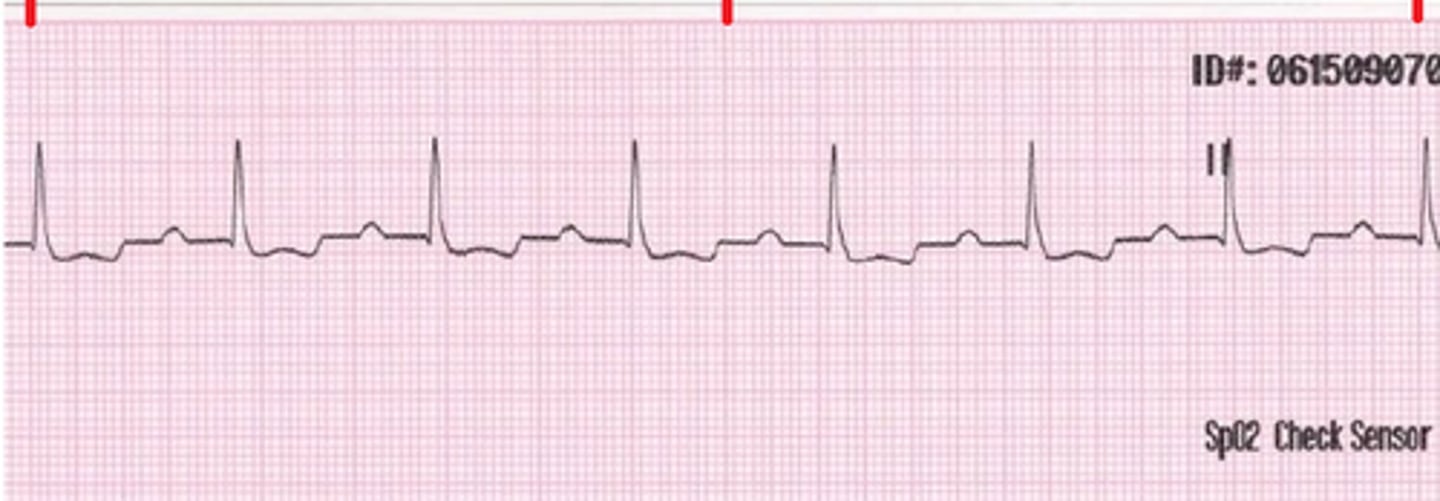
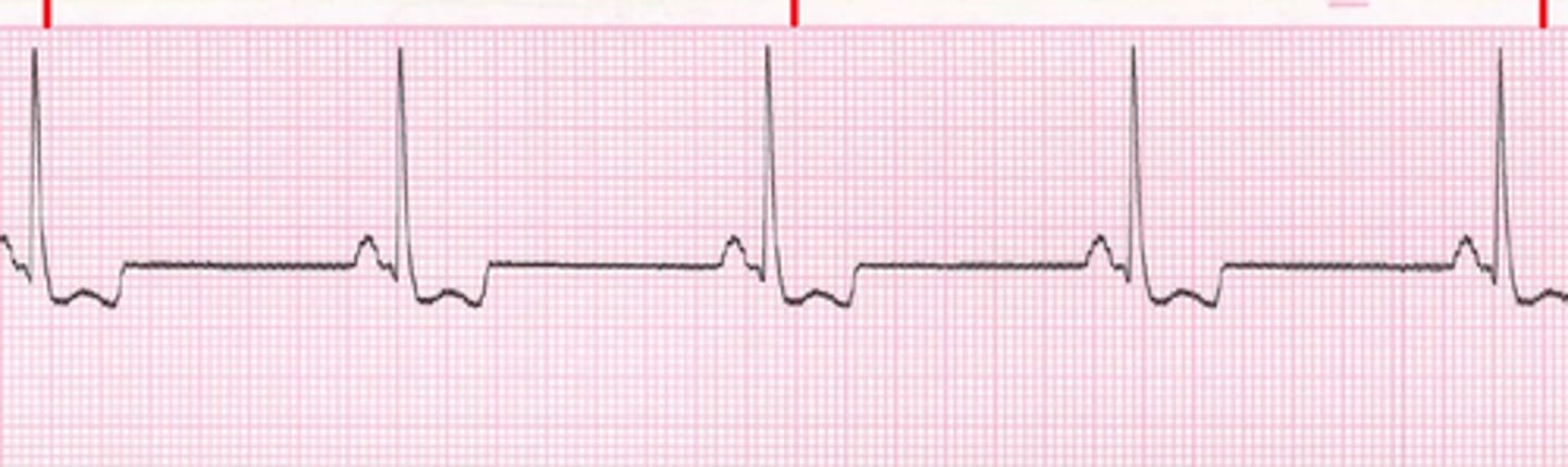
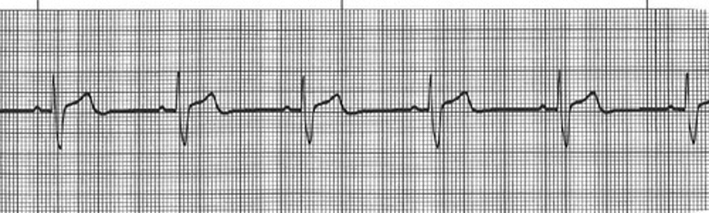
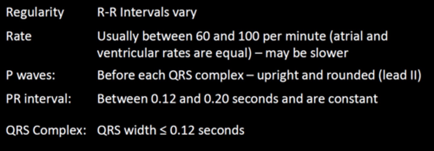
Sinus Rhythm
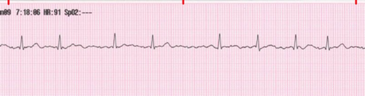
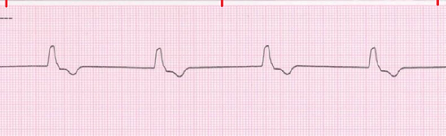
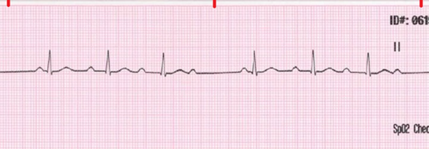
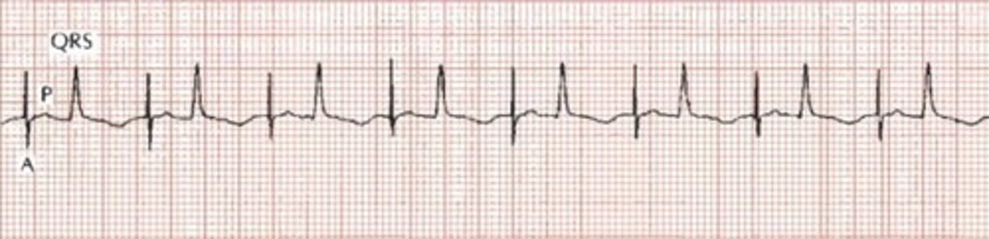
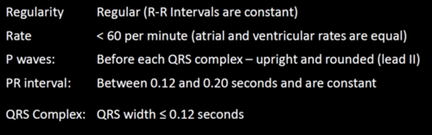
Sinus Bradycardia
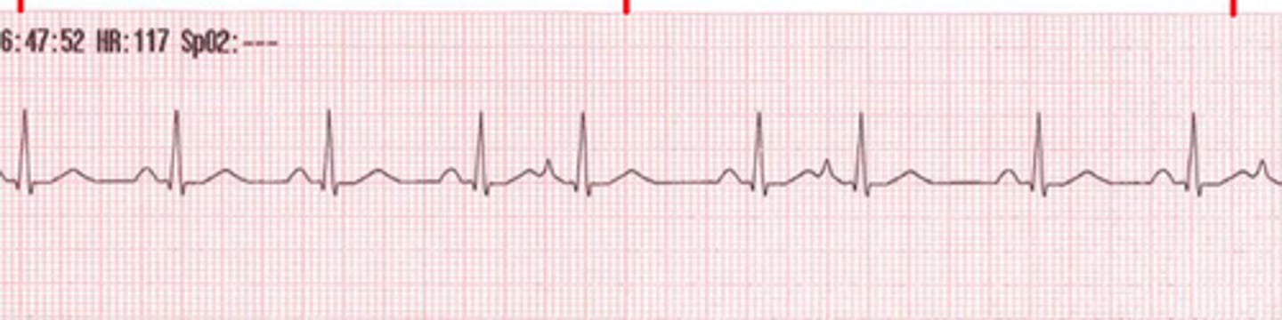
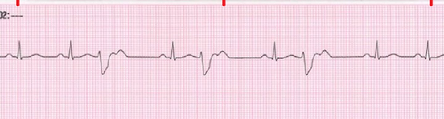
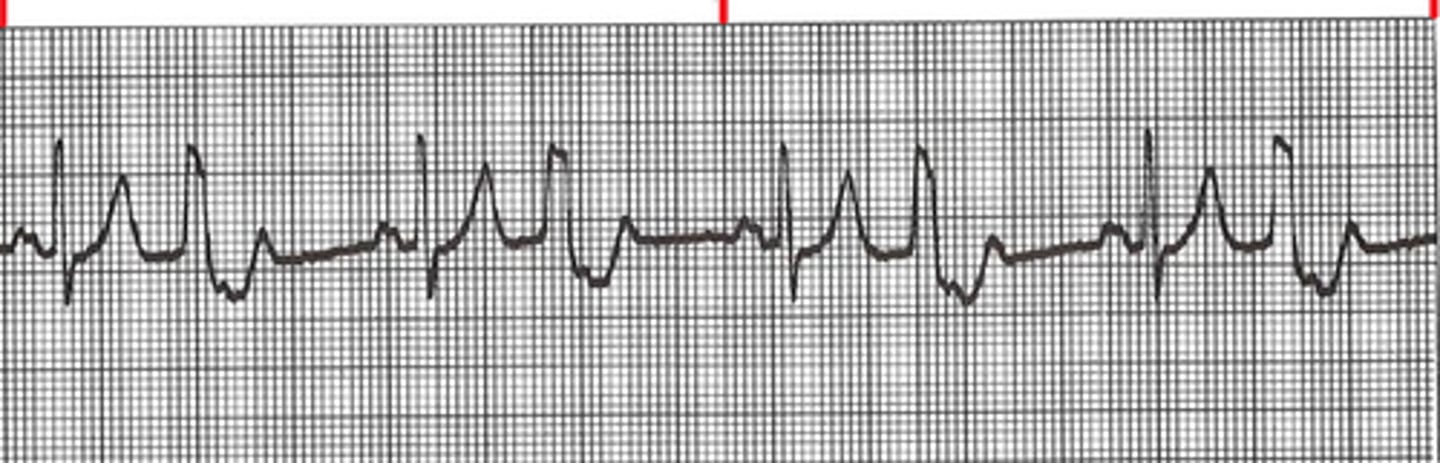
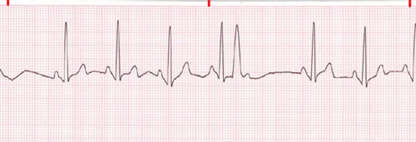
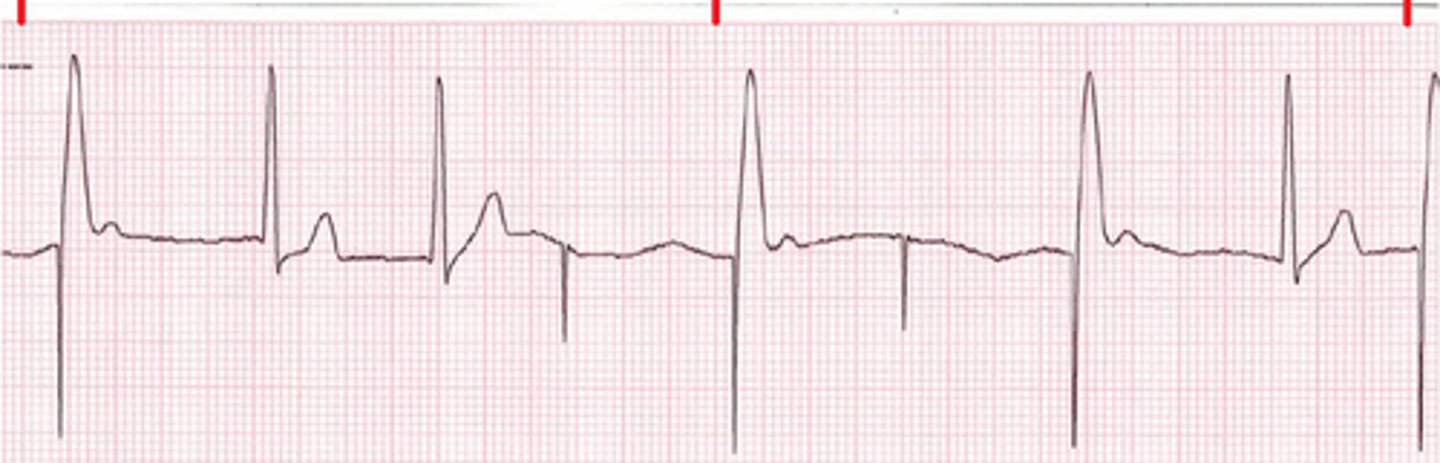
Sinus Tachycardia
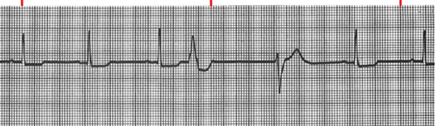
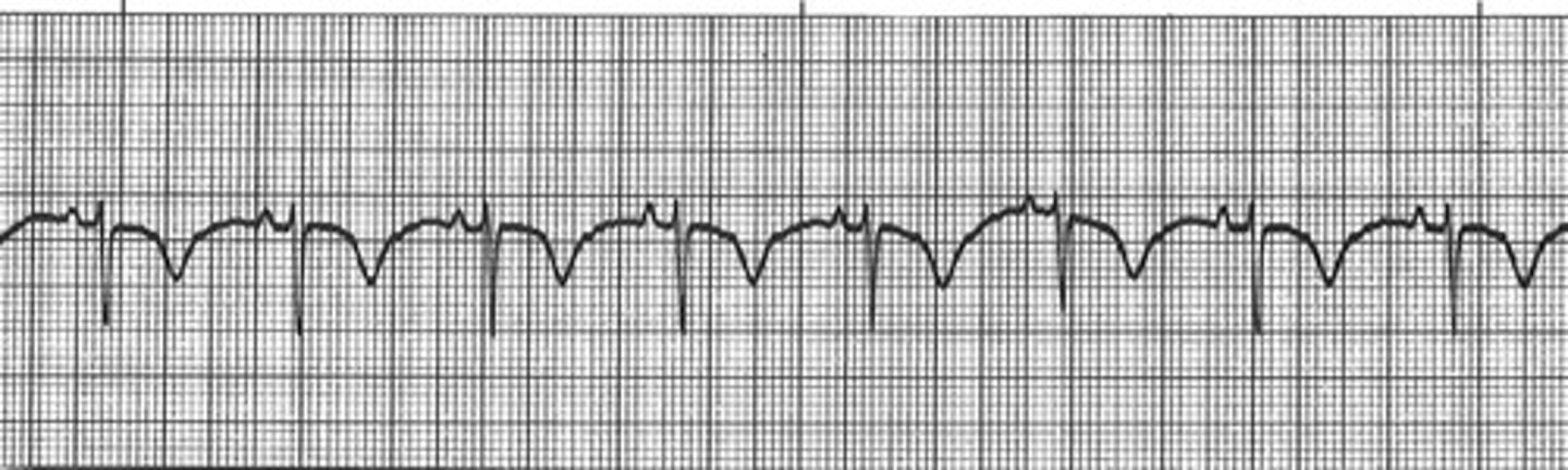
Sinus Arrhythmia
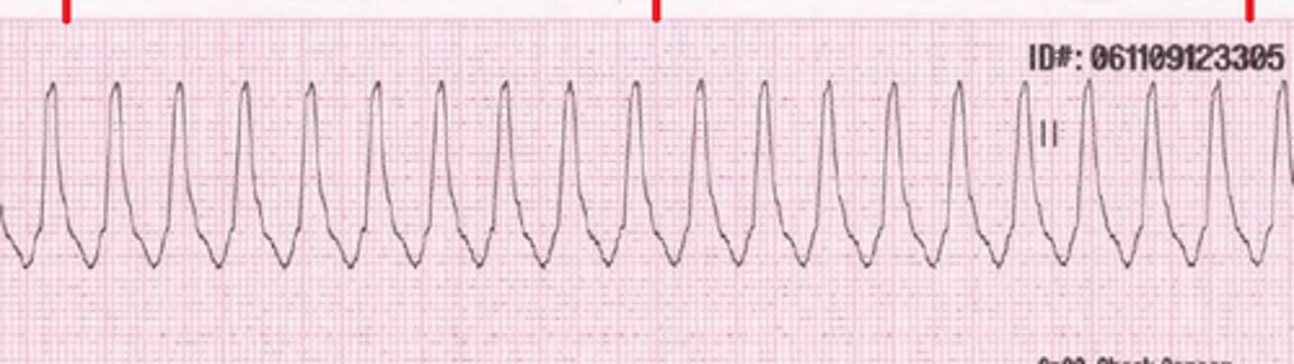
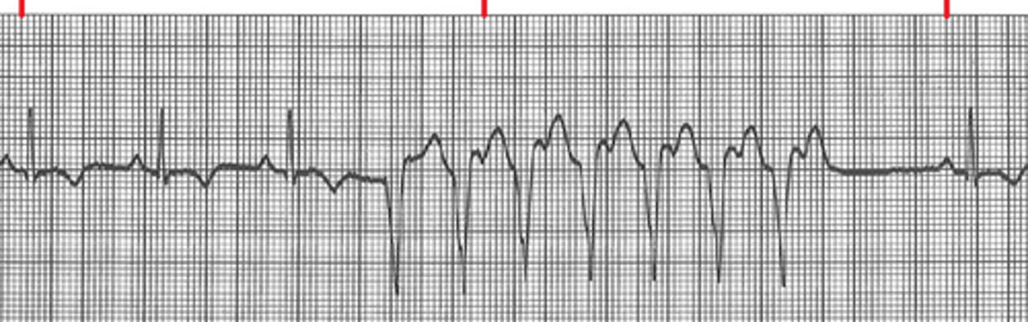
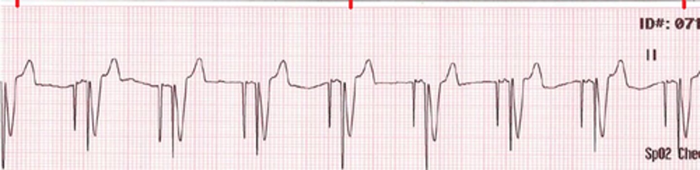
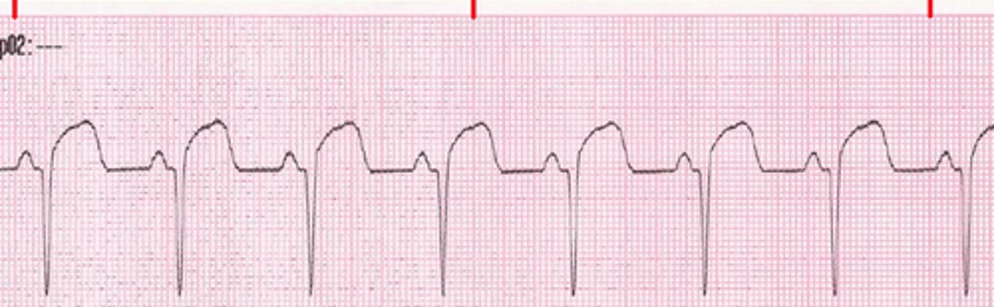
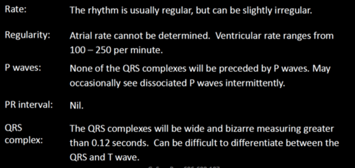
Ventricular Tachycardia
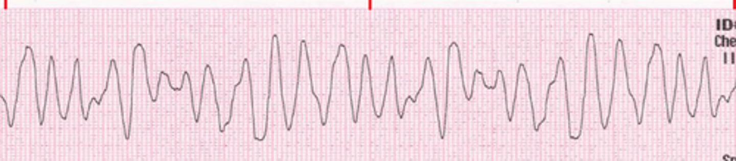
Ventricular Fibrillation - Medium Coarse
Ventricular Fibrillation - Coarse
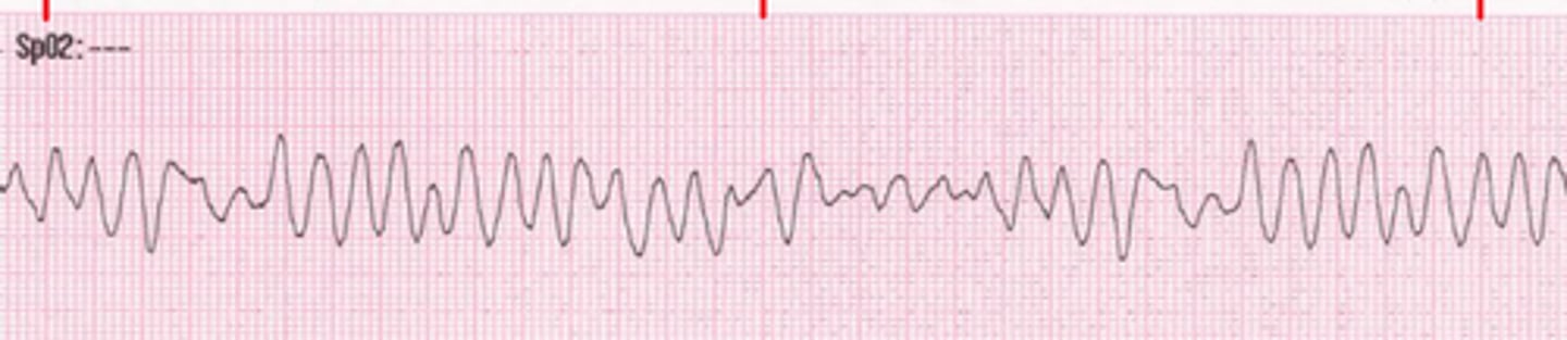
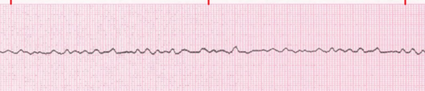
Ventricular Fibrillation - Fine
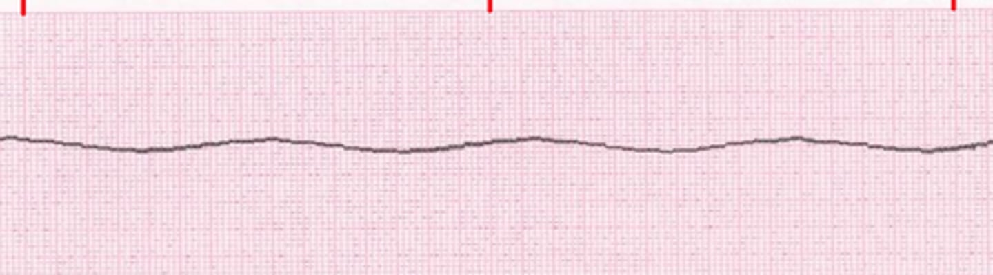
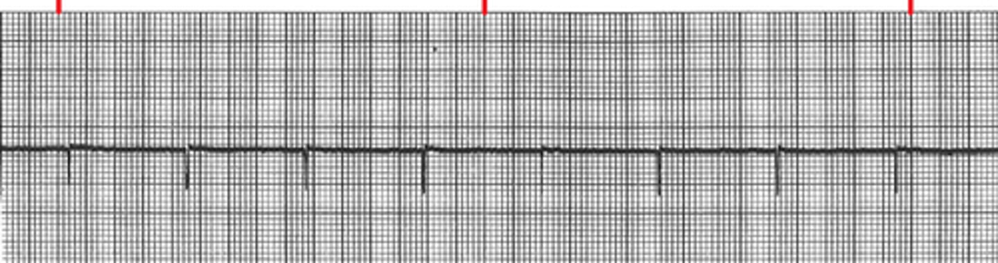
Asystole
Pulseless Electrical Activity (PEA)
Torsades de Pointes
Atrial Fibrillation
Atrial Flutter
Premature Atrial Contraction (PAC)
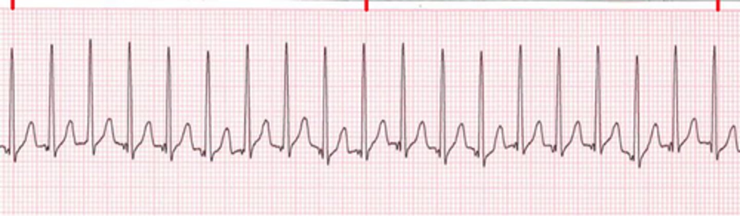
Supraventricular Tachycardia (SVT)
Junctional Escape Rhythm
Accelerated Junctional Rhythm
Junctional Tachycardia
Premature Junctional Contraction (PJC)
Idioventricular & Accelerated Idioventricular Rhythm
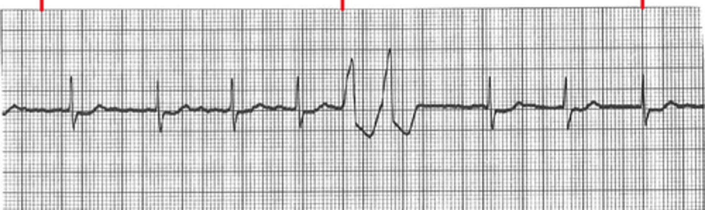
Premature Ventricular Contraction (PVC)
Unifocal PVC
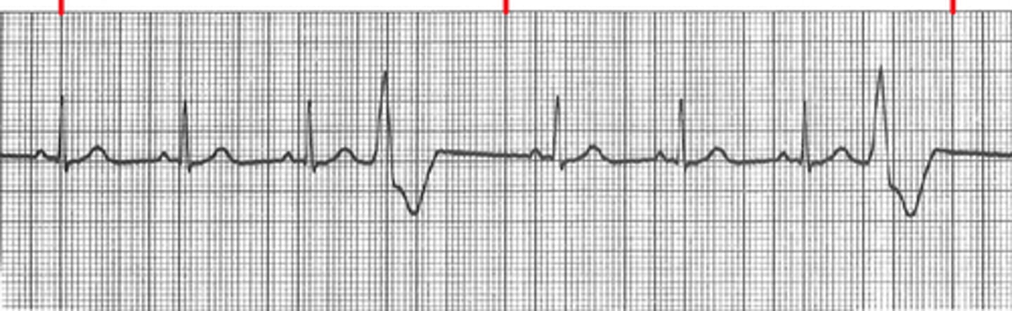
Multifocal PVC
Unifocal/Multifocal PVC Couplet
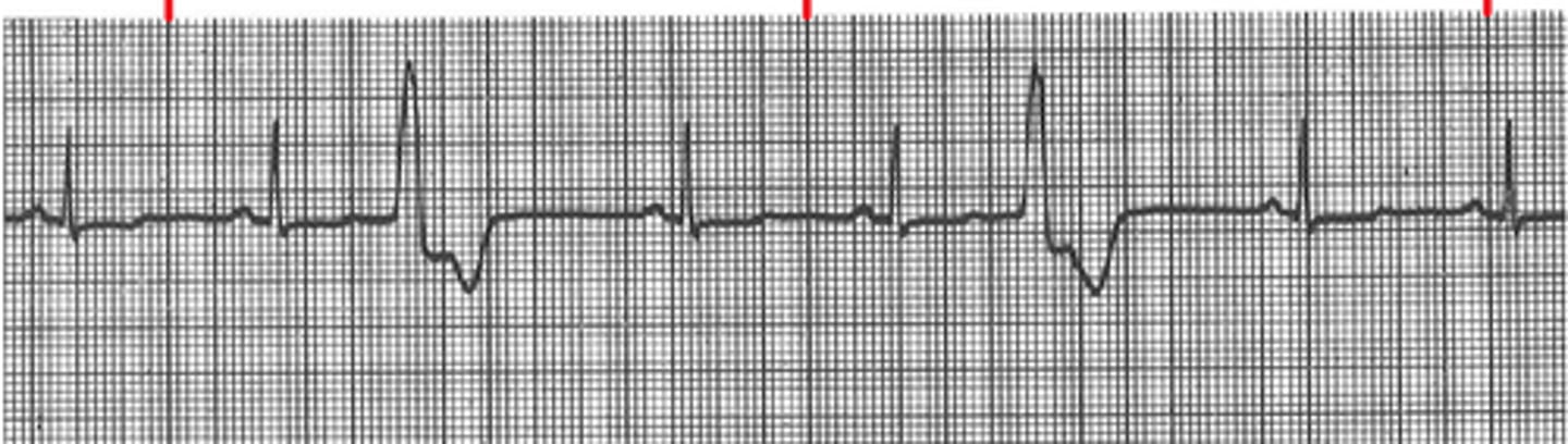
Ventricular Bigeminy PVC
Ventricular Trigeminy PVC
Ventricular Quadrigeminy PVC
Non-sustained VT
R-on-T PVC
1st Degree Heart Block
2nd Degree Heart Block Type 1
2nd Degree Heart Block Type 2
2nd Degree Heart Block with 2:1 AV Block
3rd Degree Heart Block
Ventricular Pacemaker
Atrial Pacemaker
Dual Chamber Pacemaker
Failure to Capture - Ventricles
Failure to Capture - Asystole
Undersensing
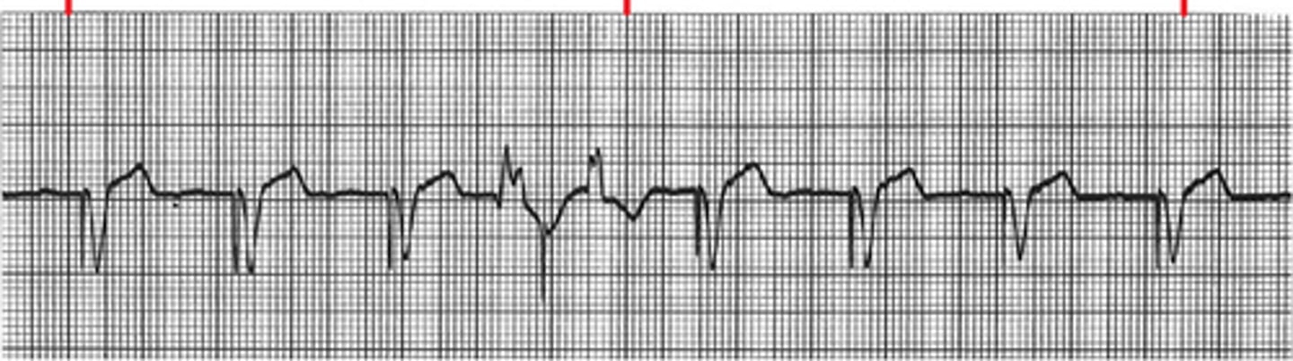
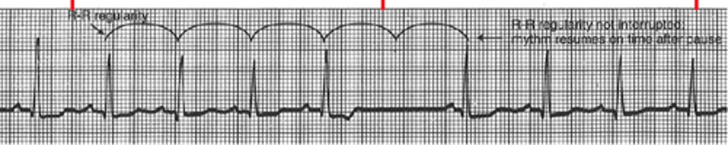
Sinus Block - Pause
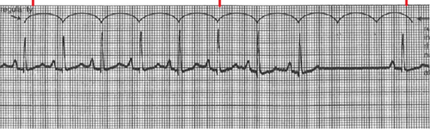
Sinus Arrest
ST Depression
ST Elevation
Inverted T Wave
Widened QRS
What is Step 1 of the Five Step Method?
Regularity
What is Step 2 of the Five Step Method?
Rate
What is Step 3 of the Five Step Method?
P Waves
What is Step 4 of the Five Step Method?
PR Interval
What is Step 5 of the Five Step Method?
QRS Complex
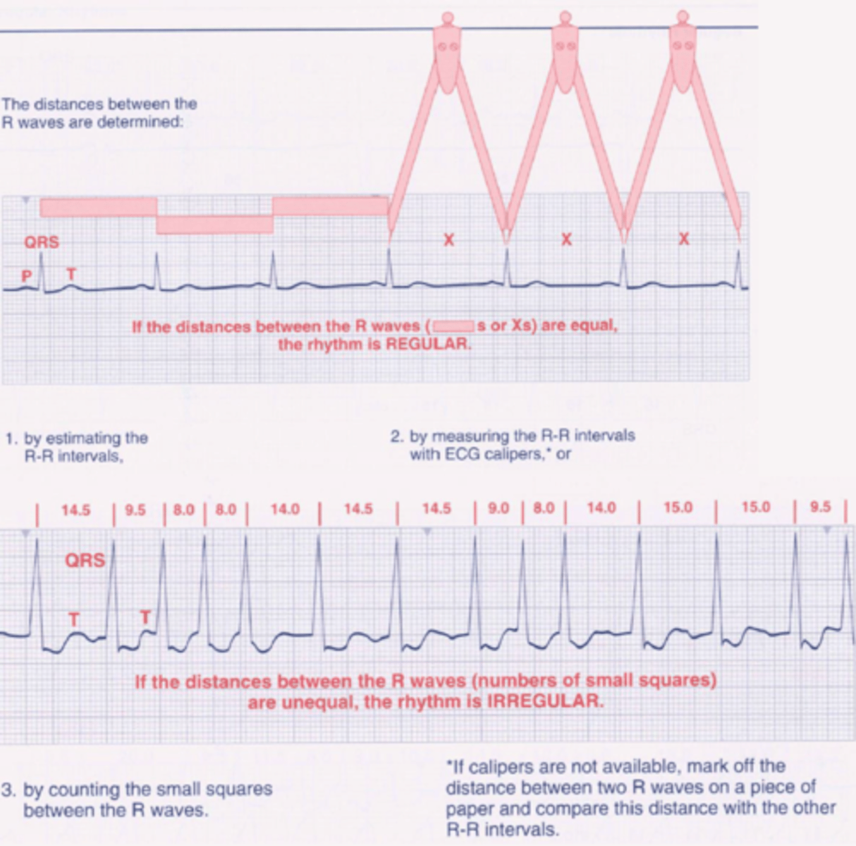
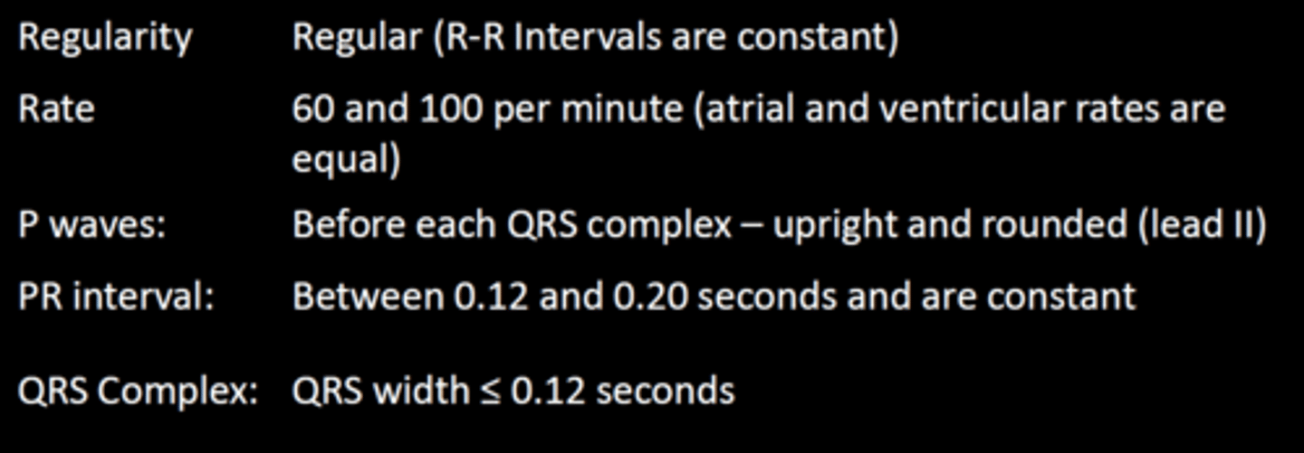
Five Step Method - Step 1: Regularity
- Is the rhythm regular of irregular?
- Are there any patterns to the regularity?
- Are there any ectopic beats, if so are they early or late?
Five Step Method - Step 2: Rate
- What is the exact rate?
- Is the atrial rate the same as the ventricular rate?
Which methods can be used to determine the Rate?
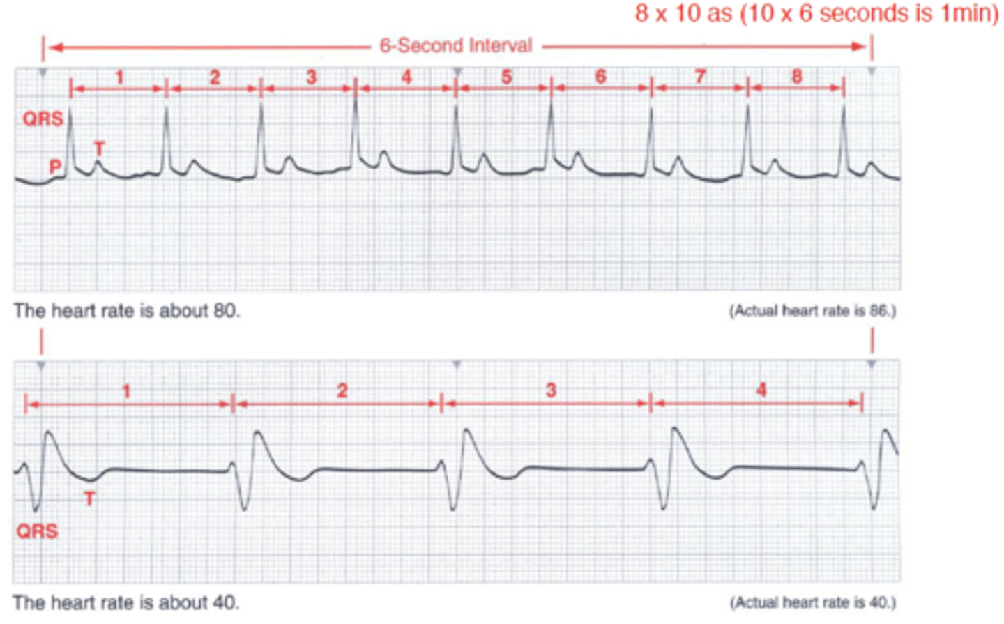
Second Strip Method
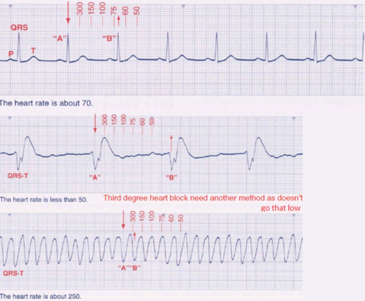
Triplicate Method
R-R Interval, including 60, 300 and 1500 Methods
Second Strip Method
Triplicate Method
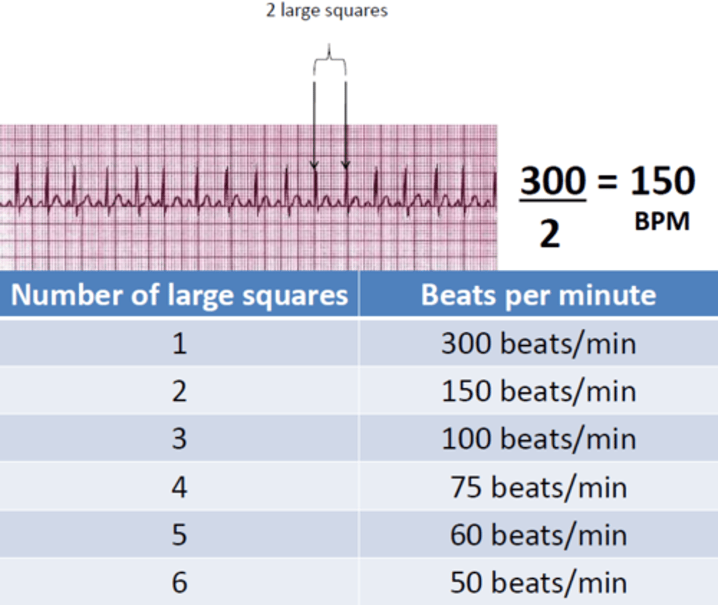
R-R Interval: 300 Method
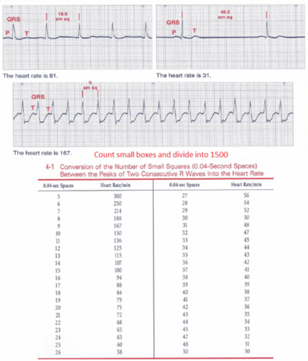
R-R Interval: 1500 Method
PQRST-U Complex
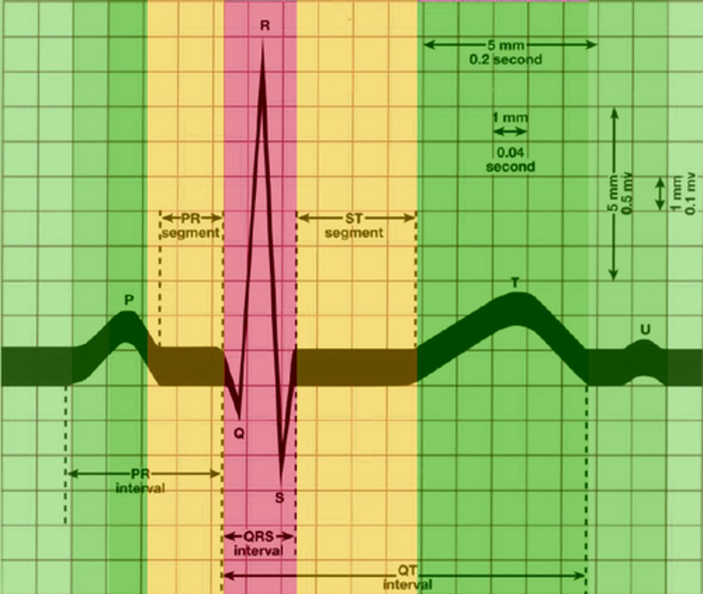
Flat Line
Isoelectric line with no electrical activity
Positive Isoelectric Line
Positive charge to positive lead
Negative Isoelectric Line
Negative charge to negative lead
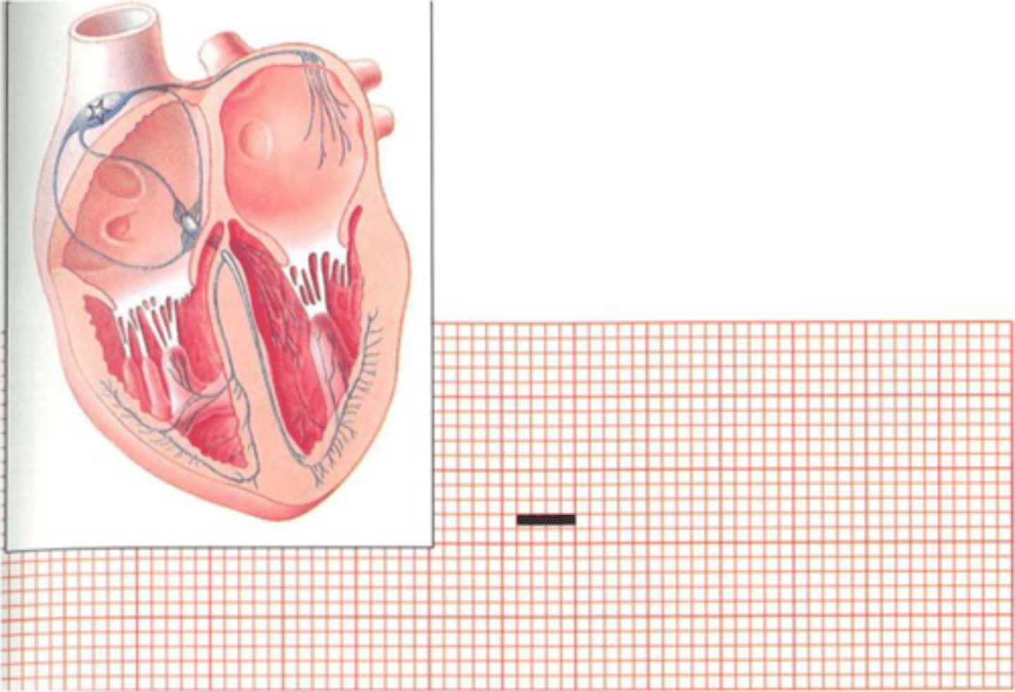
P Wave: Impulse initiated in the SA Node
Depolarisation of the atria begins
P Wave: Beginning of Atrial Excitation
Atrial depolarisation proceeds from right, through the internodal tracts to left through Bachmann's bundle
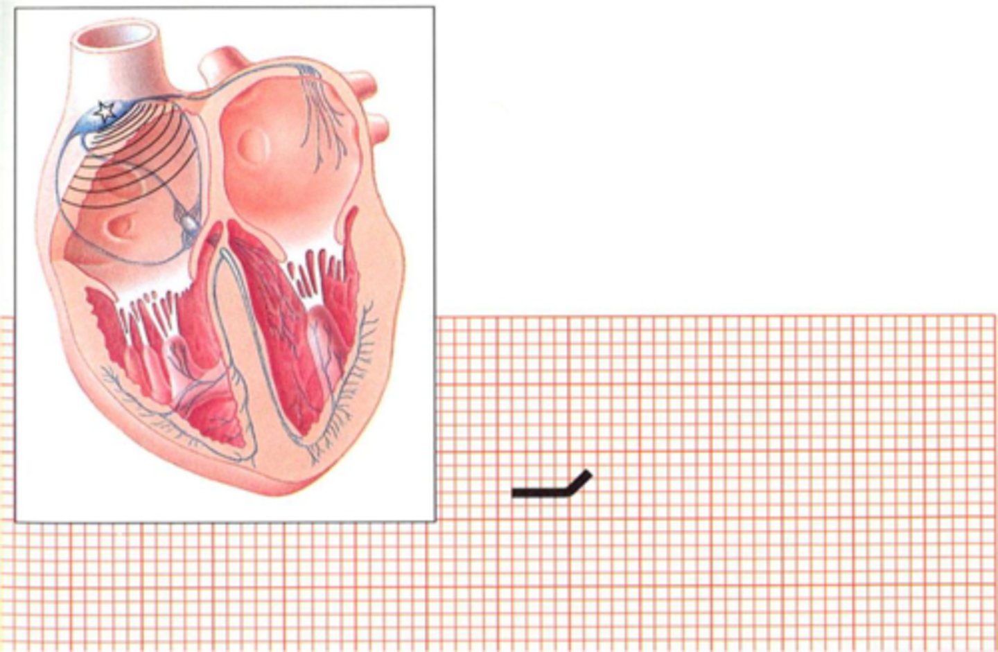
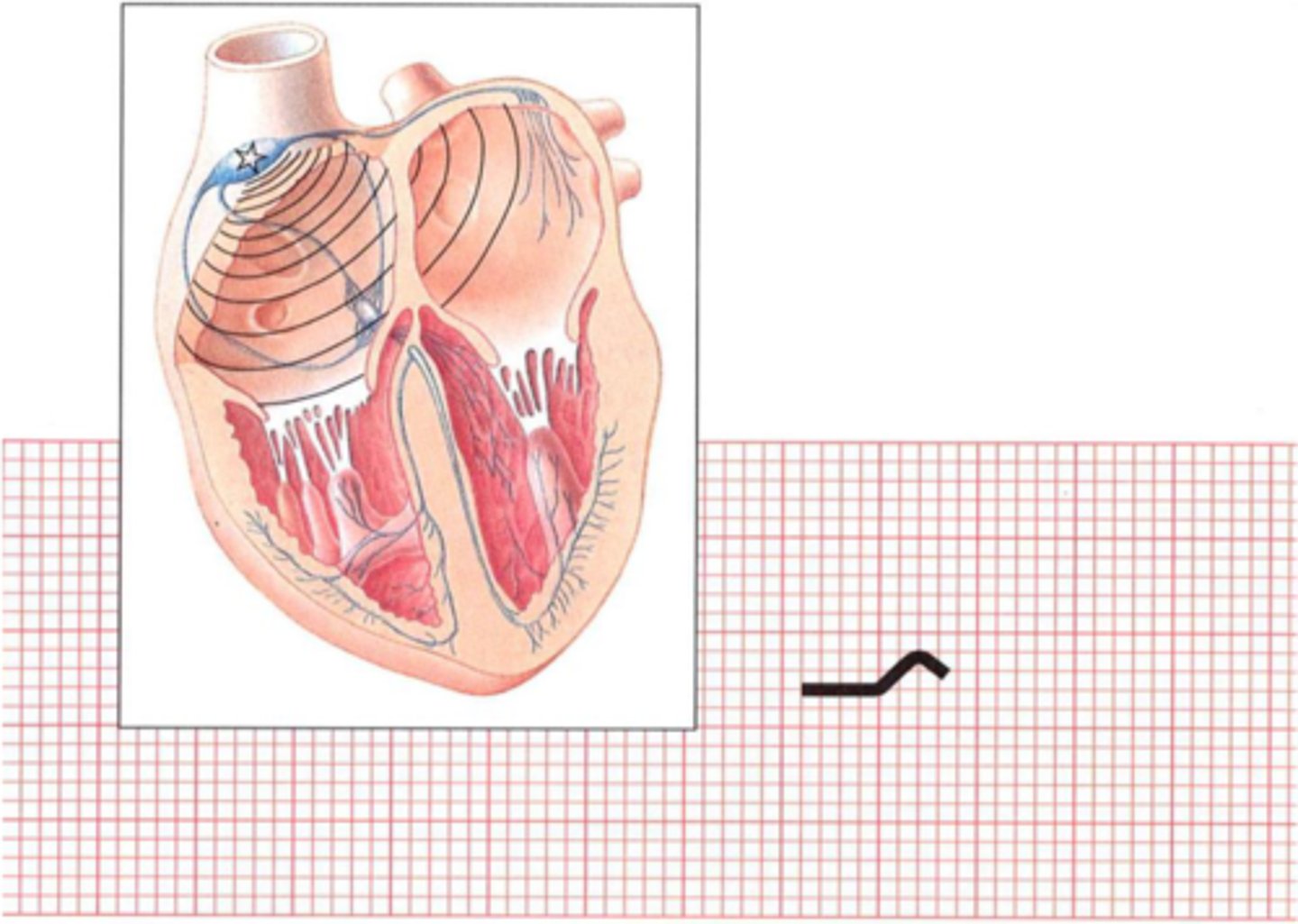
P Wave: Atrial Excitation
Atrial contraction occurs
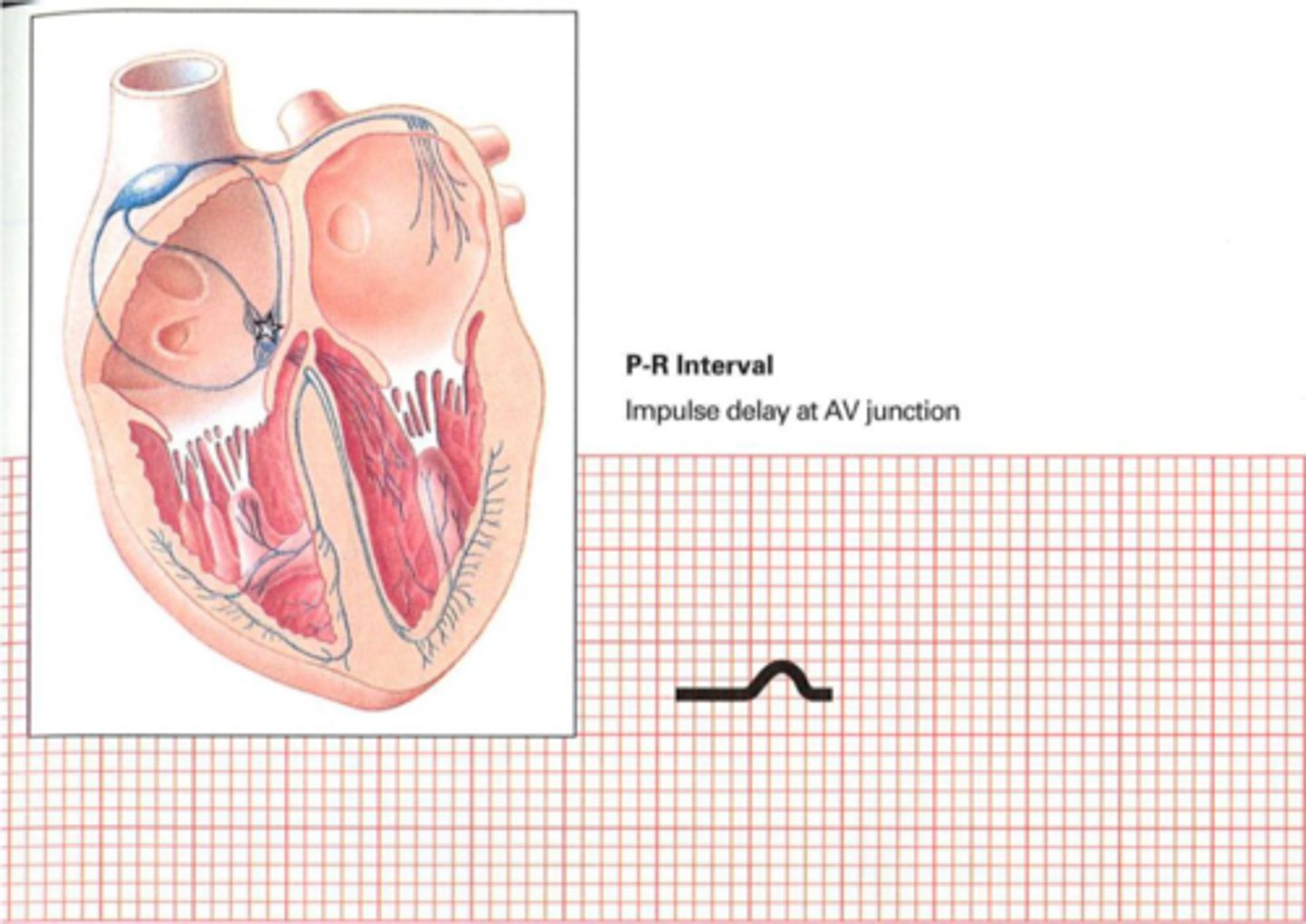
P-R Interval
Impulse reaches the AV Node
Impulse delay at AV Junction
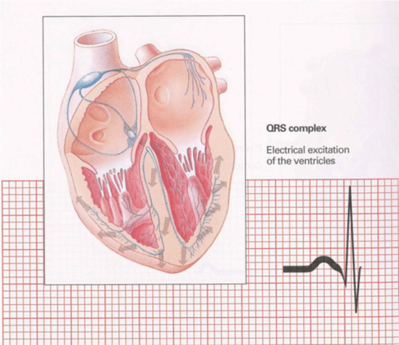
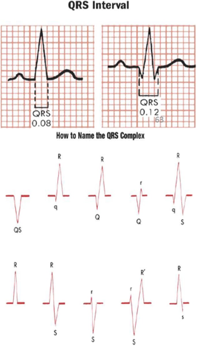
QRS Complex
Depolarisation of the ventricles through the Bundle of His, Left and Right Bundle Branches and Purkinje Fibres
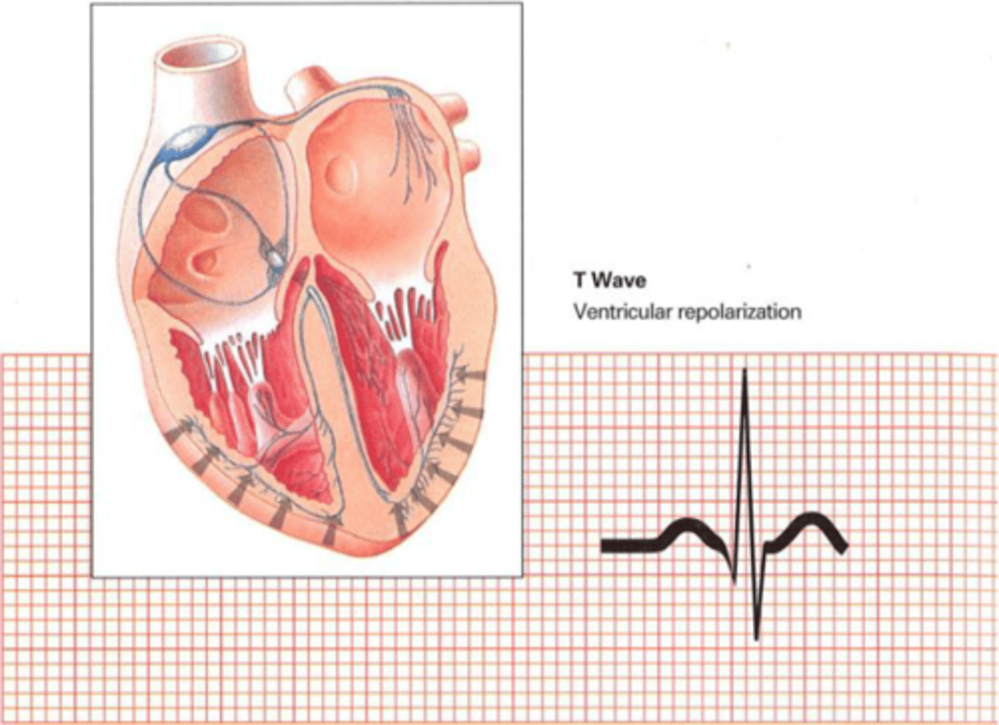
T Wave
Ventricular repolarisation occurs
Electrical charge in the pacemaker and myocyte cells return to polarised state with sodium outside and potassium inside the cell
Five Step Method - Step 3: P Waves
- Are the P waves regular?
- Is there one P wave for each QRS complex?
- Is the P wave in front of the QRS or behind it?
- Is the P wave normal and upright in Lead II?
- Are there more P waves than QRS complexes?
- Do all the P waves look alike?
- Are the irregular P waves associated with ectopic
beats?
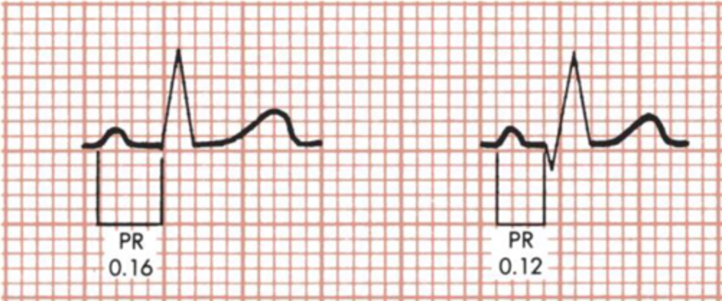
Five Step Method - Step 4: P-R Interval
- Is the PR interval measurement within normal
range? (Normally 0.12 to 0.20 seconds/3 to 5 small squares)
- Are the PRIs constant?
- If the PR interval varies, is there a pattern to the
changing measurements?
Five Step Method - Step 5: QRS Complex
- What is the width of the QRS complex? (Normally < 0.12 seconds)
- Are the QRS complexes within normal limits?
- Are all the QRS complexes of equal duration?
- Do all QRS complexes look alike?
- Are the unusual QRS complexes associated with ectopic beats?
The Cardiac Cycle - Step 1
Depolarisation wave travels from the SA node down Bachmann's bundle to the left artia and down the internodal tracts to the right atria
The Cardiac Cycle - Step 2
Right and Left Atria contract together
Pressure of contraction produces closure of the aortic and pulmonary semilunar valves
The Cardiac Cycle - Step 3
Atrial contraction pumps blood through the tricuspid and mitral atrioventricular valves into the left and right ventricles
The Cardiac Cycle - Step 4
Depolarisation wave from the SA node reaches the AV node with a 0.1 second delay before the AV node fires
The Cardiac Cycle - Step 5
Depolarisation wave travels from the AV node through the Bundle of His, down the left and right bundle branches and into the Purkinje fibres
The Cardiac Cycle - Step 6
Right and Left Ventricles contract together
Pressure of contraction produces closure of the tricuspid and mitral atrioventricular valves
The Cardiac Cycle - Step 7
Ventricular contraction pumps blood through the aortic valve into the systemic circulation and through the pulmonary valve into the lungs
The Cardiac Cycle - Step 8
Ventricular repolarisation occurs
Pacemaker Cells
Make up the electrical conduction system of the heart, including the Sinoatrial node, Bachmann's bundle, internodal tracts, Atrioventricular node, Bundle of His, left bundle branch, left posterior fascicle, left anterior fascicle, right bundle branch and Purkinje fibres
Cardiac Myocytes
Work in functional syncytium with pacemaker cells and are responsible for the mechanical contraction of cardiac muscle
Depolarisation & Repolarisation: Pacemaker Cells
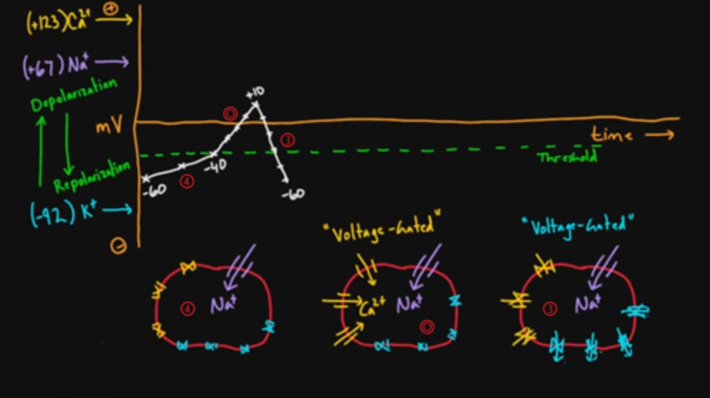
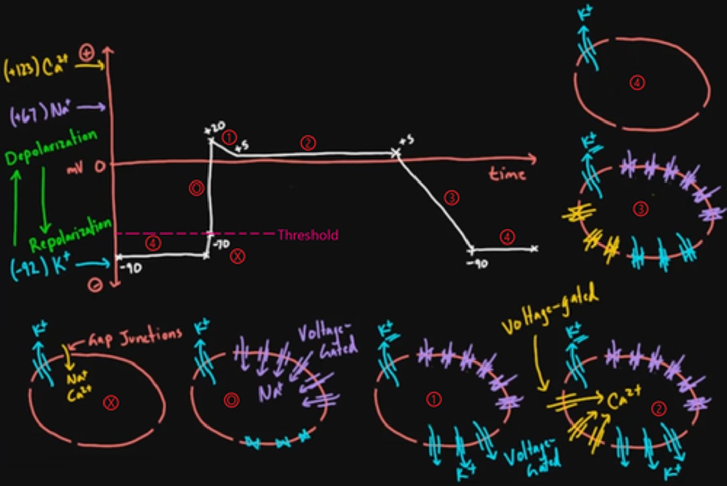
Depolarisation & Repolarisation: Cardiac Myocytes
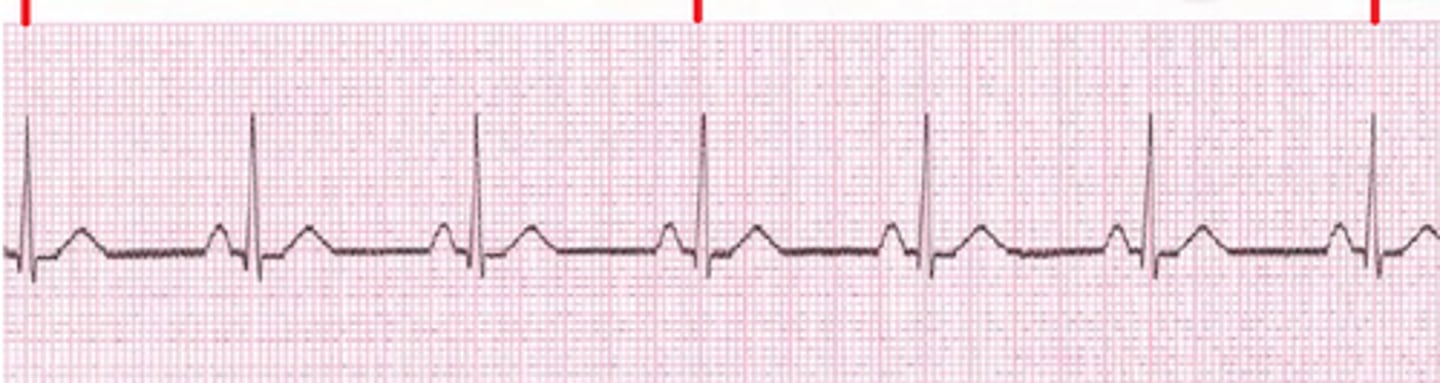
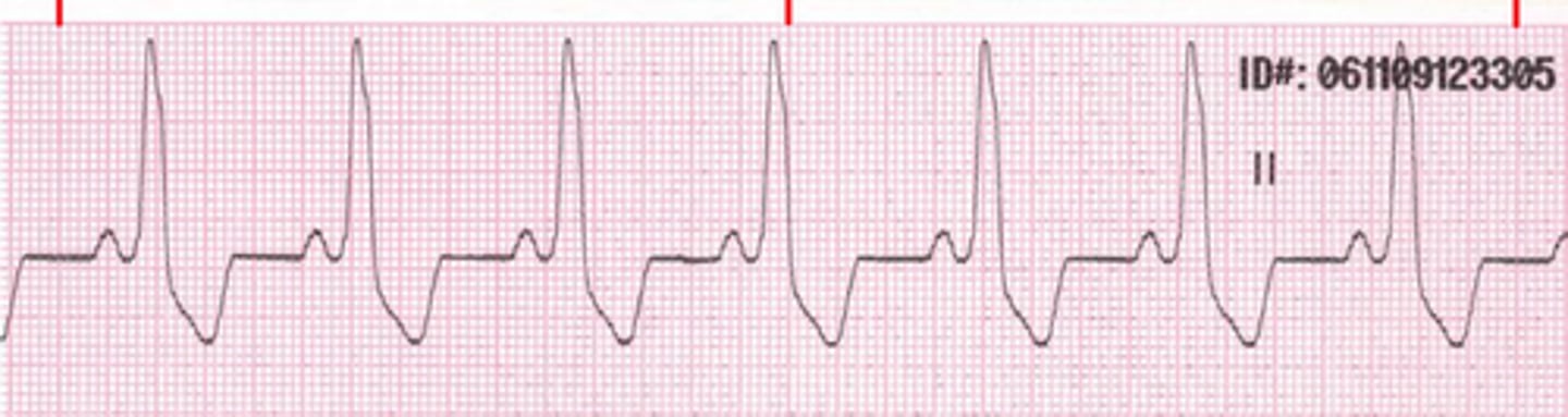
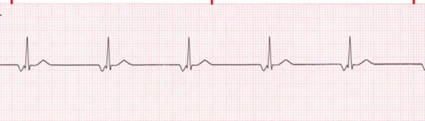
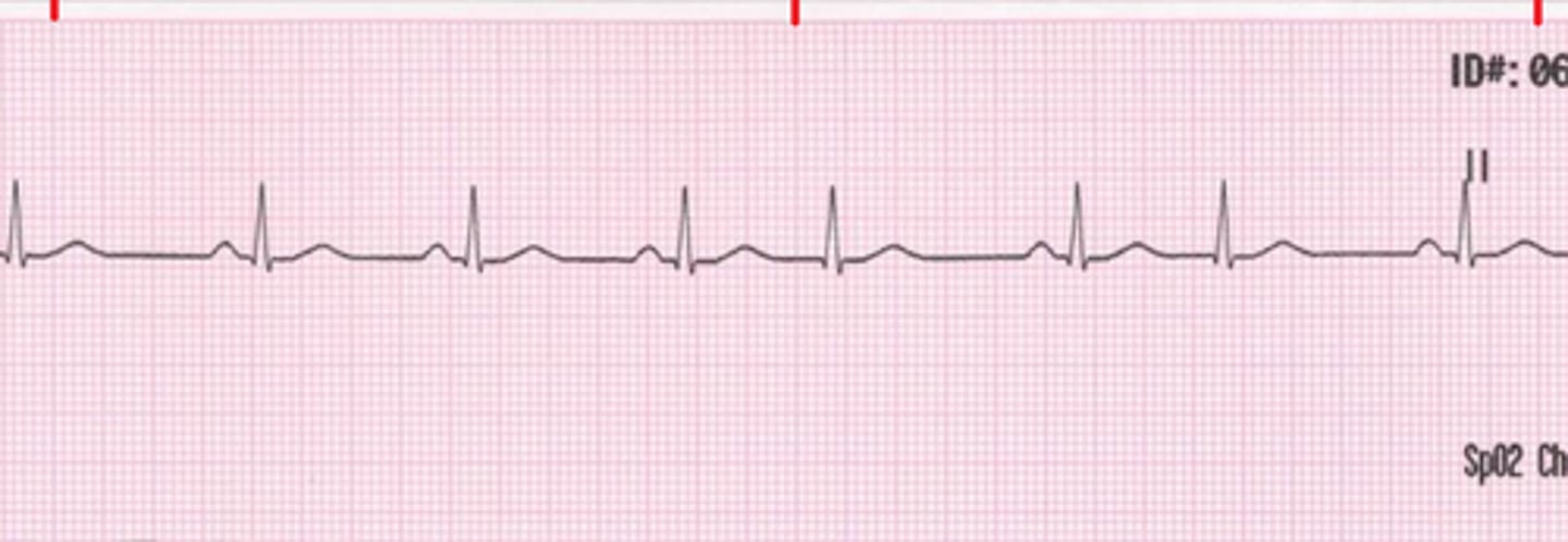
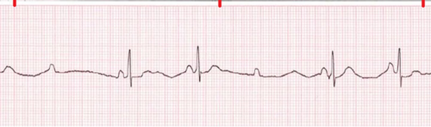
Sinus Rhythm - Interpretation Example
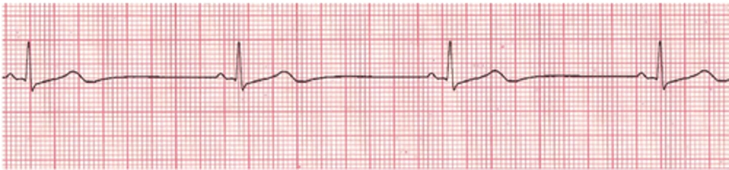
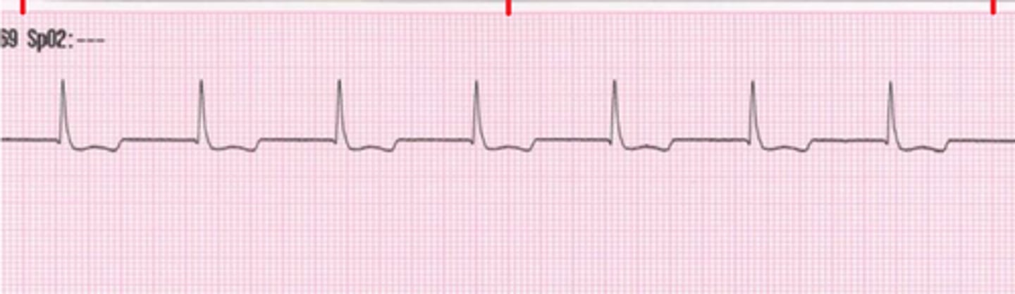
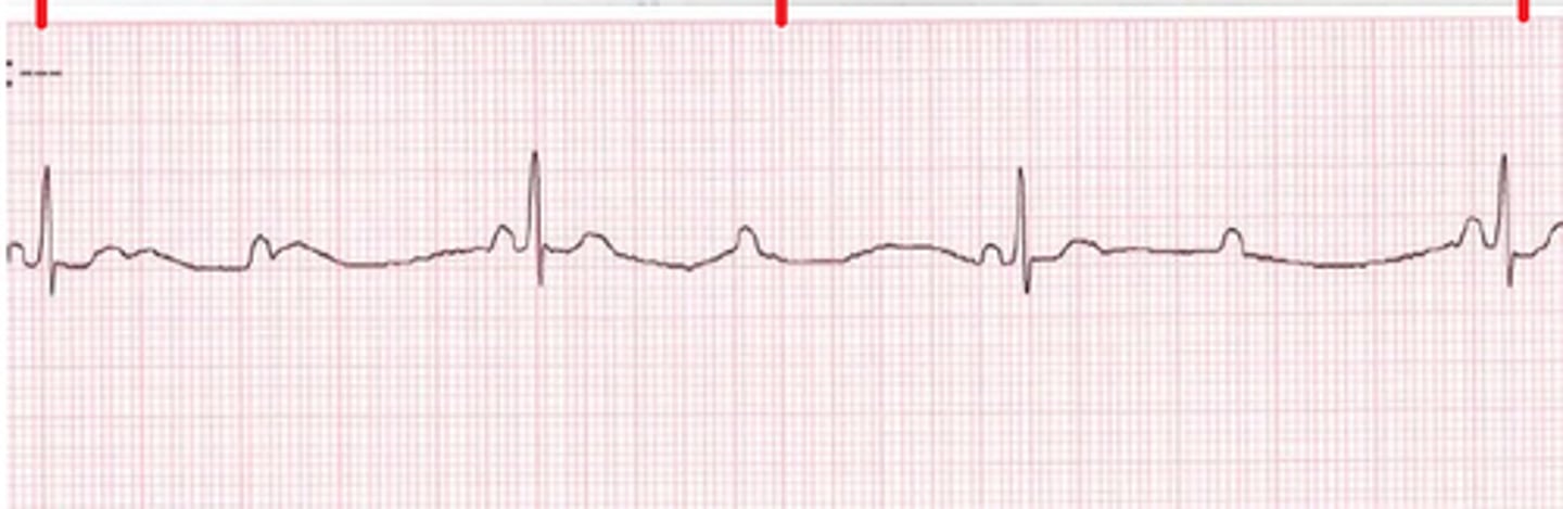
Sinus Bradycardia - Interpretation Example
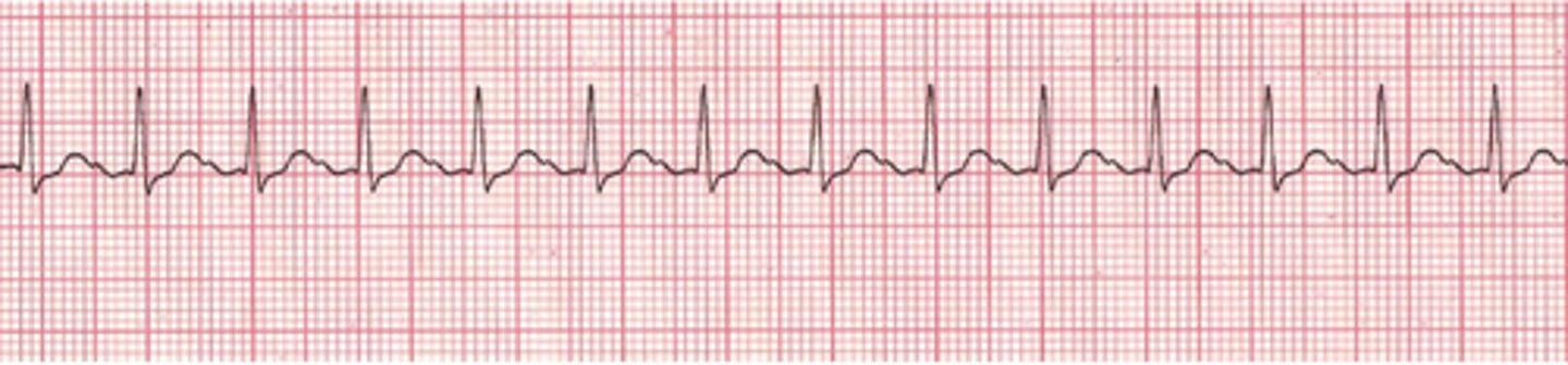
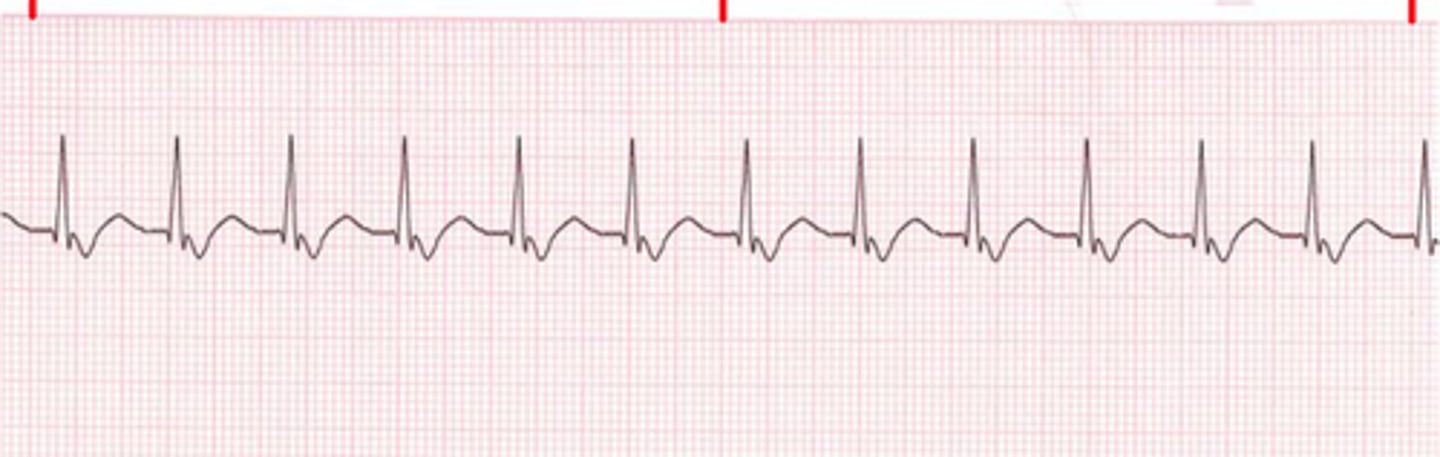
Sinus Tachycardia - Interpretation Example

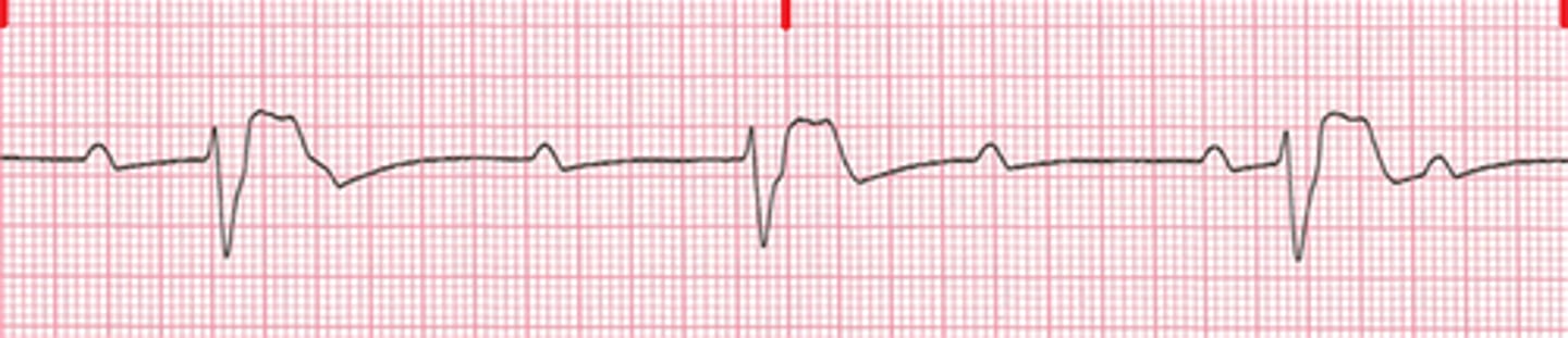
Sinus Arrhythmia - Interpretation Example
What are the four types of Sinus Rhythms?
Sinus Rhythm
Sinus Bradycardia
Sinus Tachycardia
Sinus Arrhythmia
What are the four types of 'Arrest' Rhythms?
Ventricular Tachycardia
Ventricular Fibrillation
Asystole
Pulseless Electrical Activity
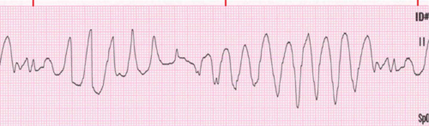
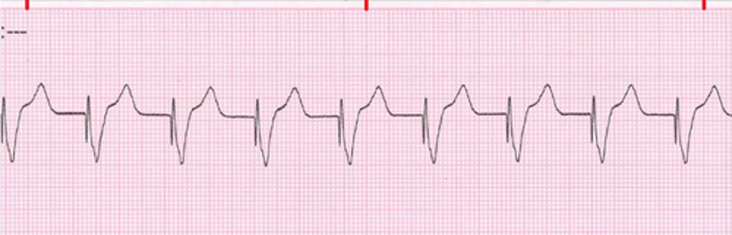
Ventricular Tachycardia - Interpretation Example
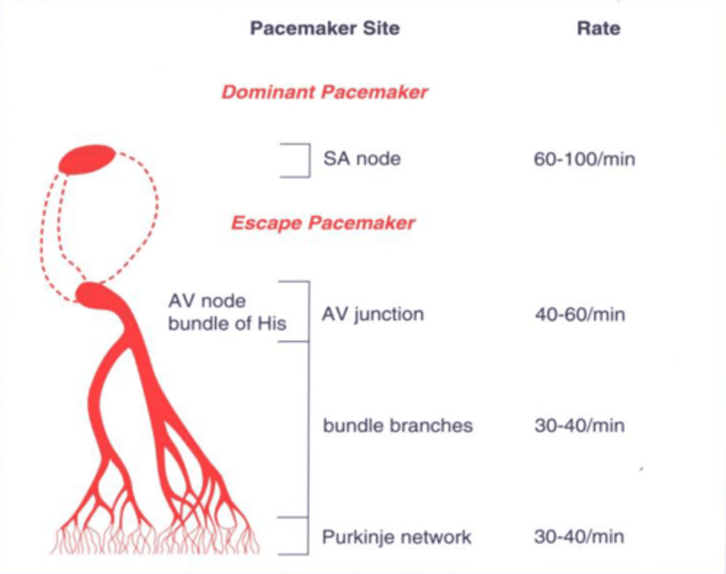
What is the Dominant Pacemaker?
Sinoatrial node
What are the Escape Pacemakers?
AV node
Bundle of His
Bundle branches
Purkinje fibres
What is the AV Junction?
Anatomical location of the AV node and Bundle of His
What is the Intrinsic Rate of the SA Node?
60-100BPM
What is the Intrinsic Rate of the AV Junction?
40-60BPM
What is the Intrinsic Rate of the Bundle Branches?
30-40BPM
What is the Intrinsic Rate of the Purkinje Network?
20-40BPM
Intrinsic Rate