pelvis
1/38
There's no tags or description
Looks like no tags are added yet.
Name | Mastery | Learn | Test | Matching | Spaced | Call with Kai |
|---|
No analytics yet
Send a link to your students to track their progress
39 Terms
what is the pelvic girdle formed by
the 2 hip bones (ossa coxae) each consisting of ilium, ischium and pubisfued at the acetabulum
the hip bones are fused in the ventral median plan at the pubic symphisis
how is the pelvis formed
2 hip bones (ossa coxae)
sacrum (fused vertevrae)
the first 2 caudal vertebrae
the sacrum articulates with the left and right ilium
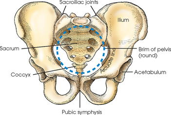
where is the pelvic cavity continuous with
the abdominal cavity at the pelvic inlet and contains the pelvic organs
digestive, urinary and reproductive tract
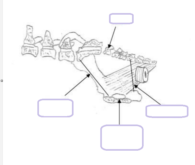
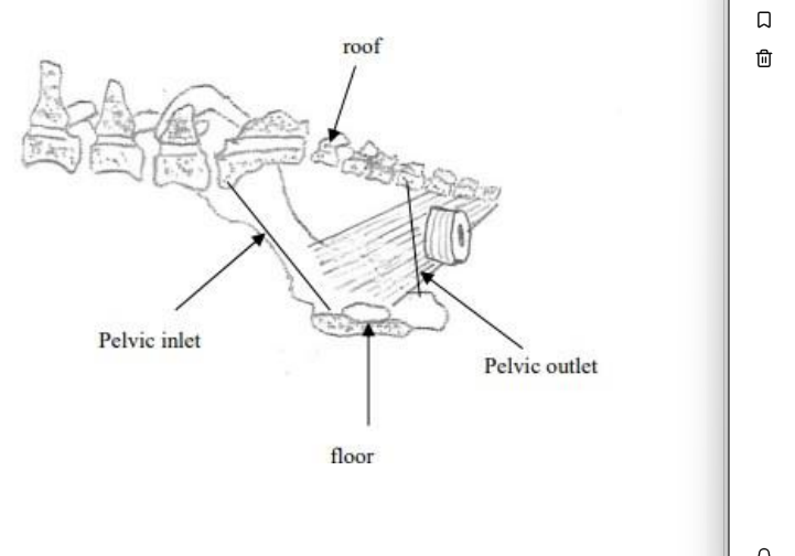
boundaries of the pelvic cavity- what are these structures defined by
pelvic inlet
roof
floor
pelvic outlet
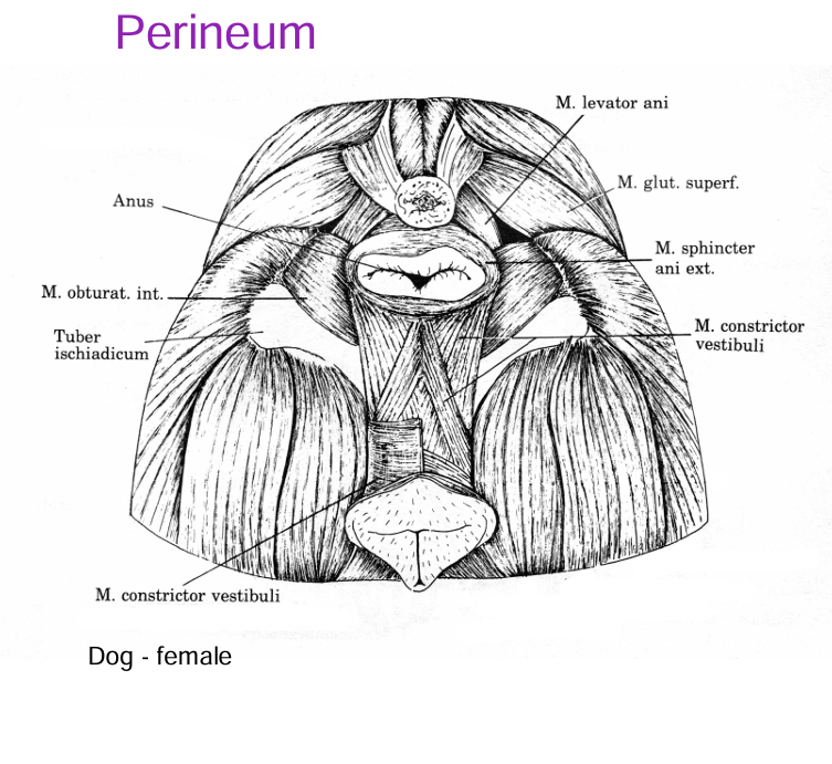
perineum
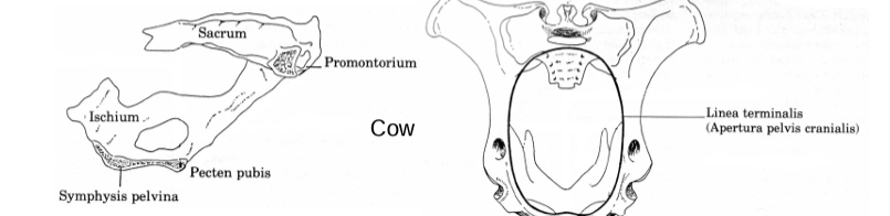
pelvic inlet is defined by the terminal line formed by the sacrum and cranial edges of the hip bones
the roof formed by the sacrum and first caudal vertebrae
the floor formed by hip bones (pubis)
the pelvic outlet is continuous with the perineum
the perineum is the region of body wall around the anus and urogenital openings
what are problems associates with the pelvic cavity
fixed diamter as a result of its bondy borders (hip bones and sacrum)
this can give rise to problems in partrition as aresult of foestal size and conformation eg braciocephaic dog
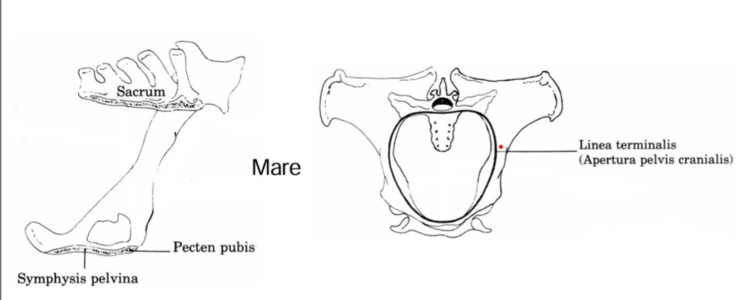
horse
pelvic inlet
ischial spine
floor of pelvis
canal
pelvic inlet is relatively wide
ischial spine and tubers are small
floor of pelvis is flat and the canal is straight
ruminant
pelvic inlet
ischial spine and tubers
floor of pelvis
pevic inlet is relatively narrow
ischial spine and tubers are larger than in the mare
floor of pelvis is concave
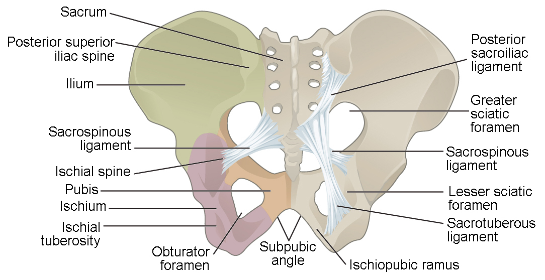
boundaries of the pelvic cavity
what forms the lateral walls
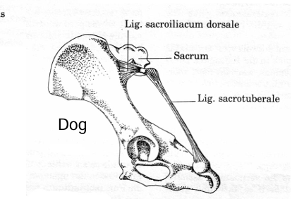
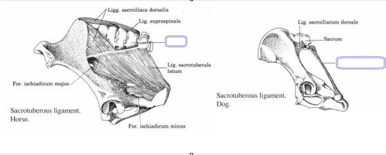
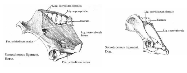
lateral walls are formed by hip bones and broad sacrotuberal ligaments
in the dog the sacrotuberal ligament is reduced to a cord
the ligament is absent in cat
dysotcia
which species is predisposed
the maternal conformation predisposes to dystocia more commonly in ruminants than in the mare which has a striaghter canal and flatter floor
other dfferent factors eg oversized foetus or ling legs of the foal
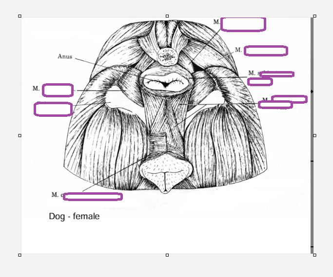
pelvic diaphragm
what muscles
formed by levator ani and coccygeus muscles
arranged as a cradle around the anus and retains the organs in the pelvis
levator ani and coccygeus muscle
perineum
is the region of the body wall around the alimentary and uorogenital openings
anus
vulva
male perineum
what does it contain
region of body wall around the alimentary and urogenital openings
contains the root (crura and bulb) of the penis
in some species the scrotum (tomcat and boar)
which arteries supply
hindlimb and body wall
pelvic region
midline structures and tauk
pelvic organ (including the artery it branches into)
wall of the pelvic caviy
paired iliac arteries supply the hindlimb and body wall
paired iliac arteries supply the pelvic region
median sacral arteries supply the midline structures and tail
internal iliac artery branches into internal pudendal artery supplying pelvic organs and caudal gluteal artery supplying the wall of the pelvic cavity
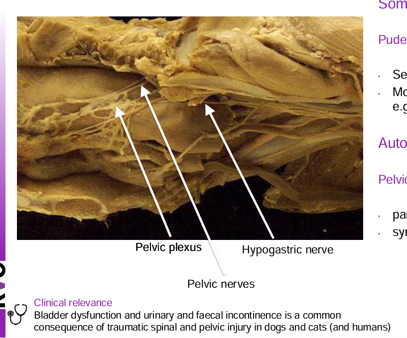
nerves
which nerve (of which origin) is sensory to the skin of the perineum and motor to striated perineum muscle
which is the autonomic
which one is parasympathetic
which one is sympathetic
pudendal nerve of the sacral spinal cord origin
sensory to skin of perineum
motor to striated msucle in perineum eg urethralis
somatic
pelvic plexus
autonomic
parasymptatheic pelvic nerves
sympathetic hypogastric nerve

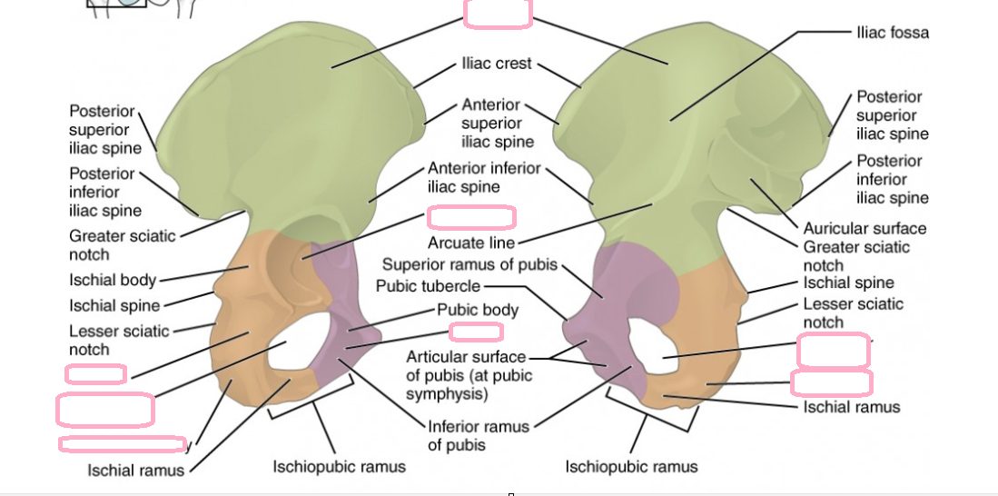
what do the os coxae consist of
ilium, ischium and pubis
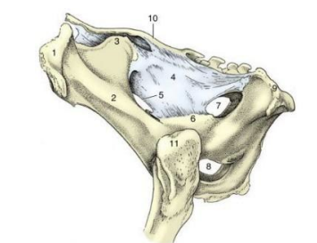
what are the deep bondy borders of the pelvic wall
dorsally
laterally
ventrally
1st caudal (or coccygeal) vertebra dorsally
the ischiadic tuberosity laterally
ischiadic arch ventrally
what is
the space lateral to the anus
what is this bordered by (ligament and muscle)
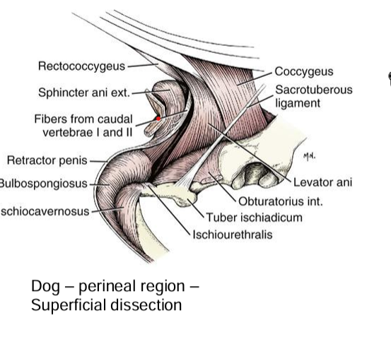
ischiorectal fossa
sacrotuberal ligament and the superficial gluteal muscle
where is each half of the diaphragm directed and where is its origin
each half of the diaphragm is directed caudomedially from its origin on the medial aspect of the spine of the ischium
causes of perineal hernias
weakening or failure of the pelvic diaphragm
may contain herniated abdominal and pelvic canal viscera
animals with perineal hernias will demonstrate a swelling adjacent to the rectum on one or more sides coupled typically with difficulty defaecating and altered tail carriage
sacrotuberal ligament
where is is the point of origin for
shape in large animals
where does it contribute to
what is the space between the ligament and the shaft of the ileum called
what is the space between the ligament and the lesser ischiatic notch called
what species is it absent in
superficial gluteal muscle
broad
lateral wall of the pelvic cavity
greater sciatic foramen
lesser sciatic foramen
cat
Q Describe the path of the ischiadic n. and its relationship to the gluteal and hamstring mm. and the os coxae
origin
where does it leave
originates from the sacral plexus
leaves pelvis through the greater sciatic foramen
passes through the gluteal but does not innervate
innervates the hamstring
sphincter ani externus
arrangement
where does it have insertions in dog
what does it cover
where does the duct of each anal sac open
circularly around the anus
coccygeal vertebrae
anal sacs (sinus paranalis)
the cutaneous zone of the anus
what is the clinical relevance of the anal sac glands
can becone inflamed, infected or impacted
tumours (most commonly apocrine gland adenocarcinoma) can invade surrounding tissues and metastasise
tumours sometimes cause hypercalcaemia
where do the internal and external obturator muscle originate and insert
the dorsal and ventral rims of the obturator foramen
and insert on the proximal femur
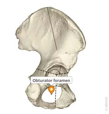
innervation of the internal obturator muscle and function
obturator internus
rotate the thigh laterally and stability to hip joint
innervation and function of the external obturator
obturator nerve
lateral rotation of the thigh, abduction of the thigh and the stability of the hip joint
clinical relevance of the obturator nerve
potential for it to be damaged during parturition as it can be crushed against the ilium by the foetus during delivery
abductor muscles will be affected
what are the
autonomic motor innervation of thee
pelvic viscera
striated muscle of the perineum
sensory innervation of the skin surrounding the anus and urogential openings
in dogs and cats what is a common consequence of spinal and pelvic injury
bladder dysfunction and faecal incontinence
what structures in the reproductive system take origin on the ischium
crura of the penis
clitora in female
ischiocavernous and ischiurethral
what surrounds the pelvic urethra
what muscle
what is it the homologue of (2)
what muscle is this a continuation of
the bulbospongiosus muscle is the homologue of the constrictor vestibuli and constictor vulvae muscle of the female
this is the continuatiopn of the urethral muscle which surrounds the pekvic urethra
coxal tuber
shaft of ileum
sacral tuber
sacrosciatic ligament
greater sciatic foramen
ischial spine
lesser sciatic foramen
right and left obturator foramina
ischial tuber
sacrum
greater trochanter