Lesson 27: Urinary Histology
1/81
There's no tags or description
Looks like no tags are added yet.
Name | Mastery | Learn | Test | Matching | Spaced | Call with Kai |
|---|
No analytics yet
Send a link to your students to track their progress
82 Terms
What is the approximate weight and size of a kidney?
About 150 g and 12 cm.
What surrounds the kidneys?
Adipose tissue.

What are the two main functional systems of the kidney?
Filtering system (blood plasma ultrafiltration) and
Tubular system (reabsorption and secretion).
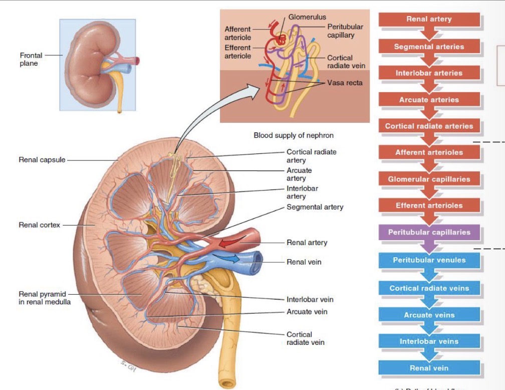
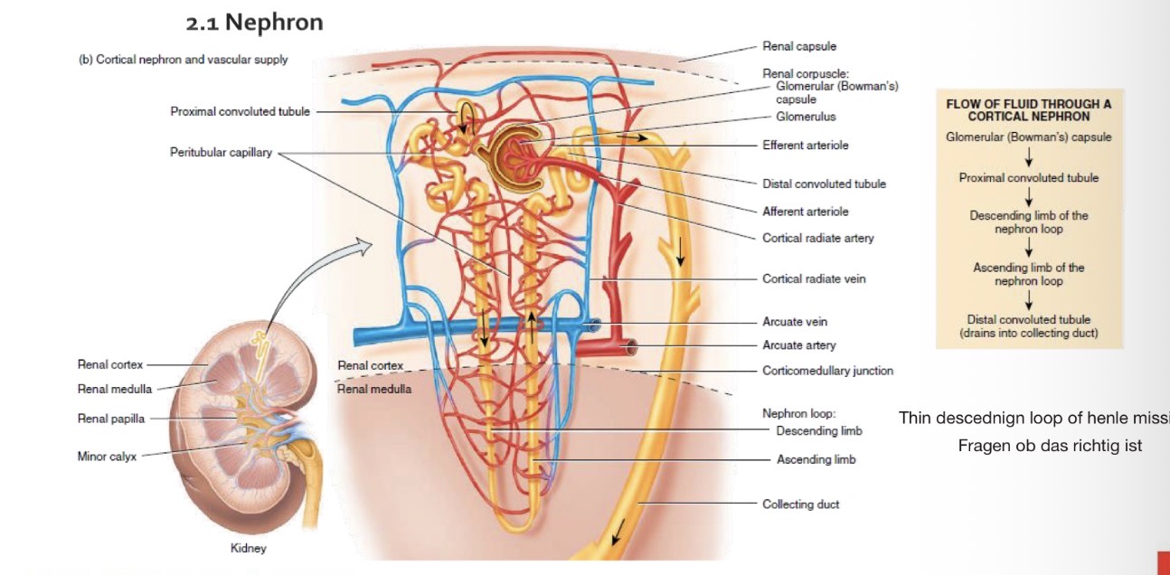
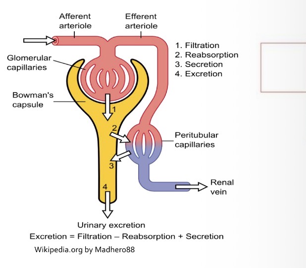
What is the Path of Blood flow?
Afferent arterioles → Glomerular capillaries → Efferent arterioles → Peritubular capillaries
What surrounds the kidneys?
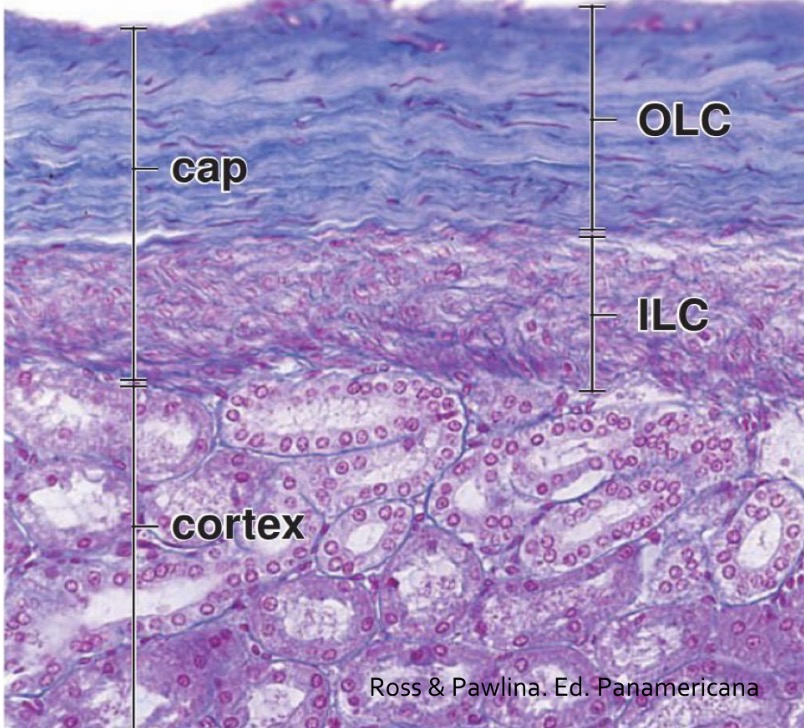
Connective tissue capsule which is thin and loosely adhered
What is the renal capsule composed of?
Outer layer: fibroblasts and collagen fiber
inner layer: myofibroblasts.
What is in the parenchyma?
Set Nephrons embedded in connective tissue.
What is the cortex of the kidney?
The outer region of the kidney.
What are medullary rays?
Structures originating in the medulla that extend into the cortex and lack renal corpuscles.
They divide the cortex in small areas
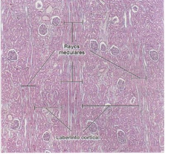
What is the renal labyrinth?
Cortical area between medullary rays containing corpuscles.
What are columns of Bertin?
Extensions of cortex that reach calyxes and separate renal pyramids.
What characterizes the renal medulla?
Inner region of the kidney with a whitish and striated appearance.
Has 8-10 renal pyramids surrounded by renal columns
Apex surrounded by renal calyx
Medullary rays depart it from base
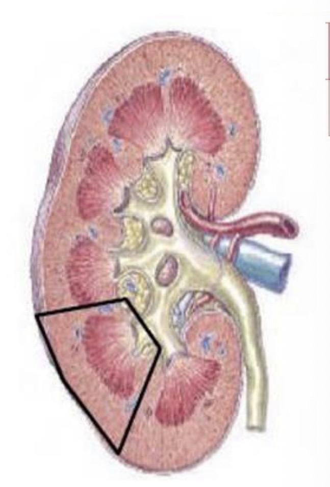
What is a renal lobule composed of?
One renal pyramid, cortex at its base, and half a column of Bertin.

What is the nephron?
The functional unit of the kidney (~1 million per kidney).
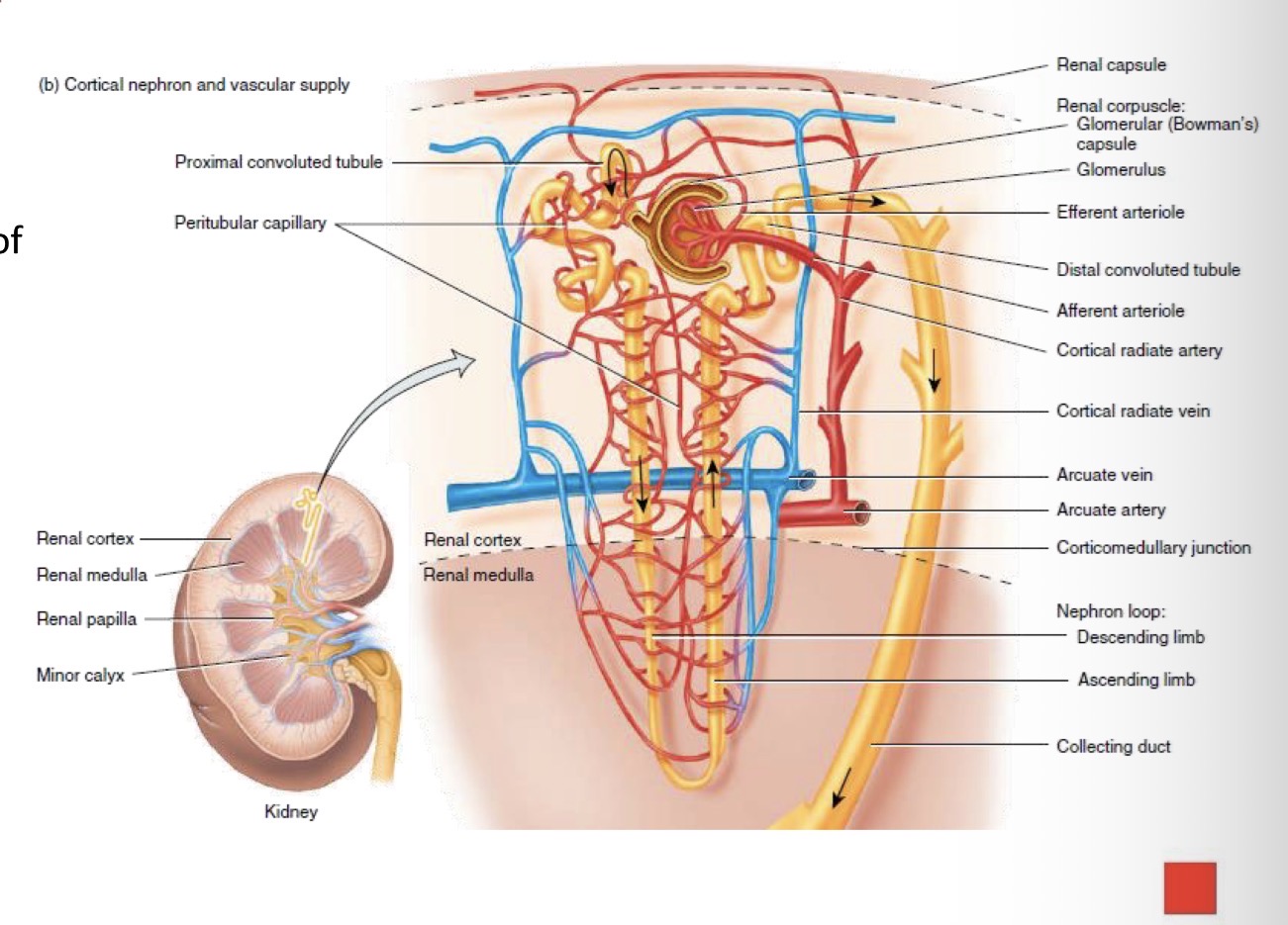

What are the two main parts of a nephron?
Renal corpuscle = where blood is filtered
renal tubule = into which the filtered fluid passes

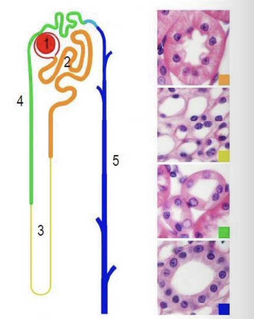
What are the parts of the nephron?
Renal corpuscle
proximal convoluted tubule
loop of Henle
distal convoluted tubule
connecting tubule
collecting duct
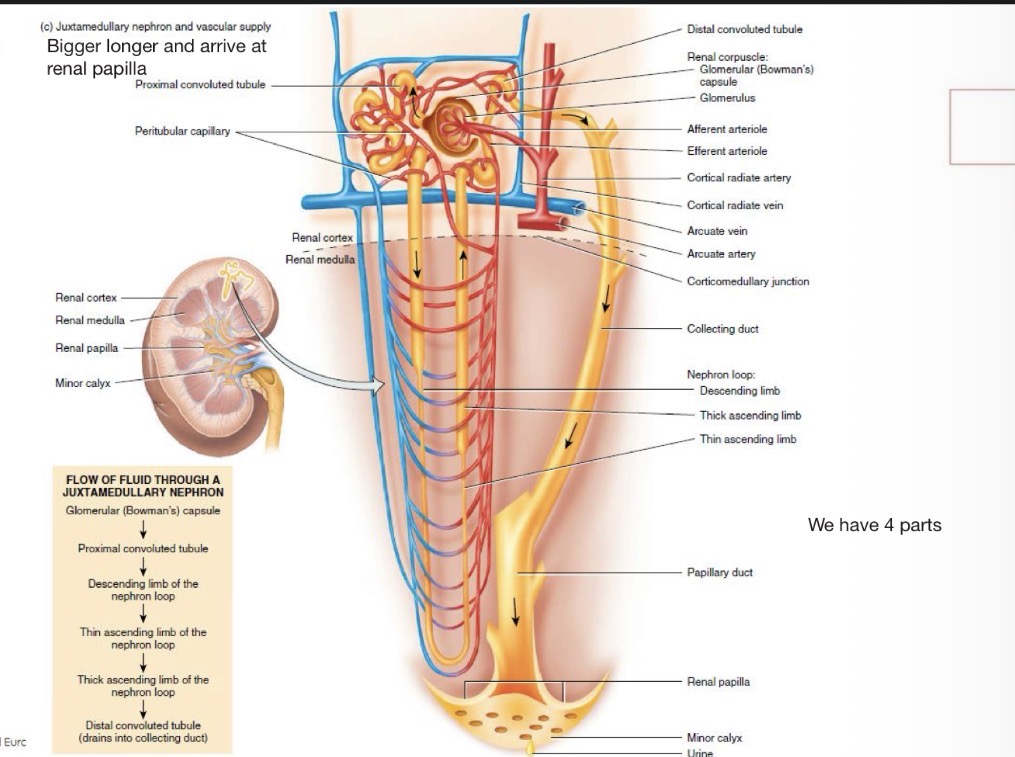
What types of nephrons exist?
Cortical and juxtamedullary nephrons.
What is the difference between a Cortical and a Juxtamedullary nephron?
Cortical nephron: The thin ascending loop of henle is missing
Juxtamedullary nephron: Bigger, longer and arrives at renal papilla
What is the flow of fluid through a cortical nephron?
Glomerular (Bowmans) Capsule → Proximal convoluted tubule (PCT) → Thick descending limb of nephron loop → Thin descending limb of nephron loop → Thick ascending limb of nephron loop → distal convoluted tubule (drains into collecting duct)
What is the flow of fluid through a Juxtamedullary nephron?
Glomerular (Bowmans) Capsule → Proximal convoluted tubule (PCT) → Thick descending limb of nephron loop → Thin descending limb of nephron loop →Thin ascending limb of nephron loop → Thick ascending limb of nephron loop → distal convoluted tubule (drains into collecting duct)
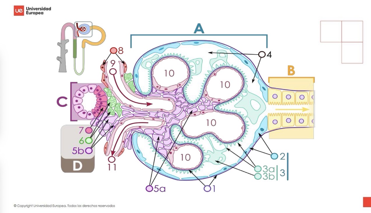
What is the function of the renal corpuscle?
Filtration of blood plasma.

Where are renal corpuscles located?
In the juxtamedullary cortex.
What forms renal corpuscles?
Glomerulus
Bowmans Capsule
Bowmans space
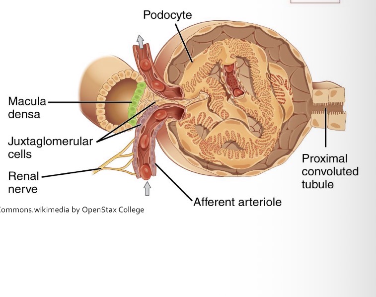
What are the poles of the renal corpuscle ?
Urinary pole and vascular pole.

What happens at Urinal pole?
Filtering out to PCT


What happens at the vascular pole?
Afferent arteriole enters and efferent arteriole exits.

What is the glomerulus?
A network of amastomosed capillaries and fenestrated capillaries surrouned by Bowmans capsule
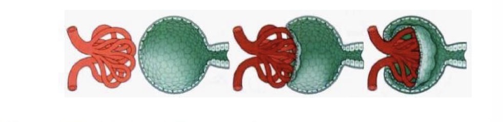
What is the Bowmans capsule?
A double layered epithelium
What forms Bowman’s capsule?
A parietal layer, a visceral layer and capsular space
What is the parietal layer of Bowman’s capsule?
Simple flat epithelium reticulin-rich lamina
What is the visceral layer of Bowman’s capsule composed of?
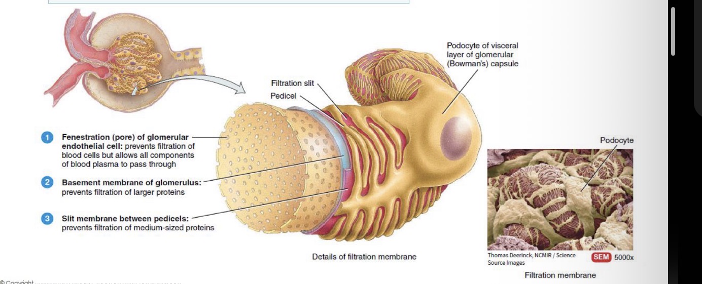
Podocytes with primary and secondary projections.
What is the capsular space?
The space that collects ultrafiltrate.
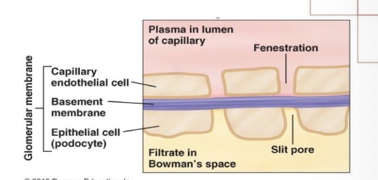
What forms the glomerular filtration barrier?
Fenestrated endothelium, basement membrane, and podocyte pedicels.

Functions of each filtration layer?
Fenestrated endothelium: prevents filtration of blood cells, but allows all components of blood plasma to pass through
Basement membrane: prevents filtration of larger proteins
Podocyte pedicels: prevents filtration of medium sized proteins
Where does the proximal convoluted tubule of a nephron begin and where is it located?
At the urinary pole of the renal corpuscle.
Only in the renal cortex, except the last part
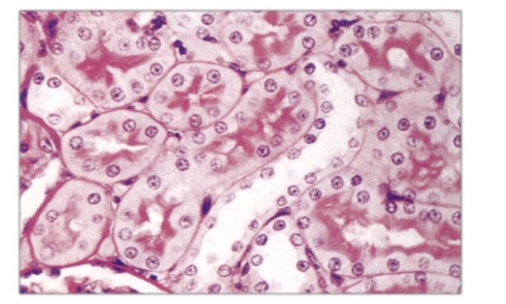
What is the function of the proximal convoluted tubule?
Reabsorption of about 75% of water and electrolytes and secretion of ions.
What epithelium lines the PCT?
Simple cuboidal epithelium with brush border (microvilli).
What are key features of PCT cells?
Microvilli, mitochondria, basal infoldings, and lateral interdigitations.
What is the shape of the loop of Henle?
U-shaped.
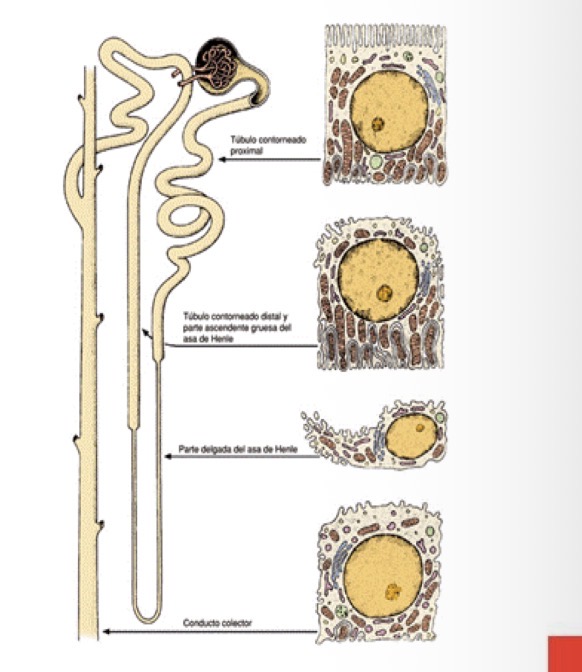
What are the parts of the loop of Henle?
Descending thick, thin descending/ascending, and thick ascending segments.

What epithelium lines the thin segments?
Simple squamous (flat) epithelium with short microvili
What epithelium lines the thick segments?
Simple cuboidal (cubic) epithelium without microvilli
What Epithelium is in the DCT?
Cubic Epithelium wiht few microvilli
What is the distal convoluted tubule a continuation of?
The thick ascending limb of Henle’s loop.
What is the DCT of a nephron located?
In the kidney cortex.
What is the macula densa?
specialized region of DCT with densely packed cells.
Differences of DCT that can be distinguished from PCT?
DCT Fewer microvilli then PCT
Larger lumen,
Tubule more
ACidophilia of cytoplasm
Similarities DCT PCT
MIcrovilli
Membrane invaginations
Abundant mitochondria
How to differenciate PCT and DCT Histology
Initial and straight area of return to the cortex (macula densa) presents
columnar epithelium and packed cells
What follows the DCT?
Connecting tubule and then collecting duct with larger diameters
Epithelium change bewteen collecting tubules to collecting ducts?
Simple cuboidal (cubic) transitioning to simple columnar (cylindrical) epithelium.
What are the cell types of the epithelium of collecting ducts?
Simple cylindrical epithelium of collecting ducts have 2 cell types:
Principal cells (clear)
Intercalated cells (dirty-looking)

What are principal cells?
Light cells with short microvilli and a central cilium
They have invaginations or folfs in the basal area
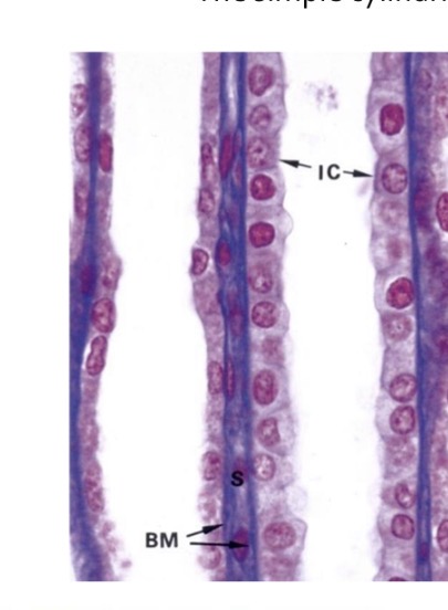
What are intercalated cells?
Dark cells with many mitochondria and microvilli.
Abundant in the proximal area until disappearing in the papilla
Scattered among light cells

What lines the renal pelvis, calyces and the proximal and of the ureter?
Urothelium
What kind of epithelium is the urothelium?
Stratified epithelium with cubic basal cells, polygonal intermediate cells and large surface cells
What does the pelvic wall contain?
Smooth muscle cells that form a continuous layer with the wall of the ureter
What is the juxtaglomerular apparatus?
Specializatrion of the afferent artiole of the glomerulus and the DCT of each nephron.
What is the function of the juxtaglomerular apparatus?
Regulation of blood pressure via RAAS mechanism
What are the components of the JGA?
Macula densa, juxtaglomerular cells, and lacis cells.
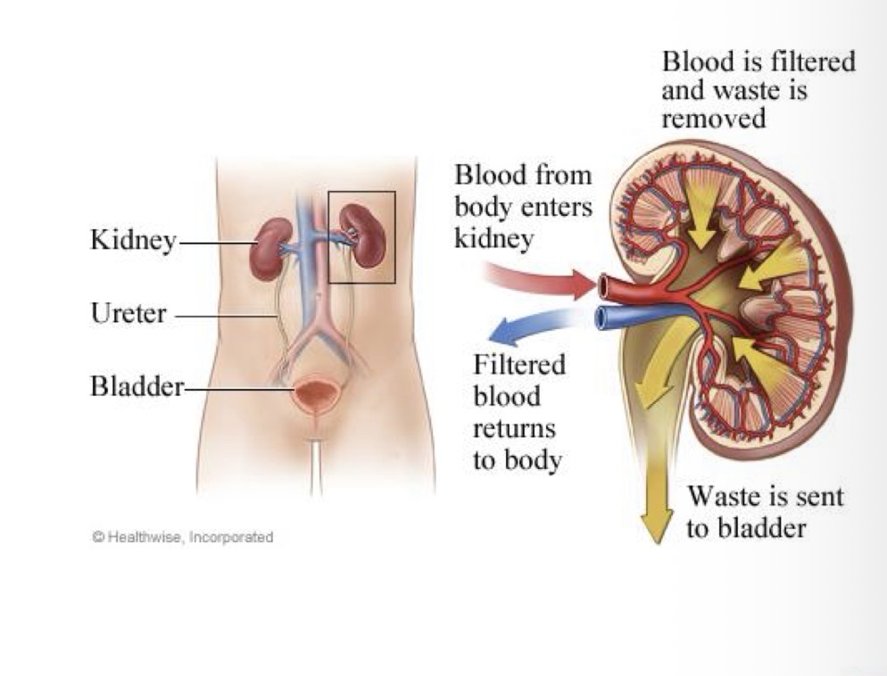
What are the ureters?
Muscle tubes that transport urine from renal pelvis of kidneys to bladder.
What epithelium lines the ureters?
Transitional epithelium (urothelium).
Laminate propia of dense ct (highly vascularized)
What layers form the ureter wall?
Mucosa, muscle layer, and adventitia.
How is the ureter muscle arranged?
2 layers of smooth muscle cells
Inner longitudinal and outer circular layers,
plus an additional outer longitudinal layer in final third of ureter
How is the Tunica adventita arranged?
Retroperitoneal adipose tissue with blood and lymphatic vessels and nerves
What is the urine bladder and what is the function of the urinary bladder?
Organ that receives and stores urine formed in the kidneys and transported by the ureters
Structure of bladder wall
Mucosa, muscle layer and adventitia
What epithelium lines the bladder?
Transitional epithelium (urothelium)
Lamina propia
What layers form the bladder wall?
What is special about bladder muscle?
Three layers of smooth muscle tissue surrounded by connective tissue with elastic fibers that contract during urination
Adventita of bladder wall
Connective tissue that contain bladder plecus with nerve fibers:
Sympathetic = supply blood vessels
Parasympathetic = participate in empyting of bladder by causing contraction of muscle layers (urination reflex)
What is the role of parasympathetic innervation?
Stimulates bladder contraction during urination.
What is the length of the male urethra?
Approximately 20 cm.
What is the function of urethra?
Carries Urine and semen.
What are the parts of the male urethra?
Prostatic, membranous, and spongy area.
How does epithelium change in the male urethra?
Transitional → pseudostratified with skeletal muscle → pseudostratified which changes to stratified flat at distal end
What is the length of the female urethra?
4–5 cm.
What is the function of the female urethra?
Transport of urine only.
What epithelium lines the female urethra?
Transition from urothelium to stratified squamous epithelium.
What are the glands of Littre?
Mucus-secreting glands that lubricate and protect the urethra.