Eukaryotic gene expression
1/55
There's no tags or description
Looks like no tags are added yet.
Name | Mastery | Learn | Test | Matching | Spaced | Call with Kai |
|---|
No analytics yet
Send a link to your students to track their progress
56 Terms
Gene expression
DNA carries the information for protein (specifically polypeptide) synthesis
information is transferred to the mRNA by transcription and is in turn transferred to the polypeptide by translation occurring on ribosomes
heredity: ability of cells to use the information in their DNA to produce particular proteins, which will affect cellular functions
gene expression: process by which the information within a gene is used, first to synthesise RNA (transcription) and then to synthesise a polypeptide (translation) eventually to affect the phenotype of an organism
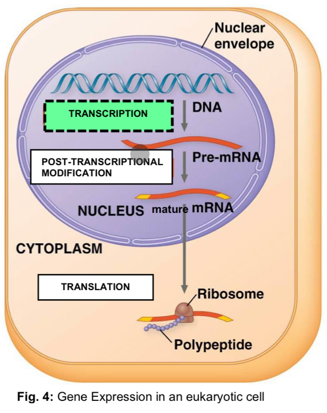
Overview of gene expression
Transcription
location: nucleus
DNA is used as a template for the synthesis of mRNA in the nucleus
Translation
location: free ribosomes in cytoplasm, bound ribosomes on rough endoplasmic reticulum (rER)
mature mRNA is used as a template for the synthesis of polypeptides at the free ribosomes or at the bound ribosomes
Post-translational modification of polypeptides
location: rER, Golgi apparatus and cytoplasm
processes
attachment of biochemical functional groups
structural changes of proteins
removal of amino acid sequence
attachment of ubiquitin for proteolytic degradation by proteosome
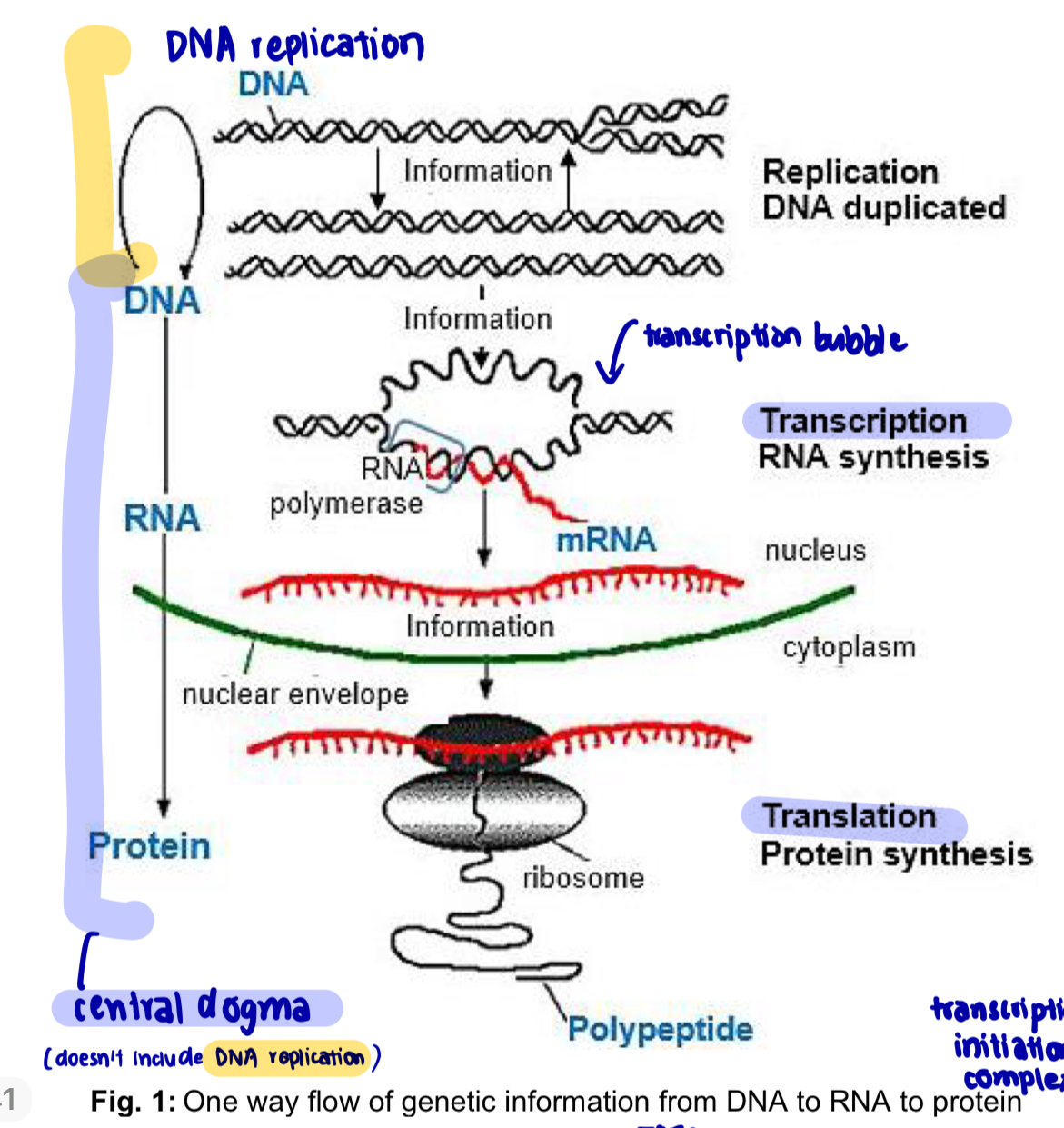
Central Dogma
flow of genetic information proceeds from DNA to RNA (t/c) to polypeptide (t/l)
unidirectional information flow —> Central Dogma
2 stages of gene expression
Transcription: DNA —> RNA
DNA is used as a template for the synthesis of mRNA in the nucleus
DNA base sequence is transcribed into a complementary mRNA base sequence
Translation: RNA —> polypeptide
mRNA is used as a template for the synthesis of polypeptides in the cytoplasm
mRNA codon sequence is translated into the amino acid sequence of. polypeptide chain
NOTE: DNA replication VS transcription
DNA replication —> both parental strands are templates
transcription —> only template strand is template
Gene expression through RNA
RNA is an intermediate in protein synthesis
its function depends on complementary base pairing, which dictates its interactions specifically with other RNA molecules
*DNA is permanent and will not leave nucleus, while RNA is temporary and leaves nucleus via nuclear pore protein complexes and enters the cytoplasm
Similarities btw RNA & DNA*****
Both RNA and DNA are polynucleotides
components of each nucleotide monomer are
phosphate group
pentose sugar
nitrogenous base
A strand of RNA and a strand of DNA has a sugar-phosphate backbone joined by phosphodiester bonds
Both RNA and DNA make use of 3 nitrogenous bases: A and G (purines), C (pyrimidine)
Both RNA and DNA sequences are determined by complementary base pairing of nucleotides with a template
Both RNA and DNA polynucleotide chains are formed via condensation reaction in which a water molecule is removed
Difference btw RNA and DNA
Molecular mass
RNA is smaller
DNA is larger
Number of polynucleotide chains
RNA: single stranded (1 chain)
DNA: double stranded (2 chains)
Secondary structure
RNA: almost always a single-stranded helical molecule, which can be folded into a complex tertiary structure
DNA: always a double-stranded helical molecule
Monomers
RNA: ribonucleotides
DNA: deoxyribonucleotides
Pentose sugar
RNA: Ribose (OH group attached to 2’ carbon)
DNA: Deoxyribose (H attached to 2’ carbon)
Chemical stability
RNA: less stable - ribose has an additional reactive 2’ OH group
DNA: more stable - deoxyribose lacks 2’ OH group
Nitrogenous bases
RNA: Adenine (A), Guanine (G), Cytosine (C), Uracil (U)
DNA: A, G, C, Thymine (T)
Ratio of bases
RNA: A:U ≠ G:C ≠ 1:1
DNA: A:T = C:G = 1: !
Basic forms
RNA: several different kinds and sizes of RNA, each with its own function
messenger RNA
transfer RNA
ribosomal RNA
small nuclear RNA (snRNA)
small interfering RNA (siRNA)
DNA: only one basic form
Location
RNA: synthesised in the nucleus but found throughout the cell
DNA: found almost exclusively in the nucleus with exceptions of mitochondria and chloroplasts
Amount per cell
RNA: amount varies from cell to cell and, within a cell according to metabolic activity
DNA: amount is constant for all somatic cells of a species
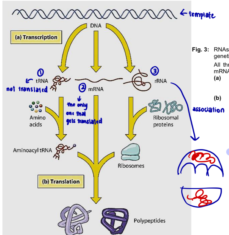
Roles of RNA
Types of RNA & their functions
Messenger RNA (mRNA)
carries information, which codes for amino acid sequences, from DNA to ribosomes
Transfer RNA (tRNA)
serves as an adaptor molecule in protein synthesis
translates mRNA codon sequence into amino acid sequence
Ribosomal RNA (rRNA)
plays catalytic and structural roles in ribosomes
*not all enzymes are proteins
Small nuclear RNA (snRNA)
plays catalytic and structural roles in spliceosomes, the complexes of protein and RNA that carry out splicing of pre-mRNA
Small interfering RNA (siRNA) & microRNA (miRNA)
involved in regulation of gene expression
Transcription
Transcription: process by which a complementary RNA copy is made under the direction of the template strand of a specific region of the DNA molecule, catalysed by the enzyme RNA polymerase
Components of the transcription machinery
Gene
RNA polymerase
General/basal transcription factors
Ribonucleotides (or more specifically ribonucleoside triphosphate )
Gene
A gene is a section of DNA that encodes information in the form of a specific base sequence to direct the synthesis of one polypeptide chain or RNA molecule
it is a unit of inheritance located in a fixed position (locus) on the chromosome which specifies a particular character of an organism
3 components of eukaryotic gene
Promoter
Coding region
Termination sequence
illustration of GTF and RNA polymerase
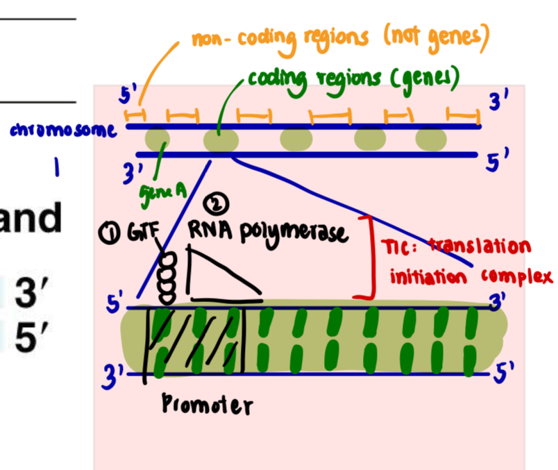
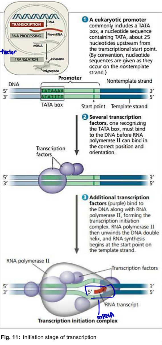
Promoter
contains TATA box and transcription start site (+1: nucleotide where RNA synthesis begins)
TATA box is binding site for a general transcription factor (GTF) i.e. TFIID —> facilitates binding of RNA polymerase
TATA box is usually located 25bp upstream of transcription start site
RNA polymerase + GTF = transcription initiation complex
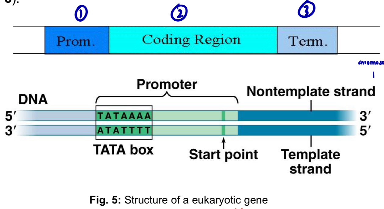
Coding region
the segment of DNA that is transcribed into a single-stranded RNA molecule (i.e. single primary transcript known as pre-mRNA or primary transcript)
bounded by the transcription start site and termination site
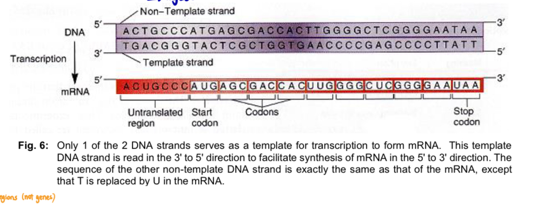
only 1 of the 2 strands serves as the template for transcription
template DNA strand is read in the 3’ to 5’ direction to facilitate synthesis of RNA in the 5’ to 3’ direction
template strand
DNA strand that is transcribed
sequence on this strand is complementary to that of the RNA
template to direct the synthesis of the RNA molecule
non-template strand
not transcribed
sequence on the DNA strand is exactly the same as that of the RNA (except thymine is replaced with uracil in RNA)
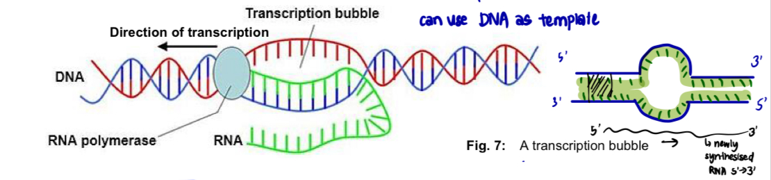
RNA synthesis occurs within a transcription bubble in which DNA is transiently separated into its single strands, one of which will be used as template
transcription bubble
comparison btw RNA polymerase (transcription) and DNA polymerase (DNA replication)
DNA pol needs helicase (to unwind and separate the DNA strands first); RNA pol requires just itself
DNA pol needs primase (to form RNA primer); RNA pol itself can start transcription
DNA pol needs SSB to keep template open; RNA pol itself
DNA pol needs topoisomerases, ligase; RNA itself
Termination sequence
at the end of a gene and it codes for a polyadenylation signal sequence (AAUAAA) in the pre-mRNA
the whole termination sequence is transcribed —> transcription termination i.e. formation of phosphodiester bonds stops
RNA polymerase
RNA polymerase is an enzyme comprising of several protein subunits and is found in the nucleoplasm
can bind the following to initiate transcription: template and nucleotide
RNA pol responsible for RNA synthesis using ribonucleoside triphosphate (NTP) as its substrate
During t/c, RNA polymerase reads the DNA template in the 3’ to 5’ direction, catalysing
the assembly of ribonucleotides, which form complementary base pairs with the template
formation of phosphodiester bond btw the free 5’ phosphate group of the incoming ribonucleotide/ribonucleoside triphosphate (NTP) and the free 3’ OH group of the growing RNA polynucleotide chain
RNA is synthesised in the 5’ to 3’ direction
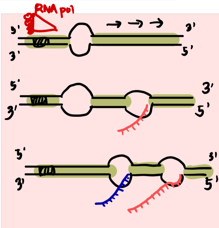
Simultaneous transcription from the same DNA template strand is possible
many RNA polymerase molecules can be transcribing different parts of the same gene simultaneously
NOTE:
in prokaryotes, there is only 1 type of RNA polymerase
in eukaryotes, there are 3 types of RNA polymerases
RNA pol 1 transcribes genes encoding rRNA (nucleus)
RNA pol II transcribes most genes including all those that encode proteins, synthesised mRNA** (nucleoplasm)
RNA pol III transcribes genes encoding tRNA (nucleoplasm)
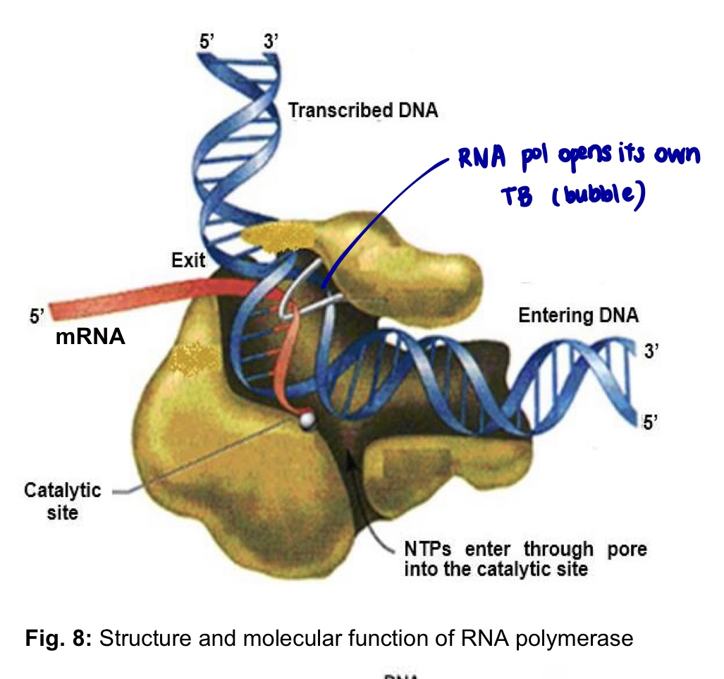
illustration of RNA polymerase catalysing formation of CBP
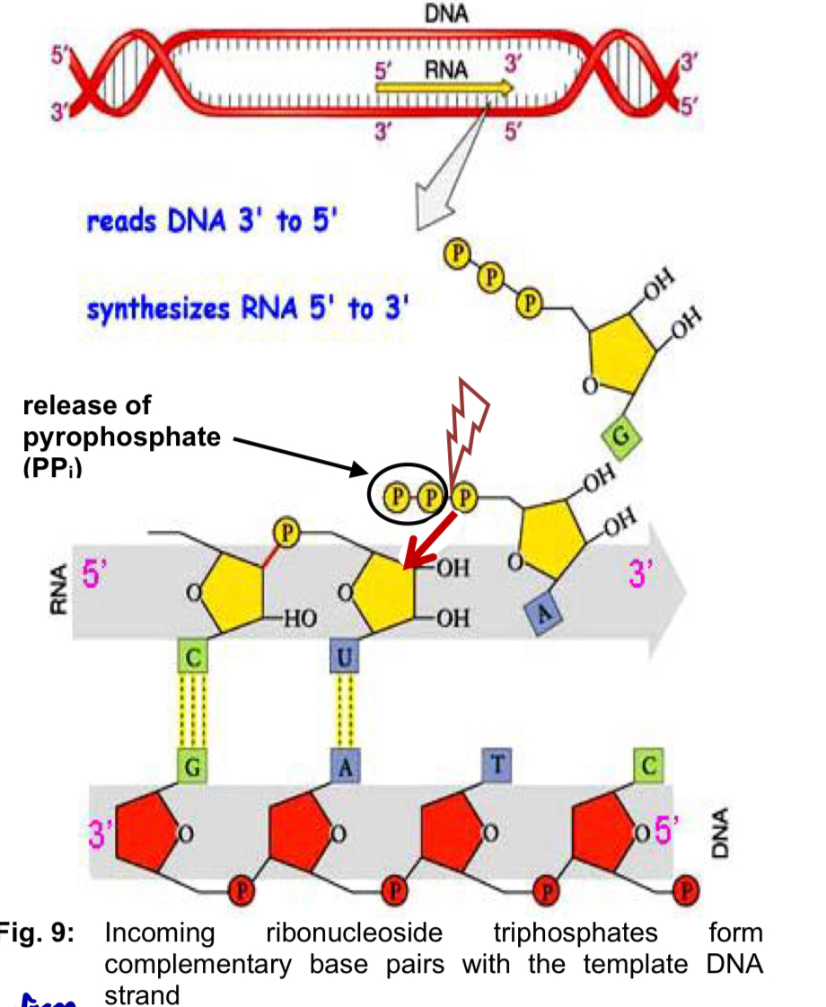
illustration of RNA polymerase catalysing formation of CBP
General transcription factors
protein that is required for an RNA polymerase molecule to bind to its promoter and initiate transcription
roles
***position RNA polymerase correctly at the promoter
release RNA polymerase from the promoter to begin elongating the RNA against the DNA template once t/c has begun
GTF bind first —> call RNA pol —> RNA pol binds —> TIC
Steps involved in t/c
Initiation
Elongation
Termination
*all are mediated by RNA polymerase itself (unwinding, initiating RNA synthesis, reforming the double helix)
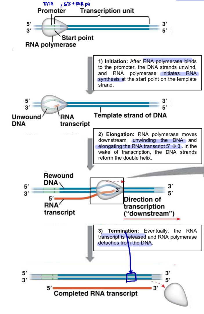
Initiation
Step 1: Formation of Transcription Initiation Complex
General transcription factors assembled along the promoter
TFIID binds to the TATA box found within the promoter
GTF mediate the binding of RNA polymerase to the promoter, forming a complex known as transcription initiation complex
Step 2: Unwinding of DNA helix and separation of 2 strands
Binding of RNA polymerase to the promoter causes the DNA double helix to unwind and the 2 strands separate
during this process
hydrogen bonds btw complementary base pairs are disrupted
transcription bubble exposing a short stretch of nucleotides on each strand is created
Step 3: Assembly of Ribonucleotides & Formation of 1st Phosphodiester Bond
one of the two exposed DNA strands acts as a template for CBP to direct the assembly of incoming ribonucleotides (nucleoside triphosphates)
RNA polymerase catalyses the formation of the first phosphodiester bond = marks the end of transcription initiation
Initiation illustration
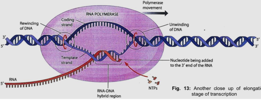
Elongation
Step 1: Movement of the transcription bubble
As RNA polymerase moves along the template DNA in the 3’ to 5’ direction, DNA double helix continues to transiently unwind
*****Step 2: Elongation of polynucleotide
Ribonucleotides (i.e. monomers) form CBP with the DNA template
As each ribonucleoside triphosphate is brought in, its two terminal phosphates are removed
the remaining free 5’-phosphate group is added to the free 3’-hydroxyl group of the growing RNA chain via the formation of a phosphodiester bond catalysed by RNA polymerase
i.e. mRNA is synthesised in the 5’ to 3’ direction
Step 3: Re-annealing of DNA and proofreading
RNA polymerase reanneals the unwound DNA behind it, dissociating the growing RNA chain from the template
RNA polymerase carries out proofreading functions and is responsible for the removal of any incorrectly inserted ribonucleotide
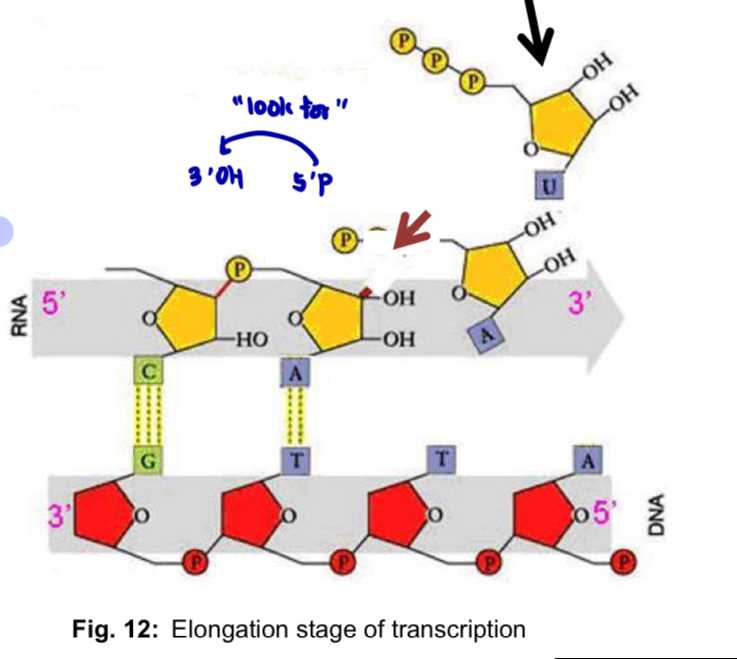
Elongation illustration
Termination
Transcription proceeds until after the RNA polymerase transcribes a termination sequence in the DNA
triggers the release of the RNA chain and the dissociation of the RNA polymerase from the DNA
the transcribed terminator - RNA sequence - codes for a polyadenylation signal sequence (AAUAAA)
in eukaryotic cell, the RNA polymerase continues transcription until at a point about 10 to 35 nucleotides downstream of the polyadenylation signal sequence
proteins bind at this point to cut and free the pre-mRNA from the RNA polymerase
cleavage site on the mRNA is also the site of addition of poly (A) tail
Post-transcriptional modification of mRNA
all the pre-mRNA transcribed in the eukaryotic nucleus must undergo processing to produce functional mature RNA molecules for export to the cytosol, where they will be used as templates for translation to form polypeptides
3 steps
pre-mRNA
addition of 5’ cap
RNA splicing
addition of 3’ poly (A) tail
mature mRNA (only mature mRNA can exit the nucleus via nuclear pores)
structure-function relationship of mature mRNA
transport across nuclear envelope
stability (yet so stable, yet so unstable)
facilitation of translation
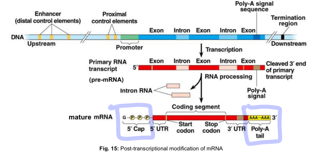
Addition of 5’ Cap
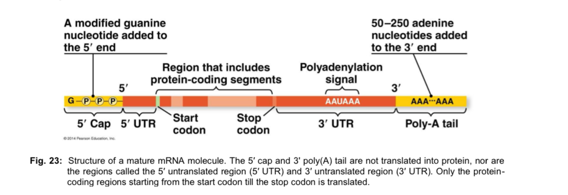
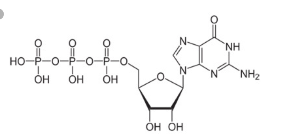
5’ end of the new RNA molecule is modified by addition of a cap that consists of a methylated guanine (G) nucleotide/methylguanosine triphosphate
Functions of the 5’ cap
5’ cap protects the mRNA from degradation by hydrolysis enzymes such as nucleases
5’ cap defines the 5’ end of the mRNA, which serves to recruit the small subunit of the ribosome for translation initiation
5’ cap distinguishes mRNAs from the other types of RNA molecules (e.g. tRNAs, rRNAs)
drawing of post-transcriptional modification
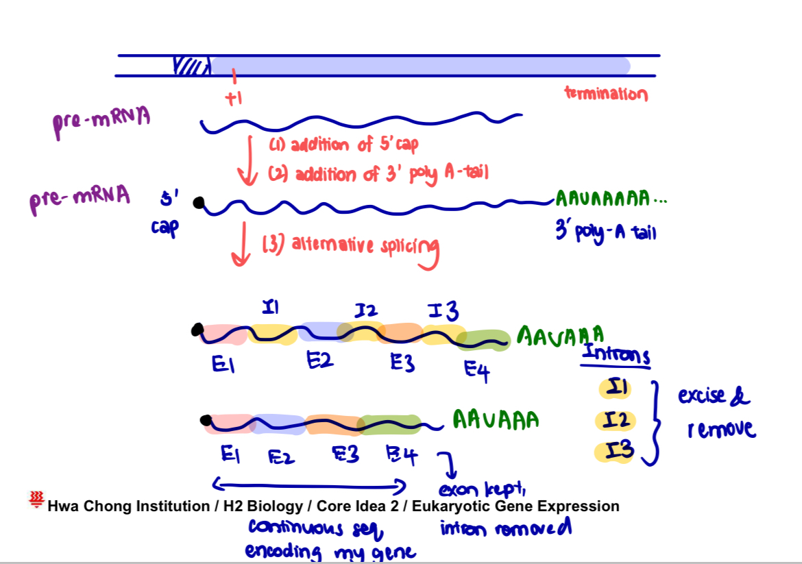
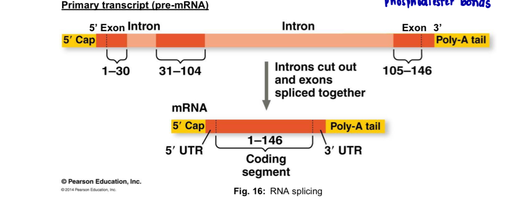
RNA splicing
eukaryotic genes contain
exons, which are protein-coding sequences in the gene
introns, which are long stretches of nucleotides inserted btw exons that do not code for any portion of the polypeptide i.e. non-coding sequences
both intron and exon sequences are transcribed into pre-mRNA
RNA splicing occurs after the release of pre-mRNA from RNA polymerase
during this process, introns are removed while the remaining exons are spliced/joined together to form mature mRNA - require hydrolysis of ATP (breaking and joining phosphodiester bonds)
splicing is carried out by spliceosome which is a large complex comprising of several subunits known as small nuclear ribonucleoproteins (snRNPs)
each snRNP contains small nuclear RNAs (snRNAs) and a set of proteins
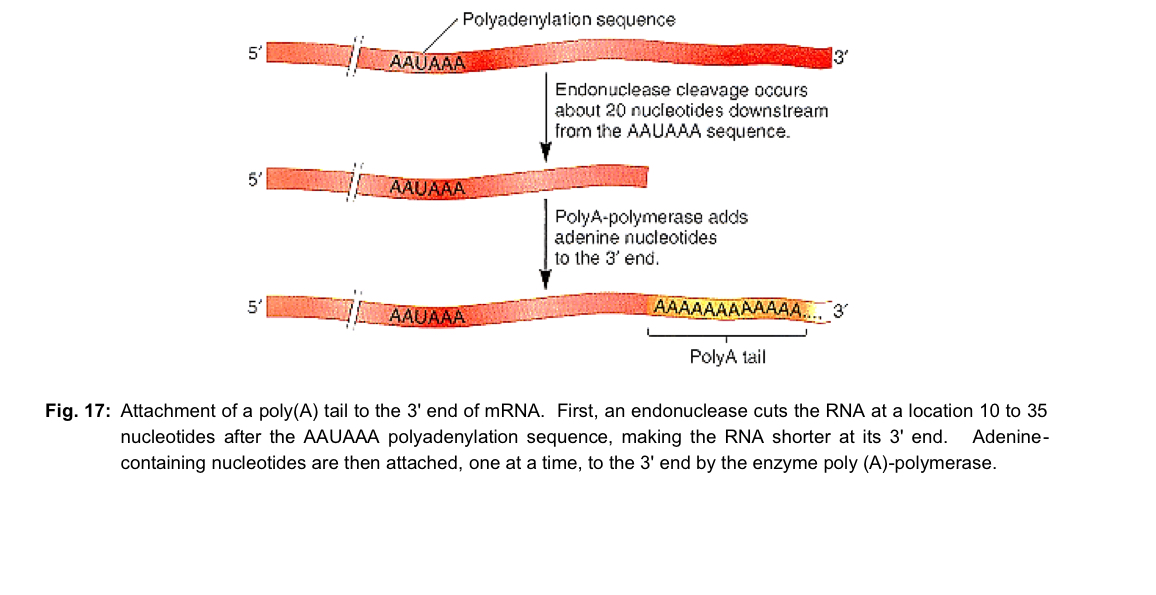
Addition of 3’ Poly (A) Tail
immediately after the pre-mRNA is cleaved by an endonuclease at a site 10-35 nucleotides after the AAUAAA polyadenylation sequence, the 3’ end of the pre-mRNA is modified by addition of a series of approximately 200 adenine (A) nucleotides - poly (A) tail
addition of adenine nucleotides at this cleavage site is catalysed by the enzyme poly (A) - polymerase
Functions
3’ poly (A) tail protects the mRNA from degradation by nucleases; 3’ poly (A) tail makes the mRNA more stable template for translation in the cytoplasm
3’ poly (A) tail is required to facilitate export of mRNA out of the nucleus via nuclear pores
The Genetic Code
proteins are linear polymers of amino acids
sequence of bases along the DNA strand determines the sequence of amino acids in polypeptides
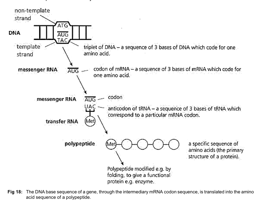
DNA base sequence of a gene, through the intermediary mRNA codon sequence, is translated into the amino acid sequence of a polypeptide by genetic code
The Triplet Code
20 different amino acids in proteins, but only 4 different bases in DNA (A, G, C and T)
each amino acid is specified by a codon, a base triplet
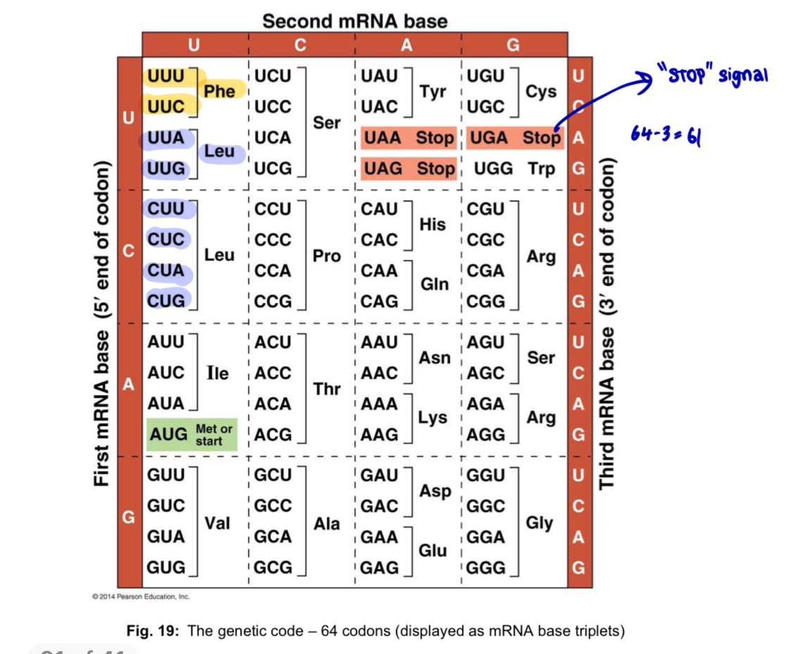
3-nucleotide codon (triplet code): 4³ = 64 different codons possible (more than enough to code for all 20 a.a)
Features of the genetic code
General features
sequence of triplet bases in the non-template/non-transcribed strand of DNA
64 possible codons
61 code for amino acids and include a start signal (start codon)
3 serve as termination signals of polypeptide synthesis (stop codons) —> do not code for any a.a
Key features
*TUND - Triple, Universal, Non-overlapping, Degenerate code
The genetic code is a triplet code
each mRNA codon that specifies an amino acid in a polypeptide chain consists of 3 nucleotide bases
The genetic code is (almost) universal
same code is used by almost all organisms
for instance, the codon AGA specifies the amino acid arginine in bacteria, human and all other organisms whose genetic code has been studied
The genetic code is continuous and non-overlapping
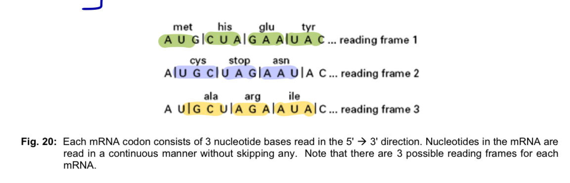
each codon is read as a triplet in the 5’ to 3’ direction
nucleotides in the mRNA are read continuously as successive groups of 3 nucleotides, one codon at a time without skipping any nucleotides
as the genetic code is read in blocks of 3 nucleotides, there can be 3 possible reading frames for every mRNA sequence
The genetic code is degenerate but unambiguous
a single amino acid can be coded by more than one different codon (e.g. UUU and UUC code for the same amino acid, phenylalanine)
in the genetic code, there are more codons than amino acids
only 2 amino acids - methionine (AUG) and tryptophan (UGG) are coded for by a single codon each
however, each codon codes for just one amino acid, thus the code is unambiguous
most of the amino acids are encoded by degenerate codons that differ in the 3rd position (3rd base) of the codon; thus mutations can arise in this position of the codon without altering amino acid sequences —> silent mutations
e.g. arginine is coded for by 4 degenerate codons - CGU, CGC, CGA, CGG (differ only in 3rd base)
Wobble base phenomenon occurs
a single tRNA can recognise 2 or more of the degenerate codons
phenylalanine tRNA with the anticodon AAG recognises not only the complementary mRNA codon UUC but also UUU
violation of the usual rules of base-pairing at the 3rd nucleotide of a codon is called wobble
base pairing at the 3rd base is not so specific - change in the 3rd base by a mutation may still permit the correct incorporation of a given amino acid in a polypeptide
The genetic code has punctuation codons - start and stop codons
start codon
within an mRNA message, the start signal for protein synthesis is the start codon AUG, which codes for the incorporation of methionine
start codon defines
the first amino acid of the polypeptide chain
reading frame used from that point on
stop codon
3 stop codons UAA, UAG and UGA are stop signals in the mRNA message marking the end of the protein synthesis - do not code for any amino acid
there is no tRNA with an anticodon complementary to these 3 codons
Translation
Translation: Process in which a polypeptide chain is synthesised by ribosomes using genetic information encoded in a mature mRNA template
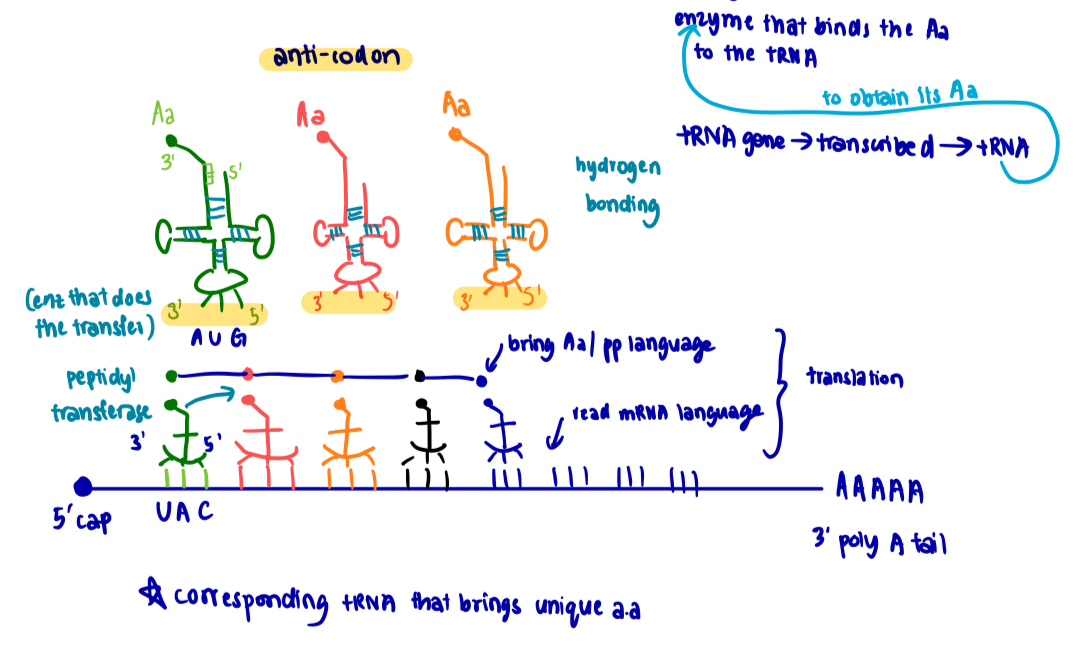
mRNA contains a series of codons that interact with the anticodons of aminoacyl-tRNAs so that a corresponding series of amino acids is incorporated into a polypeptide chain
ribosomes provide the environment for controlling the interaction via CBP btw mRNA and aminoacyl-tRNA
ribosome travels along the template mRNA in the 5’ to 3’ direction, engaging in peptide bond synthesis
polypepide is assembled by the sequential addition of amino acids beginning from the N-terminal to the C-terminal
Components of the translation machinery
Components
mature messenger RNA (mRNA)
transfer RNA (tRNA)
amino acids
aminoacyl-tRNA synthetase
ribosomal RNA (rRNA) and ribosome
translation factors
Mature messenger RNA (mRNA)
Structure
mature mRNA is a single RNA strand that exists for a relatively short time as it is continuously being synthesised and degraded
mature mRNA is obtained after the pre-mRNA undergoes post-transcriptional modification (5’ cap, 3’ poly A tail, RNA splicing)
then mRNA is transported to cytoplasm via nuclear pores
mature mRNA contains two regions
protein-coding region: consists of a series of codons representing the amino acid sequence of the polypeptide, starting with the start codon AUG and ending with a stop codon UAA/UAG/UGA
untranslated regions (UTRs): additional sequence at the 5’ end, preceding the start codon, known as the leader sequence or 5’ UTR; and following the stop codon - trailer sequence or 3’ UTR
*only the protein-coding regions starting from the start codon till the stop codon is translated
Role of mRNA
mature mRNA serves as an intermediate that carries the copy of DNA sequence information that encodes proteins
each codon within the coding region of the mRNA represents an amino acid in the corresponding amino acid sequence in the protein
mature mRNA acts as a template for translation i.e. guides the assembly of amino acids into a polypeptide chain
Transfer RNA (tRNA)
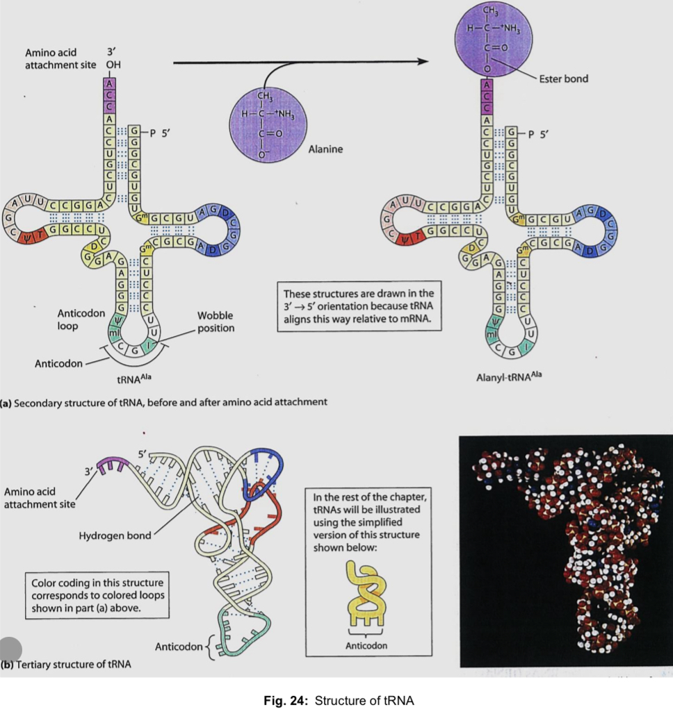
Structure
tRNA molecules are small, containing only about 80 nucleotides
the secondary structure takes the form of a 2D cloverleaf, held by CBP within the single-stranded molecule
3 loops
on the anticodon loop, 3 unpaired bases form an anticodon, which binds to a specific mRNA codon via CBP
tertiary structure is the result of actual twisting and folding of the secondary structure into a compact 3D L-shaped structure maintained by hydrogen bonds
3’ end (CCA stem) of a tRNA molecule is the attachment site for a specific amino acid
around 45 different tRNAs in a typical eukaryotic cell, thus each amino acid can be carried by more than 1 type of tRNA
3D structure of tRNA is recognised by the enzyme aminoacyl-tRNA synthetase that catalyses the formation of an ester linkage (covalent bond) btw the CCA stem and the specific amino acid
when a tRNA has its 3’ CCA stem attached to the amino acid corresponding to its anticodon —> aminoacyl-tRNA
Structure of tRNA illustration
Role of tRNA
tRNA serves as an adaptor molecule in the translation of an mRNA nucleotide sequence into the amino acid sequence in a polypeptide
they are used to bring in specific amino acids in a sequence corresponding to the sequence of codons in mRNA
*a.a corresponds to the codons (not the anticodons)
tRNA’s ability to act as an adaptor is due to
anticodon being able to determine the specific amino acid attached to the CCA stem
anticodon being able to form complementary base pairs with the mRNA codon
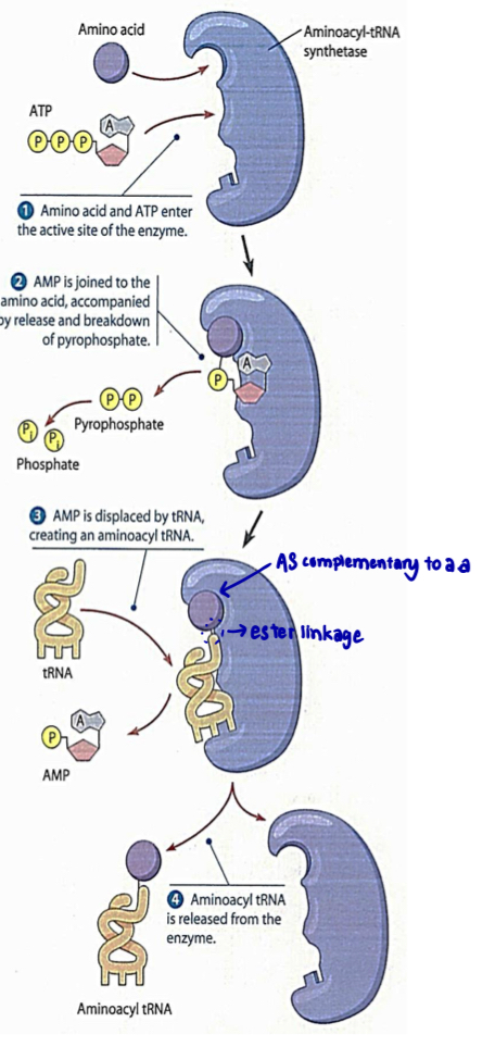
Amino acid activation by Aminoacyl-tRNA synthetase
before a tRNA molecule can bring its amino acid to the ribosome, that amino acid must be attached covalently to the tRNA
enzyme responsible for linking a.a to their corresponding tRNAs —> aminoacyl-tRNA synthetases
cells produce 20 different synthetase enzymes, one for each of the 20 distinct amino acids
one particular synthetase enzyme attaches glycine to all tRNAs that recognise codons for glycine
each of the 20 different synthetase enzymes must recognise the specific anticodon on a tRNA as well as a specific amino acid
each of the 20 different synthetase enzyme covalently attaches a specific amino acid to the 3’ CCA stem of its appropriate set of tRNA molecules via an ester linkage, (between carboxyl group of a.a and 3’ OH of tRNA) forming aminoacyl-tRNA, requires hydrolysis of ATP (basically enzyme joins a.a to tRNA via ester linkage)
active site of each aminoacyl-tRNA synthetase must be complementary to 3D conformation of the specific amino acid and specific anticodon sequence of the tRNA in order for them to bind
the resulting aminoacyl-tRNA is released from the synthetase enzyme and delivers its amino acid to a growing polypeptide chain on a ribosome
Ribosomal RNA (rRNA)
Structure
the most abundant RNA in cells are rRNA (80% of RNA in rapidly dividing cells)
rRNA genes are transcribed (by RNA pol I) and the rRNA is processed and assembled with proteins imported from the cytoplasm in the nucleolus - completed ribosomal subunits are exported via nuclear pores to the cytoplasm
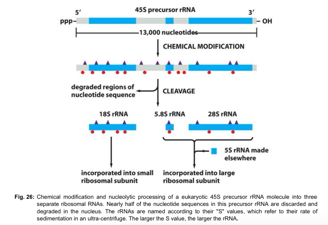
4 types of eukaryotic rRNA (1 copy of each type per ribosome)
3 of the 4 rRNA (18S, 5.8S and 28S) are made by chemically modifying and cleaving a single large precursor rRNA
each modification is made at a specific position in the precursor rRNA, function of modification probably regarding folding and assembly of final rRNA
5S rRNA is synthesised from a separate cluster of genes (by RNA pol III), does not require chemical modification
Role of rRNA
rRNA forms the core of the ribosome - it is the main constituent of the A and P sites and of the interface between the large and small ribosomal subunits
rRNA in the large ribosomal subunit has peptidyl transferase activity
catalyses the formation of peptide bonds btw amino acids
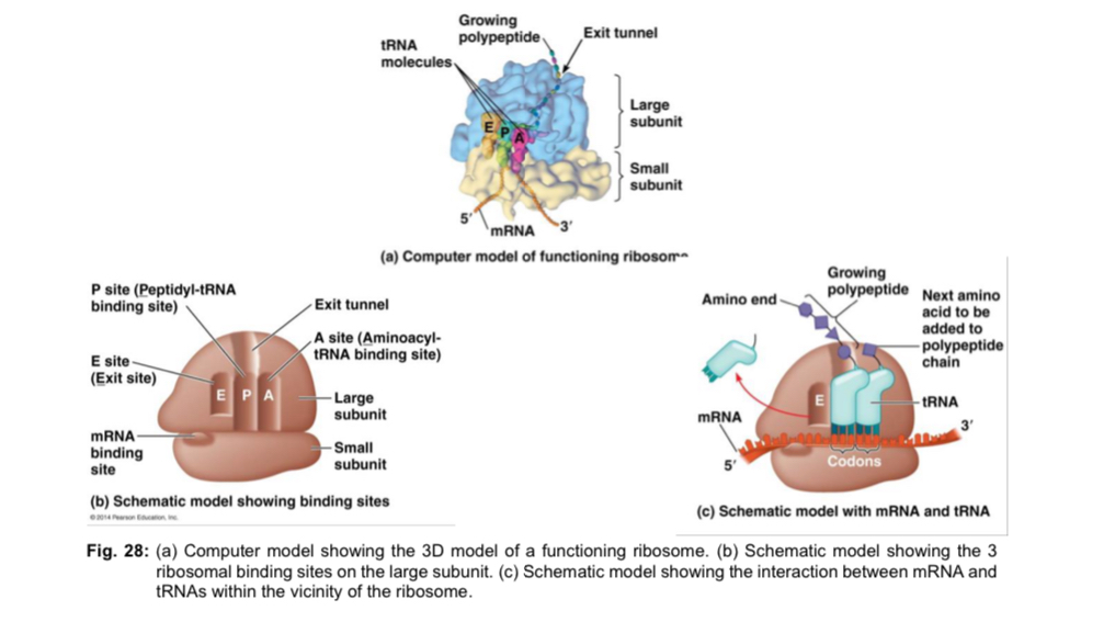
Ribosome
Structure
ribosome is a large ribonucleoprotein complex composed of ribosomal proteins and ribosomal RNA (rRNA)
bacteria ribosomes are ~70S while eukaryotic ribosomes are larger at ~80S
each eukaryotic ribosome consists of 2 subunits
small subunit (40S) contains an mRNA binding site, where the mRNA binds - mRNA binding site is associated along the surface close to the junction of the subunits
large subunit (60S) - 3 binding sites for tRNA
A site (aminoacyl-tRNA site): holds the incoming tRNA carrying the next amino acid to be added
P site (peptidyl-tRNA site): holds the tRNA carrying the growing polypeptide chain
E site (exit site): site of release of the deacylated tRNA (tRNA without amino acid attached)
role of the ribosomes
ribosomes are the organelles where the synthesis of polypeptides under the direction of mRNA occurs
in protein synthesis, the ribosome provides an environment for specific recognition between a codon of mRNA and an anticodon of tRNA
the ribosome holds the tRNA and mRNA in close proximity - the ribosome positions the new amino acid for addition to the growing polypeptide
rRNA in the large ribosomal subunit has peptidyl transferase activity - catalyses the formation of peptide bonds btw amino acids
ribosome begins translation at the 5’ end of the coding region of mRNA and proceeds towards the 3’ end (5’ —> 3’ direction)
Translation factors
initiation factors: required for assembly of mRNA, the first tRNA and ribosomal subunits
elongation factors: required for synthesis of polypeptide chains
release factors: required for recognition of the stop codon and disassembly of the translation machinery
several translation factors use GTP (guanosine triphosphate) as an energy source to carry out their functions
Steps involved in translation
Initiation
Elongation
Termination
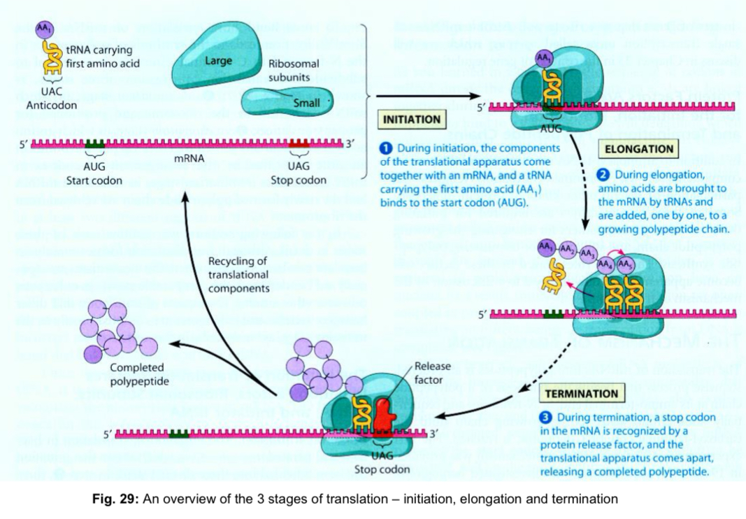
Stage 1: Initiation
initiation stage of translation brings together mRNA, the initiator tRNA and the 2 subunits (large and small) of a ribosome
involves the reactions preceding the formation of the 1st peptide bond
Step 1A: Binding of initiation factors to small subunit
eukaryotic initiation factors (eIFs) bind to small subunit of ribosome and position the initiator tRNA which carries a methionine to its P site
requires GTP
Step 1B: Binding of small subunit to mRNA
small subunit binds to the mRNA by recognition of its 5’ cap
small ribosomal subunit then moves downstream in the 5’ to 3’ direction along the mRNA in search of the start codon AUG (start site of translation)
Step 1C: Association of tRNAMet
and formation of initiation complex
anticodon on initiator tRNA associates with the start codon on mRNA through CBP
methionine is always the first amino acid in a newly formed polypeptide
initiator tRNA has a unique anti-codon loop that is distinct from that of the tRNA that normally carries methionine
followed by the dissociation of eIFs (hydrolysis of GTP), which allows for the binding of the large ribosomal subunit, completing a eukaryotic 80S translation initiation complex
initiator tRNA sits in the P site of the ribosome, and the initial methionine forms the N-terminus of the polypeptide
A site is vacant, waiting for entry of the next aminoacyl-tRNA complementary to the second codon of the mRNA

Elongation and Translocation
each amino acid is added to the C terminal end of the growing polypeptide by means of a cycle of 3 sequential steps
codon recognition and aminoacyl-tRNA binding
peptide bond formation
translocation
elongation: synthesis of the 1st peptide bond to the addition of the last amino acid
Step 2A: Codon recognition and aminoacyl-tRNA binding
after the initiation complex has formed, an aminoacyl-tRNA carrying the 2nd amino acid in the chain binds to the ribosomal A site via CBP btw its anticodon and the codon in the mRNA exposed at the A site
held in place by hydrogen bonds
tRNAs are brought in by elongation factors
energy is expended with hydrolysis of GTP
Step 2B: Peptide bond formation
when the second tRNA is bound to the ribosome, its amino acid is placed directly adjacent to the initial methionine
peptidyl transferase in the large ribosomal subunit catalyses the formation of a peptide bond btw carboxyl end of methionine and the amino group of the 2nd amino acid
methionine is transferred to the 2nd amino acid carried by the aminoacyl-tRNA at the A site
ester bond btw the initial methionine and the tRNA is broken to release the initial methionine (P site)
deacylated tRNA (lacking an amino acid) lies in the P site, while the new peptidyl-tRNA (tRNA carrying the growing polypeptide ) has been created in the A site
Step 2C: Translocation
ribosome is translocated one codon or 3 nucleotides at a time in the 5’ to 3’ direction, guided by elongation factors, with the hydrolysis of GTP to provide energy
this movement
relocates the initial deacylated tRNA (from the P site) to the E site from where it diffuses out of the ribosome
repositions the peptidyl-tRNA at the P site (from the A site)
exposes the next codon (3rd codon) on the mRNA at the A site
Steps 2A to 2C are repeated with a new incoming aminoacyl-tRNA entering A site, cycle will be repeated until a stop codon is encountered at the A site
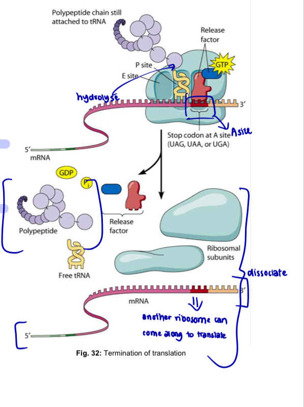
Termination
Termination occurs when a stop codon in the mRNA reaches the A site of the ribosome
base triplets UAG, UAA and UGA are stop codons that do not code for amino acids but act as signals to stop translation
Release factor (protein) binds directly to the stop codon in the A site
release factor causes the addition of a water molecule instead of an amino acid to the polypeptide chain
this reaction frees the carboxyl end of the completed polypeptide from the tRNA in the P site by hydrolysis
polypeptide is released through the exit tunnel of the ribosomal large subunit
ribosome then releases the mRNA and separates into large and small subunits i.e. the translation complex comes apart
tRNA molecules may then be recycled and used to form new aminoacyl-tRNA
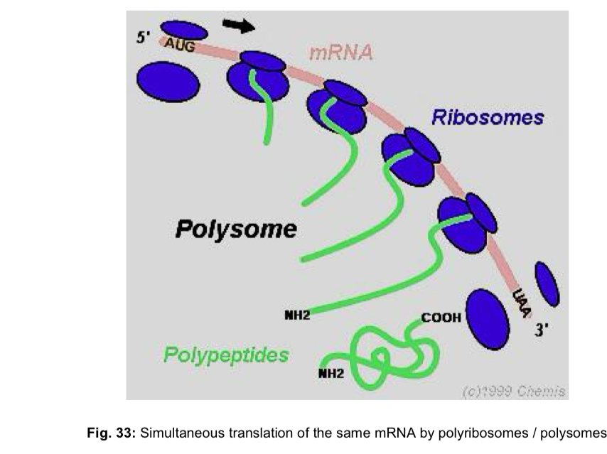
Polyribosomes/Polysomes
ribosomes are often seen to occur in clusters known as polyribosomes or polysomes
when ribosomes occur as such aggregates, they are simultaneously translating polypeptides from the same mRNA strand
each ribosome in the polysome independently synthesised a single polypeptide during its translation of the mRNA sequence
advantage: these multiple initiations mean that many more polypeptide molecules can be made in a given time than would be possible if each had to be completed before the synthesis of the next could start
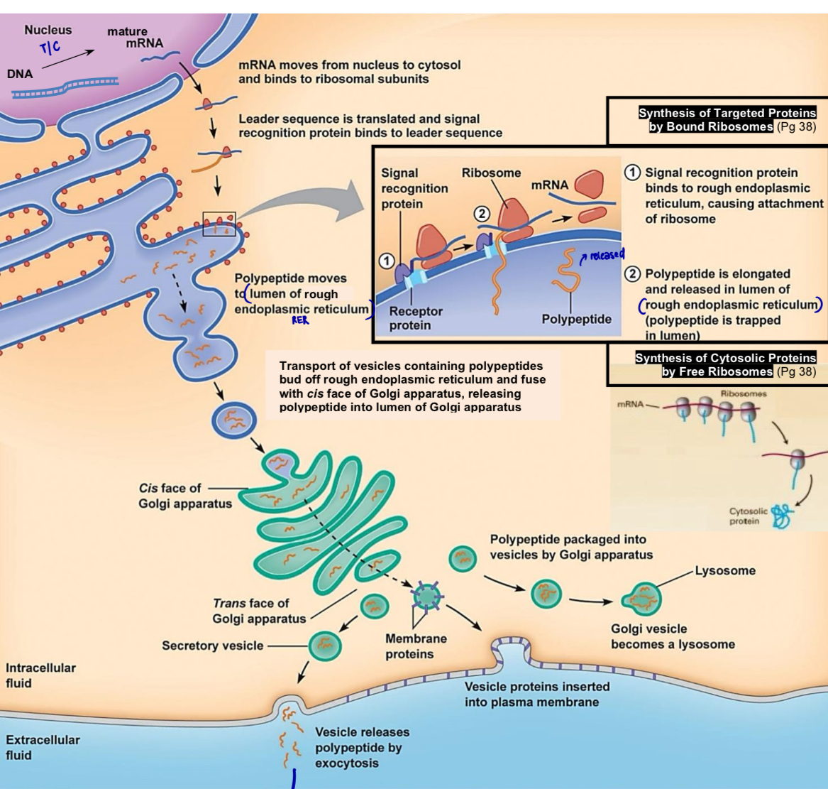
Free ribosomes
free ribosomes are suspended in the cytoplasm
mostly synthesise proteins that dissolve in and exert their effects in the cytosol
Bound ribosomes
attached to the cytoplasmic side of the endoplasmic reticulum (ER)
synthesise proteins of the nuclear envelope, ER, Golgi apparatus, lysosomes, vacuoles, and plasma membrane, as well as proteins to be secreted from the cell
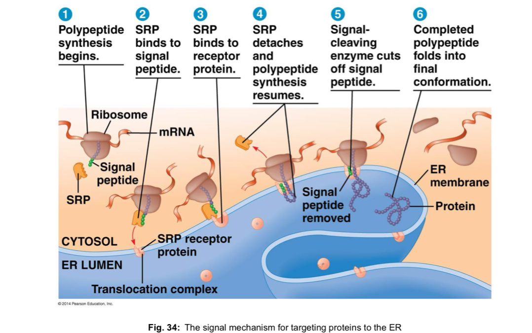
polypeptides of proteins destined for the endomembrane system or for secretion are marked by a signal peptide, which targets the polypeptides to the ER
signal peptide - sequence of about 20 a.a at or near the N-terminal of the polypeptide, is recognised as it emerges from the ribosome by a protein-RNA complex called a signal-recognition particle (SRP)
SRP functions as an adaptor that brings the ribosome to a receptor protein built into the ER membrane
polypeptide synthesis continues there, and the growing polypeptide snakes across the ER membrane into the cisternal space via protein pore
Post-translational modification of polypeptides
immediate product of translation is actually not a protein, but a polypeptide chain
to become a functional protein, one or more such linear polypeptide chains must coil and fold in a precise, pre-determined manner to assume the specific three-dimensional conformation either in the cytoplasm or in the lumen of the RER
subsequent transport to the Golgi apparatus allows for the modification of these proteins —> necessary for biological activity
range of functions of the protein may be modified by
attaching to it biochemical functional groups e.g. acetate, methyl, phosphate, various lipids and carbohydrates
glycosylation - addition of specific short-chain carbohydrate/oligosaccharide is very common in membrane proteins
glycoproteins - important in e.g. signal recognition, and activation of an immune response
reversible phosphorylation of threonine, serine, or tyrosine residues by enzymes called kinases (which add a phosphate) and phosphatases (which remove the phosphate) plays an important role in the signal transduction processes regulating growth and cell cycle control
Phosphorylation may occur sequentially from one protein to another, resulting in a series of activations called a “phosphorylation cascade”
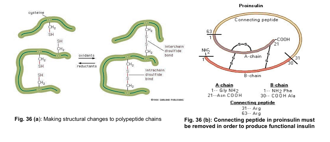
Making structural changes, like the formation of disulfide linkages
Removing a sequence of amino acids from the protein, or cutting the peptide chain in the middle
e.g. peptide hormone insulin is cut twice after disulfide bonds are formed, and a connecting peptide is removed from the middle of the chain —> proteolytic cleavage
resulting protein consists of two polypeptide chains connected by disulfide bonds
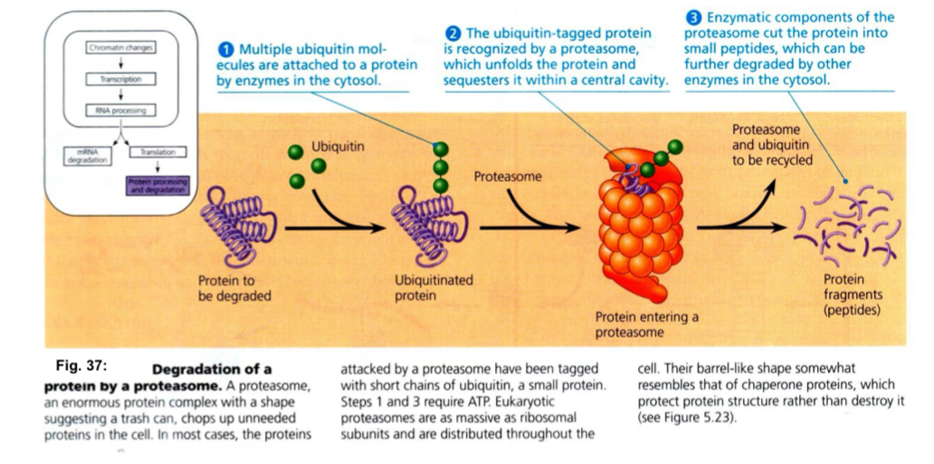
attaching to it ubiquitin
ubiquitin marks proteins for proteolysis by the proteasome
at least 4 ubiquitin are required on the substrate before a proteasome can bind to it
polyubiquitin chain on a target protein is recognised by a specific receptor in the proteasome
selective degradation of proteins allows for the control of the length of time in which a protein can function
Degradation of a protein by a proteasome