Cerebellum:
1/24
There's no tags or description
Looks like no tags are added yet.
Name | Mastery | Learn | Test | Matching | Spaced | Call with Kai |
|---|
No analytics yet
Send a link to your students to track their progress
25 Terms
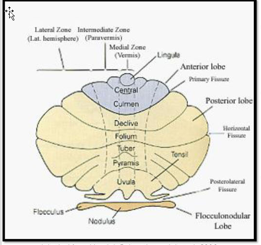
Describe lobes of Cerebellum and fissure associated
Anterior Lobe
primary fissure
Posterior Lobe
posterolateral (prenodular) fissure
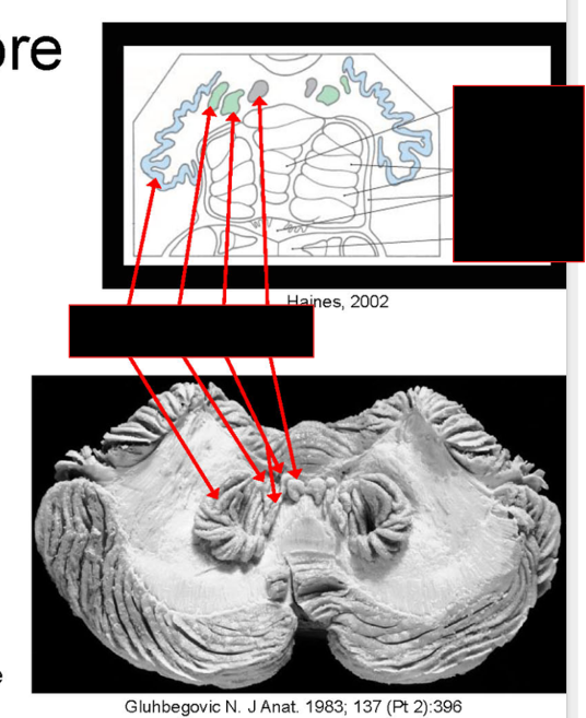
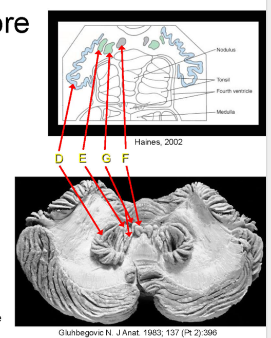
Flocculonodular Lobe
nodulus
midline flocculus (paired) — lateral
What are Lobules
What are they?
Composed of?
Locations?
Lobules:
Divides lobes of the cerebellum
Composed of:
cortical ridges called folia.
Locations:
Anterior Lobe: Lobules I-V
Posterior Lobe: VI-IX
floculonodular lobe: X
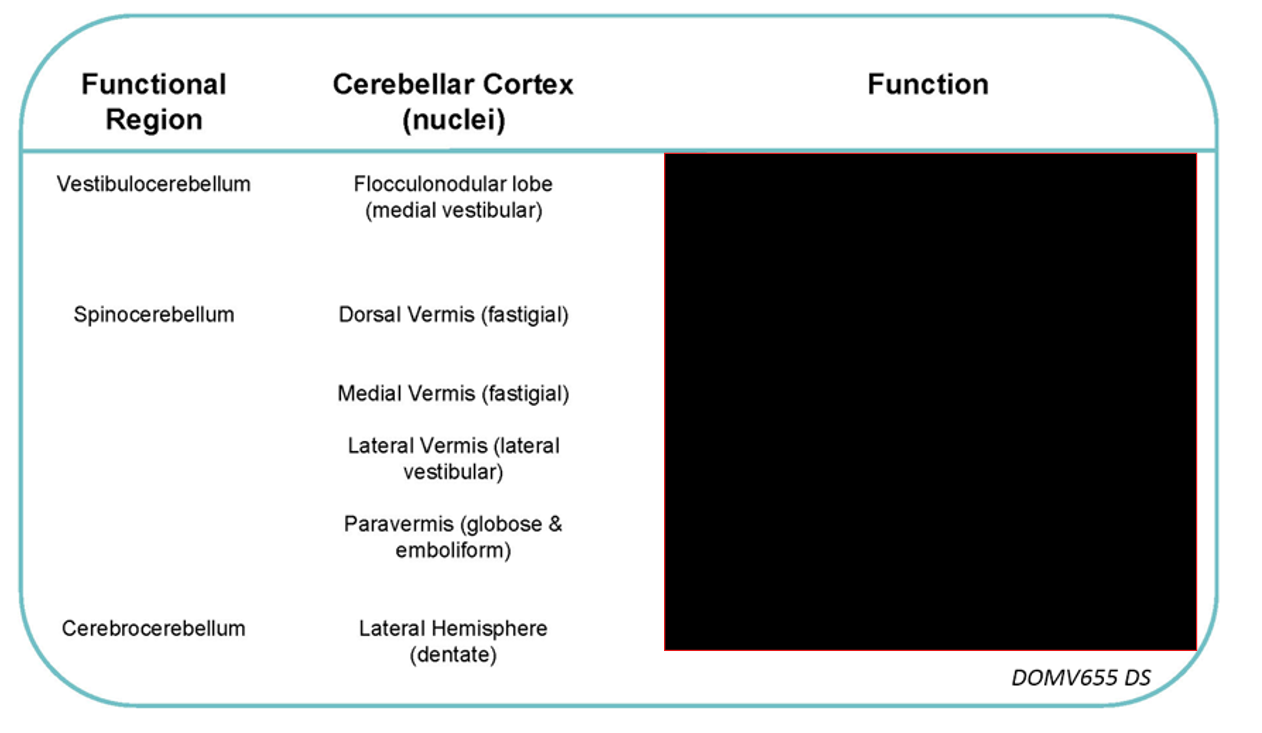
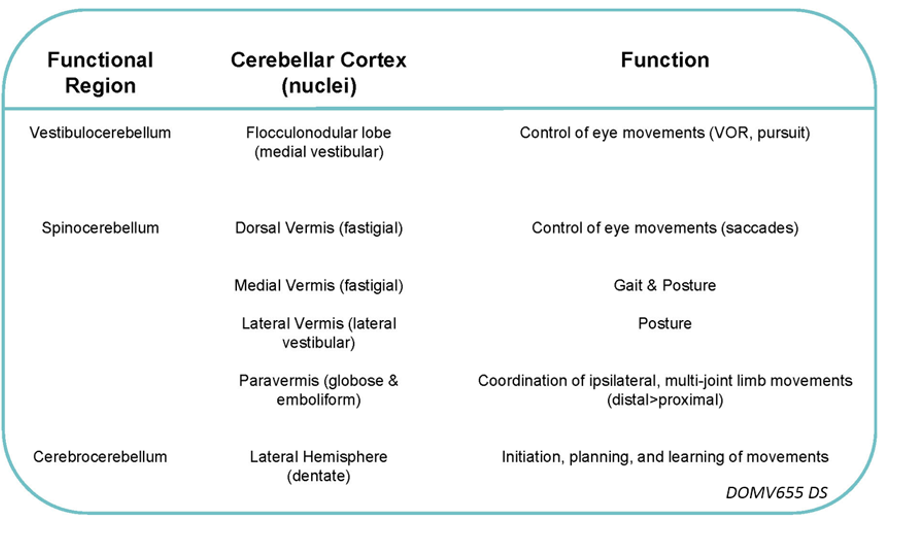
What are the functional divisions?
Locations?
Function?
Vestibulo-cerebellum
(flocculonodular lobe)
Control of eye movements
Spino-cerebellum
(vermis and paravermis)
Controls muscle tone and ongoing axial/limb movements
Cerebro-cerebellum
(lateral hemisphere)
Role in planning and initiation of movements
Regulation of discrete upper limb movements
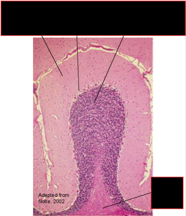
Describe the Cytoarchitecture
Layers? (3)
Types of neurons? (5)
Cytoarchitecture
three layers:
Molecular Layer (outer)
Purkinje Cell Layer (intermediate)
Granule Cell Layer (inner)
***Memorization: Move Please Grandma***
five types of neurons:
principal neuron:
Purkinje Cell
intrinsic neurons
Basket (molecular layer)
Stellate (molecular layer)
Granule (granule cell layer)
Golgi (granule cell layer)
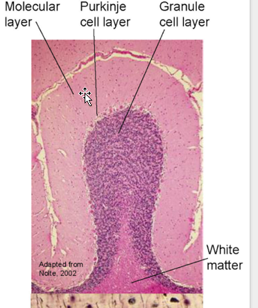
Describe the White matter Core
Contains?
Pairs of Nuclei? (4)
White Matter Core
Contains:
Incoming and outgoing fibers
Four Pairs of Deep Cerebellar Nuclei (gray matter)
Dentate
Emboliform
Globose
Fastigial
emboliform and globose = Interposed nuclei
Describe the Source of Input
SC
Vestibular System
Cerebral Cortext
Describe the Somatotopic Organization of Inputs
Afferents:
SC:
Spinocerebellar tracts (dorsal, ventral)
Cuneocerebellar tract
Vestibular System:
Vestibulocerebellar tract
Direct to cerebellum (primary vestibulocerebellar fibers from vestibular apparatus)
Via vestibular nuclei (secondary vestibulocerebellar fibers)
Cerebral Cortex
Cortico-pontocerebellar
Cortico-reticulocerebellar
Cortico-rubro-olivocerebellar*
Cortico-olivocerebellar*
NOTE: *Olivocerebellar input corresponds to the aforementioned longitudinal zones (corticonuclear projection zones)
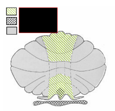
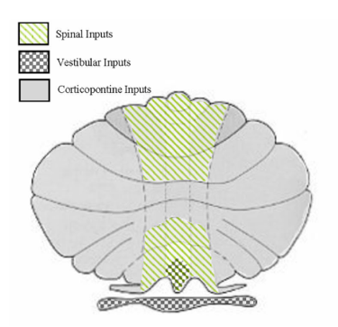
Somatotopic Organization of Inputs
Anterior Lobe (one homunculus)
Leg anterior
Face posterior
Posterior Lobe (two homunculi)
Face posterior
Legs anterior
Trunk: midline
Extremities: lateral
Dorsal Vermis:
receives auditory and visual input,
overlaps w/ head region of homunculi
Describe the Fiber Systems
1. Transmits?
2.
3. Content? Termination? Function?
Fiber Systems:
Climbing Fiber System:
Transmits olivocerebellar input
Mossy Fiber System:
All other input except those listed below:
Multilayered Fiber System
Content:
Hypothalamus
select cell groups w/in brainstem
Terminatation:
deep cerebellar nuclei
diffusely throughout cerebellar cortex
Function:
Decrease spontaneous and evoked activity of purkinje cells
(LC & Raphe n. , particularly)
Describe the Afferent/Efferent to Cerebellar Peduncles (Inferior,Middle,Superior)
Inferior (restiform/juxtarestiform body) (A/E)
Olivocerebellar tract (major component)
Dorsal Spinocerebellar tract
Cuneocerebellar tract
Reticulocerebellar & Cerebelloreticular tracts
Vestibulocerebellar and cerebellovestibular tracts
Arcuatocerebellar tract
Trigeminocerebellar tracts (spinal and main)
Middle (brachium pontis) — Afferent fibers
—Pontocerebellar tract
Superior (brachium conjunctivum) (A/E)
Cerebellothalamic (dentatothalamic, interpositothalamic)
Cerebellorubral (dentatorubral, interpositorubral)
Ventral Spinocerebellar tract
Trigeminocerebellar (mesencephalic)
Describe the Circuitry:
Climbing
Mossy Fibers
Climbing Fiber Circuit
Climbing fiber → Purkinje cell + Deep Cerebellar nuclei
Excitatory (aspartate)
Mossy Fiber Circuit
Mossy fiber → Granule cell + Deep Cerebellar Nuclei
Excitatory (glutamate)
Granule cell axons (parallel fibers) → Purkinje cell + Intrinsic neurons
Excitatory (glutamate)
Intrinsic neurons → Purkinje cell
Inhibitory (GABA)
Purkinje cell → Deep Cerebellar Nuclei
Inhibitory (GABA)
Describe how Climbing fiber (CF) input is highly focused
Mech
Function (2)
Coincident activation?
Climbing fiber (CF) input is highly focused
Mech:
1 CF → 10 purkinje cells → 1000-2000 synapses with each cell
Note: Each purkinje cell receives input from only 1 CF.
Function:
Produces Complex Spike in purkinie cell @ irregular intervals (low spike frequency)
Alters Purkinje cell sensitivity to parallel fiber input
CF input + parallel fiber-purkinje synapse → long term depression @ parallel Tiber- purkinje cell synapses,
Describe the Extensive divergence in mossy fiber- granule cell input
Mech
AP generation
Function (2)
Extensive divergence in mossy fiber- granule cell input
Mech:
Parallel fibers run along long axis of folium → hundreds purkinje cells.
Each purkinje cell: 100 -200 thousand parallel fiber synapses
AP generation:
Requires summation of Parallel fiber input
Function:
Produce Simple Spikes in purkinje cell (high spike frequency)
High Frequency → encode magnitude + duration of peripheral stimuli or central input.
Evokes high level of tonic activity in cerebellar cortex
Describe how Intrinsic inhibitory neurons increase the resolution of mossy fiber- parallel fiber input
Stellate/Basket Cells:
Axons
Function
Result? What is this call
GIVE SIGNALS?
Golgi Type II neurons
What happens to it?
Result?
GIVE SIGNALS?
What does the Purkinje fibers do
Stellate/Basket Cells:
Axons project laterally from Parallel Fibers’ Plane
Function: Inhibits purkinje cells
→ longitudinal patches of purkinje cell excitation bounded by Fences of Inhibition
(Center Surround Antagonism)
Gives Signal SPATIAL RESOLUTION
Parallel fiber excitation of Golgi Type II neurons
Result:
feedback inhibition to granule cell (& mossy fiber)
→ Controls gain of granule cell input and shortens duration of parallel fiber bursts
→ Gives Signal TEMPORAL RESOLUTION
Purkinje Cells:
Actively shape (inhibition) excitatory output of tonically active deep cerebellar nuclei
Describe the Inferior Olivary Nucleus (ION)
Contains?
Function? What happens when adaption is needed?
Describe the pathway of the two Recurrent olivo-cerebellar loops
ION:
Contains:
spiny excitatory neurons
coupled via gap junctions
Function: important role in motor learning
sends error signals related to motor commands or timing to the cerebellum
When Adaption is needed:
→ climbing fiber activity Increases → complex spikes increases → progressive improvement
Recurrent olivo-cerebellar loops:
Olivocerebellar mesenencephalic-olivary loop
CF → (Directly + or Indirectly (purkinje) -) Deep Cerebellar N → (+) contra parvocellular red nucleus → (+) Ipsilateral ION via CTT → Increases synchrony of ION discharge
Olivocerebellar nucleo-olivarv loop
Deep Cerebellar Nuclei → (-) contra ION → Decreases synchronous discharge of ION
***NOTE: Deep cerebellar Nuclei: Glutamatergic (+) or GABAergic neurons (-)***
List out the cerebellar output (4)
Fastigial N Pathway:
Globose and Emboliform N. Pathway:
Dentate N. Pathway:
Flocculonodular lobe (vestibulocerebellum) Pathway
Cerebellar Output
Spino-cerebellum
Medial Vermis → Fastigial n
Lateral Vermis → Lateral Vestibular n.
Paravermis → Globose and Emboliform n.
Cerebro-cerebellum
Lateral Hemisphere→ Dentate n.
****Note: there is overlap between the spinocerebellum and the vestibulocerebellum. ****
Fastigial N Pathway:
→ Vestibular bilateral) and Reticular Nuclei (Contra)
→ SC (Medial Motor Systems)
vestibulospinal
reticulospinal
→ nuclei involved in EOM (MLF)
Related to posterior vermal (occulomotor vermis) and vestibulocerebellar input to Fastigeal Nucleus
***Note: Axons also project to the contralateral superior colliculus (tectospinal tract) and ventrolateral thalamic nucleus (MI anterior corticospinal tract) via the uncinate fasciculus. These are functionally related to vermal input to the fastigial n. **
Globose and Emboliform N. Pathway:
→ Red Nucleus (caudal, magnocellular portion)
→ SC (rubospinal tract)
***Note: There are also projections to the ventrolateral thalamic nucleus (MI *lateral corticospinal tract).***
Dentate N. Pathway:
→ Thalamus (VL)
→ Cerebral Cortext
Primary Motor (MI) and Premotor Cortex
Prefrontal and Posterior Parietal Cortex
These cortical areas project back → cerebellum via pontine nuclei forming cerebrocerebellar loops.
***Note: there are also projections to the red nucleus (rostral, parvocellular portion). Rubroolivary projections originate here.***
Flocculonodular lobe (vestibulocerebellum) Pathway
→ ipsilateral vestibular nuclei via juxtarestiform body
(sparse input to the lateral vestibular nucleus)
→ caudal fastigial nucleus via Nodulus purkinje cells
→ vestibular nuclei (bilaterally)
EOM (MLf)
Balance (VST)
Complex overlap with spinocerebellum
List out the Function of the cerebellum:
Motor (2)
Non-Motor (2)
Clinical?
Main Function (motor):
Error detection and Movement correction
Matches Info for Cortext and Peripheary regarding movement
Planning and Initiation of movement
Via Deep Cerebellar Nuclei discharge perior to movement
provides processed sensory info
Non-Motor Function
Autonomic:
Stimulation of vermis → cardiovascular function (e.g., carotid sinus reflex, BP) + pupillary diameter.
May play a role in higher brain function, cognition, and behavior
Clinical
Flushing and dialated pupils noted in patients with cerebellar lesions.
What are the symptoms of Cerebellar Disorder
A: ataxia
Lack of coordination resulting in unsteadiness of movement
H: hypotonia
Decrease in muscle tone — upon passive movement
A: adiadochokinesia
Inability to perform rapid successive movements
N: nystagmus
Rhythmic involuntary oscillatory movements of the eyes
D —dsymetria
Can’t stop movement as the target IS approached
→ overshoot of target
May be combined w/ kinetic tremor (see video)
D —dyssynergia
Decomposition of movement
→ Jerky and tremulous movement of extremity
D —dysarthria (speech disturbance)
Slurred hesitant speech w/ inappropriate emphasis of pitch and loudness
Describe Tremor
Kinetic Vs postural Vs titubation Vs Gate Ataxia (truncal ataxia)
Intention (kinetic) Tremor
slow, course shaking movement during attempted voluntary movement
amplitude increases during terminal portion of movement
Static (postural) Tremor
occurs during maintenance of position against gravity;
oscilating movements of shoulders and arms when arms are outstretched.
Titubation
oscillatory movements of the head or trunk;
front→ back, side → side, or rotatory
Gate Ataxia (truncal ataxia)
wide based, clumsy, staggering gait,
similar to that seen with inebriation;
Severe = difficulty sitting upright due to truncal instability
How can you test if someone has Cerebellar Disorders (3)
How can different lesions have different effects
Removal of pressure on flexed forearm → unchecked flexion
Tapping outstretched arm → oscillation of the arm around initial position,
ie., rebounding past the position.
Pendular reflexes:
patellar tendon reflex Tap → leg continuing to swing back and forth.
Different Lesions Different Symptoms:
Ipsilateral signs and symptoms with unilateral lesions
Due to double crossed circuitry
Hemispheric lesions: limb
Lesions of vermis: trunk
Lesion of deep nuclei or SCP
more severe signs than lesion of cerebellar cortex
Describe Symptoms/ Area Affected Of:
Midline Syndrome
Lateral Syndrome
Pancerebellar Syndrome.
Midline Syndrome:
Symptoms:
Disequilibrium,
(truncal ataxia),
titubation
head tilt
Nystagmus,
saccadic dysmetria,
smooth pursuit deficits
Involvement of:
floculonodular lobe and vermis (vestibulocerebellum and spinocerebellum)
Lateral Syndrome:
Symptom: A HAND3 Tremor
Involvement of
intermediate and lateral zones (spinocerebellum and cerebrocerebellum)
Pancerebellar Syndrome.
Combination of midline + lateral (hemispheral) syndrome
Symptom:
Bilateral signs of cerebellar dysfunction involving trunk, limbs, and eyes
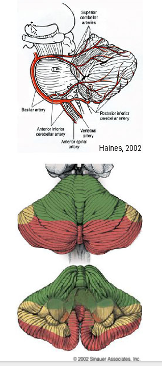
Describe the Blood Supply
Superior Cerebellar Artery
Rostral half of hemisphere and vermis;
deep cerebellar nuclei;
superior and rostral middle cerebellar peduncle;
lateral tegmentum of rostral pons
Anterior Inferior Cerebellar Artery
Anterolateral part of caudal cerebellum (including flocculus);
caudal middle cerebellar peduncle;
lateral tegmentum of caudal pons
Posterior Inferior Cerebellar Artery
Caudal cerebellum (hemisphere, inferior vermis, tonsil and nodulus);
choroid plexus of 4th ventricle;
inferior cerebellar peduncle; dorsal lateral medulla (rostral part)