written arrhythmia identification and ECG
1/29
There's no tags or description
Looks like no tags are added yet.
Name | Mastery | Learn | Test | Matching | Spaced | Call with Kai |
|---|
No analytics yet
Send a link to your students to track their progress
30 Terms

P wave shows
atrial depolarization

PR interval shows
atrial contraction
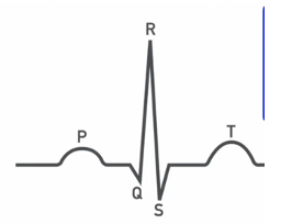
QRS complex shows
ventricle depolarization and atria repolarization

ST interval shows
ventricle contraction

T wave shows
ventricle repolarization

QT interval shows
whole ventricular action potential
hypo or hyperkalemia manifests on ECG as
T wave changes, due to repolarization effect
Hypocalcemia manifests on ECG as
prolonged QT interval
hypercalcemia manifests on ECG as
shortened QT interval
increased sympathetic activity does what to automaticity and HR
increases automaticity, increased HR
increased parasympathetic activity does what to automaticity and HR
decreased automaticity and HR
What are the two mechanisms of automatic tachycardia?
increased slope of phase 4 OR decreased threshold of non SA nodal tissues
What are some causes of automatic tachycardia?
digitalis toxicity
hypokalemia
hypoxia
increased SNS or decreased PSNS
What phase is disrupted during early afterdepolarizations and why?
phase 3; due to Na channels opening
What phase is disrupted during delayed afterdepolarizations and why?
phase 4, Ca influx/release early
what are the two mechanisms/models of reentrant tachycardia?
anatomic and functional
What is the difference between A fib and atrial flutter?
a fib - faster HR, disorganized ECG, irregularly irregular pulse
a flutter - fast but slower HR, organized ECG, atrial to ventricular depolarizations occur in a pattern (1:1, 1:2, etc)
What is the major complication of a fib or a flutter?
embolic stroke due to blood stasis in atria
What are some causes of ventricular tachycardia?
ischemia
MI
metabolic abnormalities (hypokalemia)
hypoxemia
drugs (digitalis toxicity)
ventricular tachycardia is defined:
3 or more ventricular premature beats with rate >100bpm
What are the causes of chronic or reccurent ventricular tachycardia?
underlying heart diseases or left ventricular dysfunction and aneurism
What is the difference btw monomorphic and polymorphic ventricular tachycardia?
monomorphic - single foci, QRS similar beat to beat
polymorphic - multiple foci with varying QRS size and shape
Torsades de pointes is an example of what?
polymorphic ventricular tachycardia
Torsades de pointes is characterized by
long QT interval and QRS complexes undulating around a central axis
Which fibrillation is LIFE THREATENING and why?
V fib; blood is not filling or being pushed through the ventricles and there is no blood or oxygen getting to the rest of the body
What is a pro-arrhythmia?
a new or worsened arrhythmia due to antiarrhythmic drugs
what is an atrioventricular block?
a delay or interruption in the conduction from atria to ventricles
describe a first degree AV block
prolonged PR interval, but the impulse still reaches the ventricles
site of delay is the AVN
describe a second degree AV block
2 types:
Mobitz type I: progressive PR prolongation and QRS is sometimes dropped, usually due to AV nodal fatigue
Mobitz type II: no PR prolongation, dropped QRS, block is at the AVN or below
describe a third degree AV block
complete block, the atria and ventricles beat independently. ventricular escape rhythm is very slow