Exam 3 Histology Practice
1/154
There's no tags or description
Looks like no tags are added yet.
Name | Mastery | Learn | Test | Matching | Spaced | Call with Kai |
|---|
No analytics yet
Send a link to your students to track their progress
155 Terms

Name the structure indicated by 1.
Epidermis

Name the structure indicated by 2.
Dermis
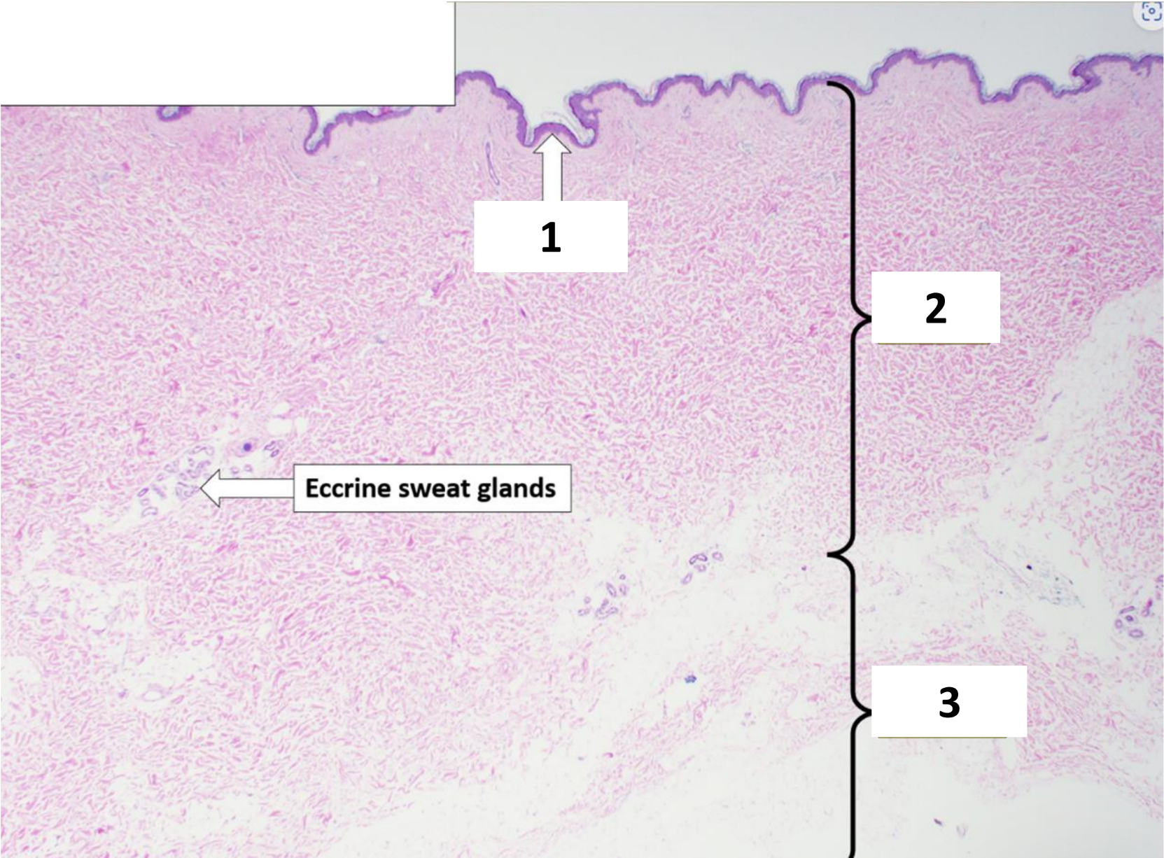
Name the structure indicated by 3.
Hypodermis

Name the structure indicated by 1.
Stratum corneum

Name the structure indicated by 2.
Stratum granulosum
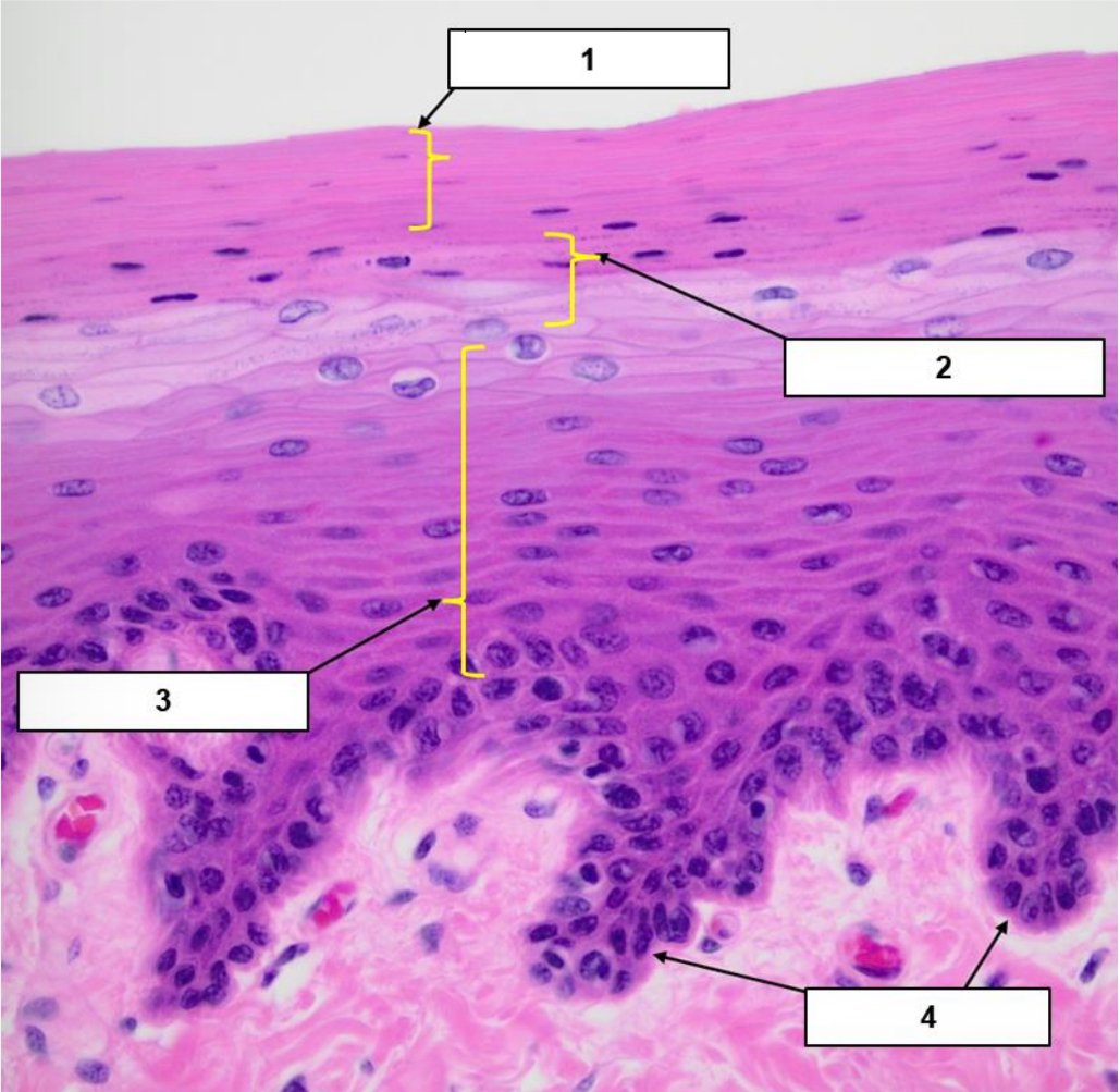
Name the structure indicated by 3.
Stratum spinosum

Name the structure indicated by 4.
Stratum basale
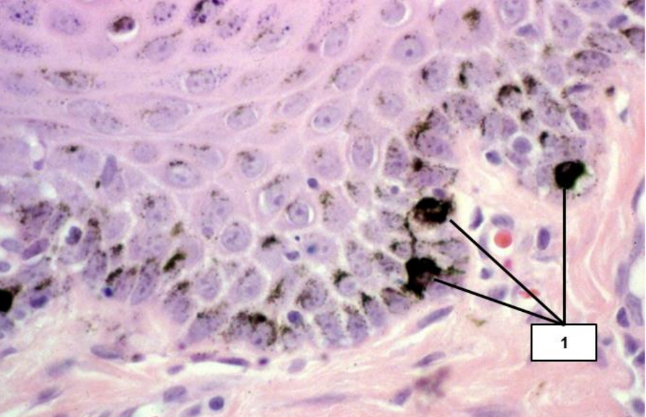
Name the cell type indicated by #1.
Melanocytes

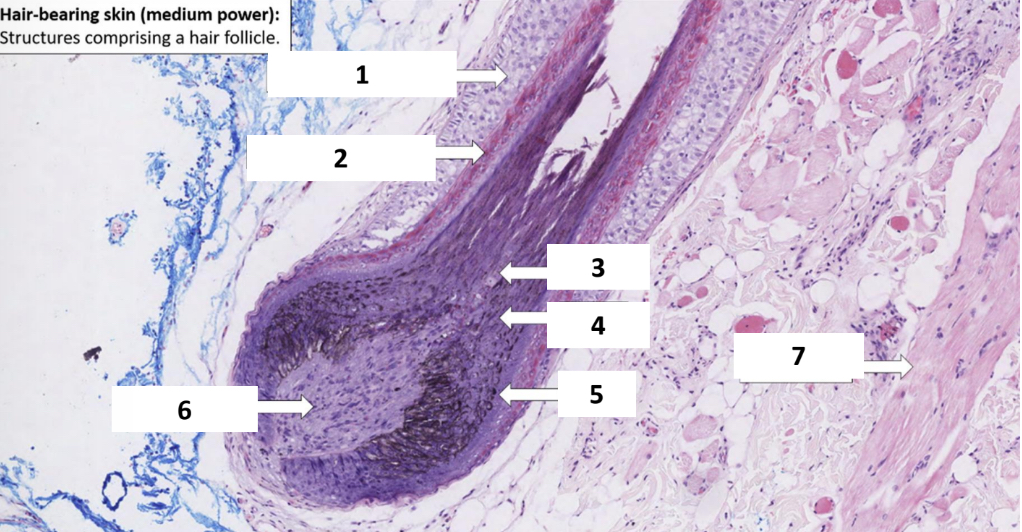
Name the structure indicated by 1.
Dermis

Name the structure indicated by 2.
Epidermis
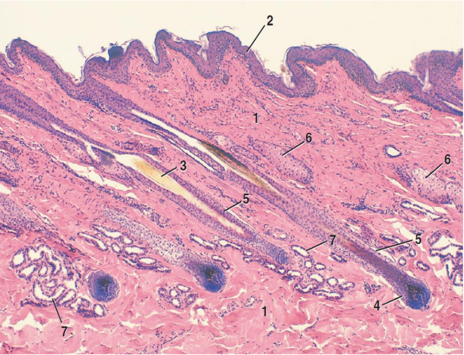
Name the structure indicated by 3.
Hair shaft

Name the structure indicated by 4.
Dermal papilla

Name the structure indicated by 5.
External root sheath

Name the structure indicated by 6.
Sebaceous glands

Name the structure indicated by 1.
External root sheath

Name the structure indicated by 2.
Internal root sheath

Name the structure indicated by 3.
Medulla

Name the structure indicated by 4.
Cortex

Name the structure indicated by 5.
Cuticle

Name the structure indicated by 6.
Dermal papilla
Name the structure indicated by 7.
Arrector pili muscle
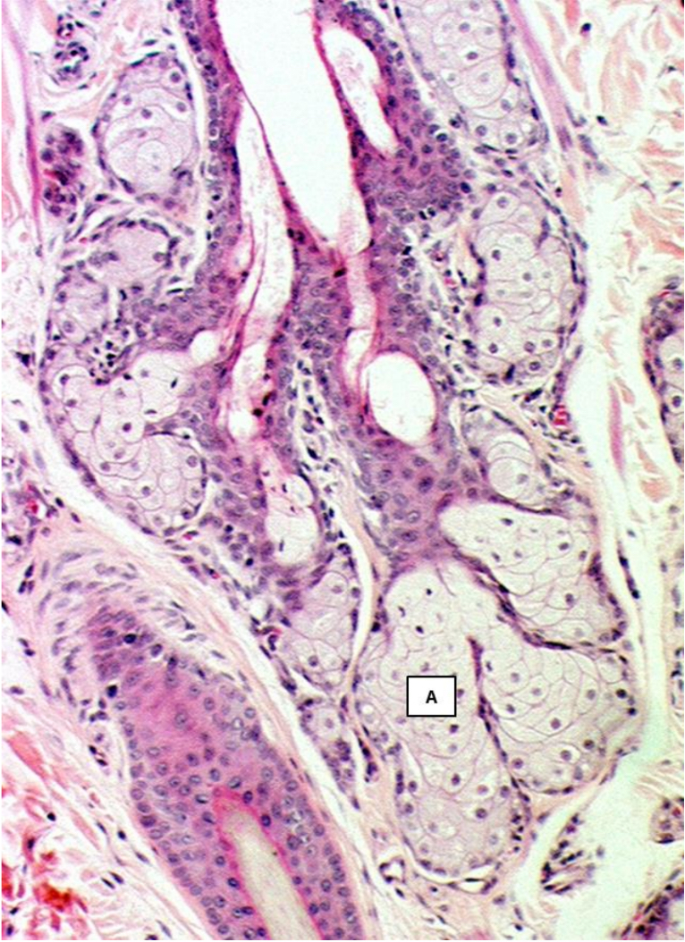
What type of glands are indicated by A?
Sebaceous gland
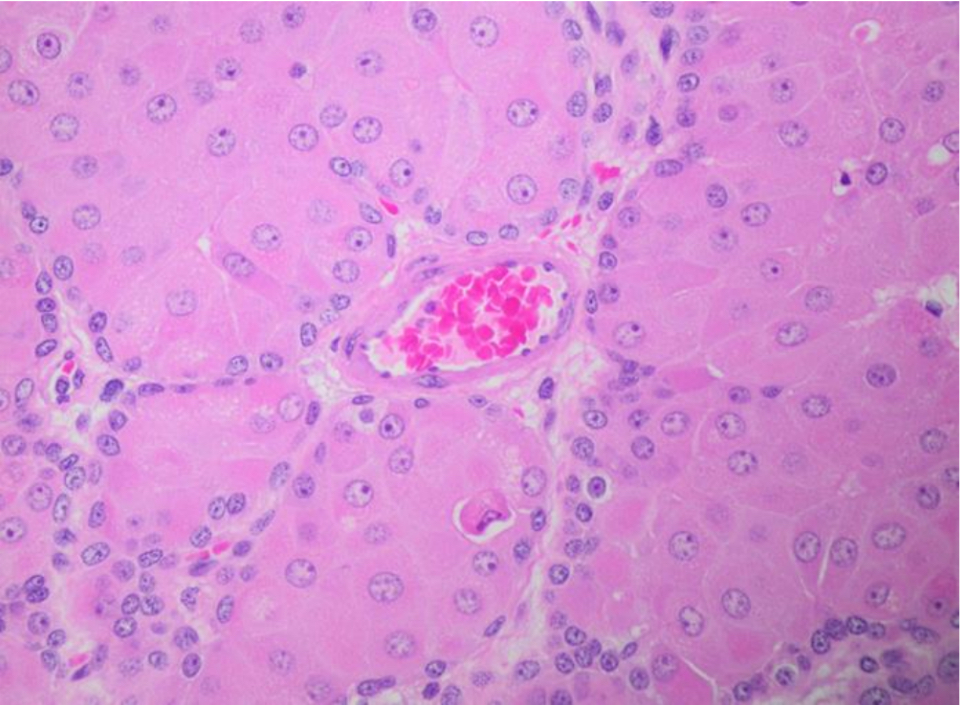
What type of gland is represented in the picture?
Hepatoid gland
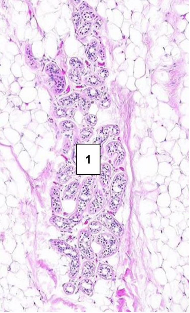
What type of gland is represented in the picture?
Sweat gland
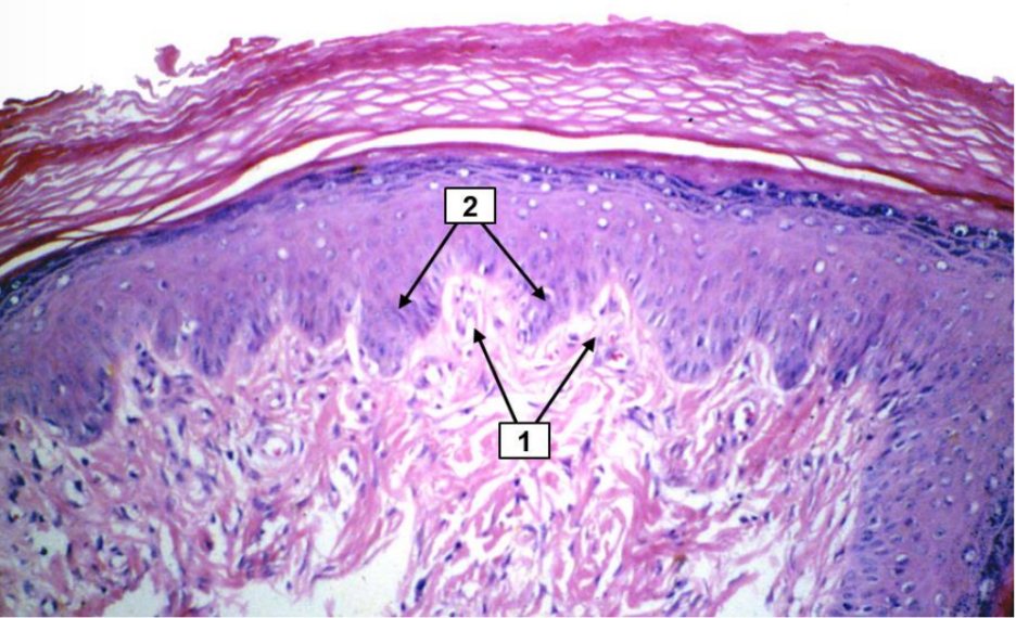
Name the structure indicated by 1.
Dermal papilla

Name the structure indicated by 2.
Rete ridges

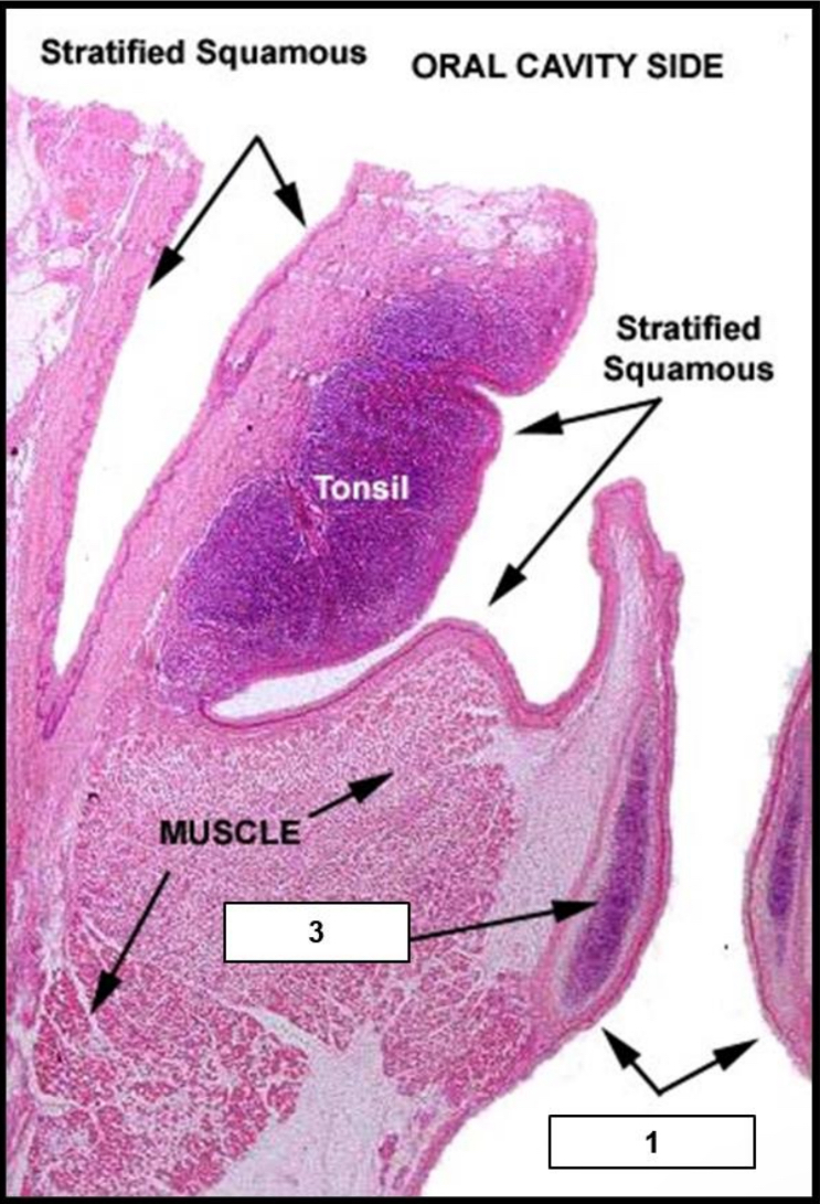
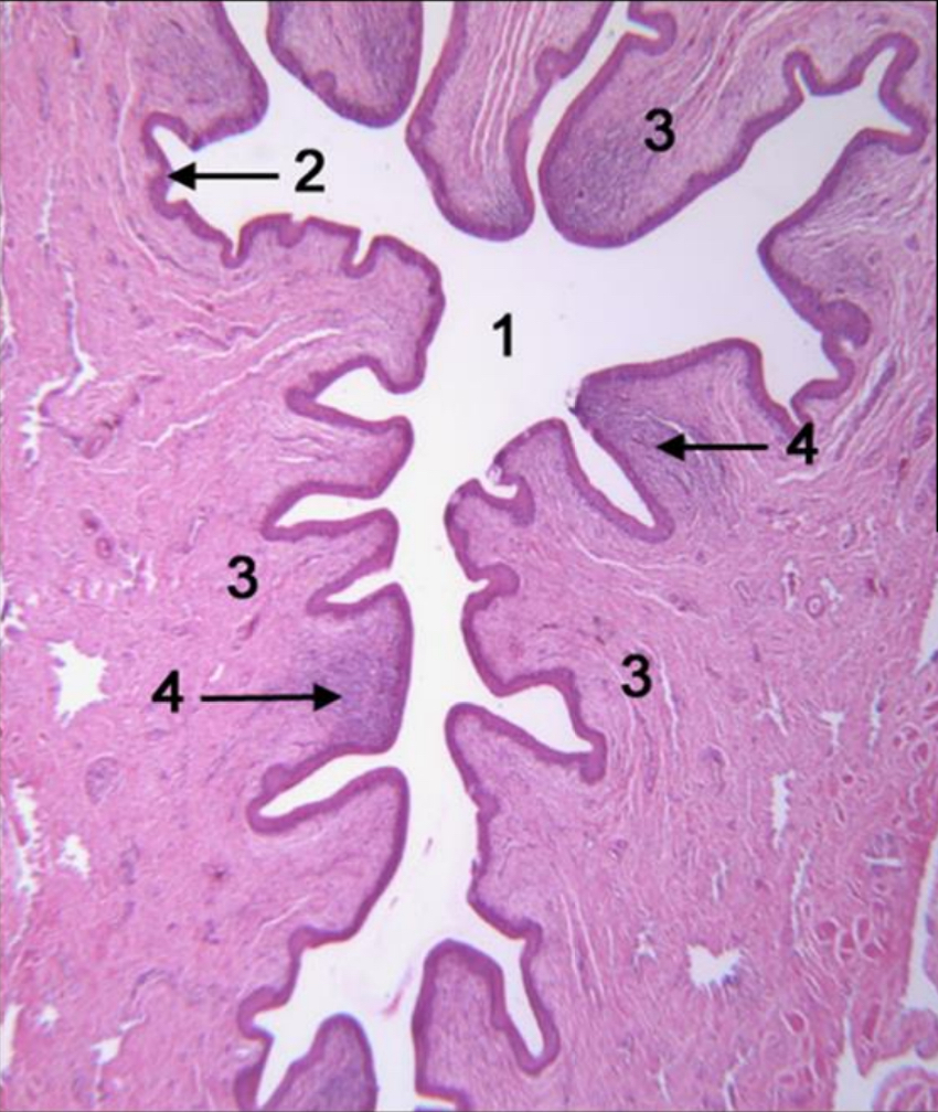
Name the structure indicated by 1.
Nasal septum
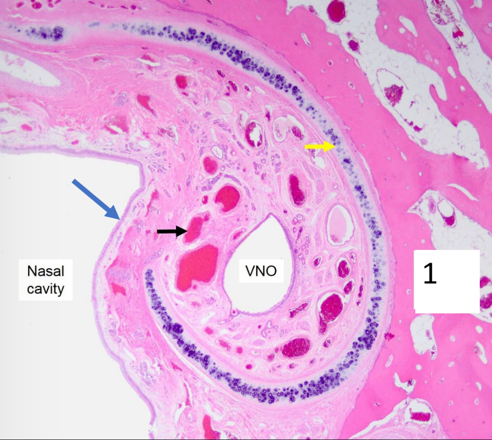
Name the structure indicated by the yellow arrow.
Cartilage

Name the structure indicated by the black arrow.
Blood vessel in laminate propria

Name the structure indicated by the blue arrow.
Respiratory epithelium

Name the structure indicated by 1.
Olfactory and sensory neuron
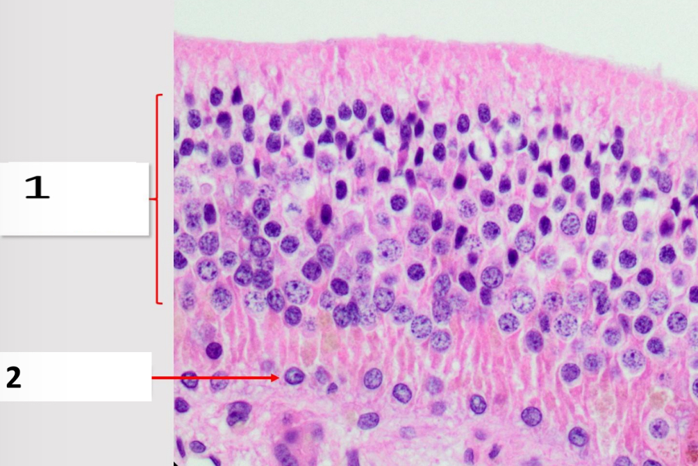
Name the structure indicated by 2.
Basal cell nuclei

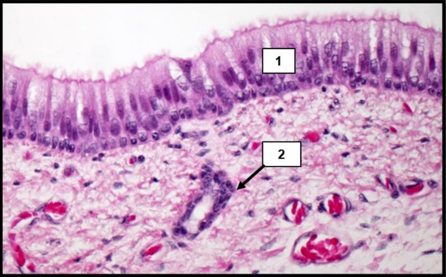
Name the structure indicated by 1.
Goblet cell
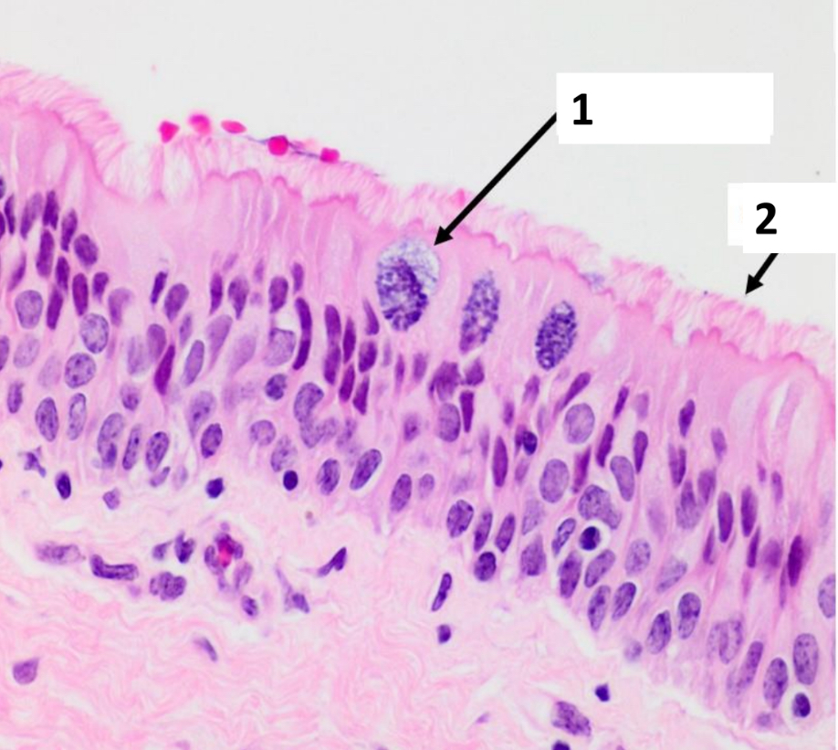
Name the structure indicated by 2.
Cilia

Name the structure indicated by 1.
Respiratory epithelium
Name the structure indicated by 2.
Tracheal gland

Name the structure indicated by 1.
Ciliated pseudostratified columnar epithelium with goblet cells
Name the structure indicated by 3.
Cartilage
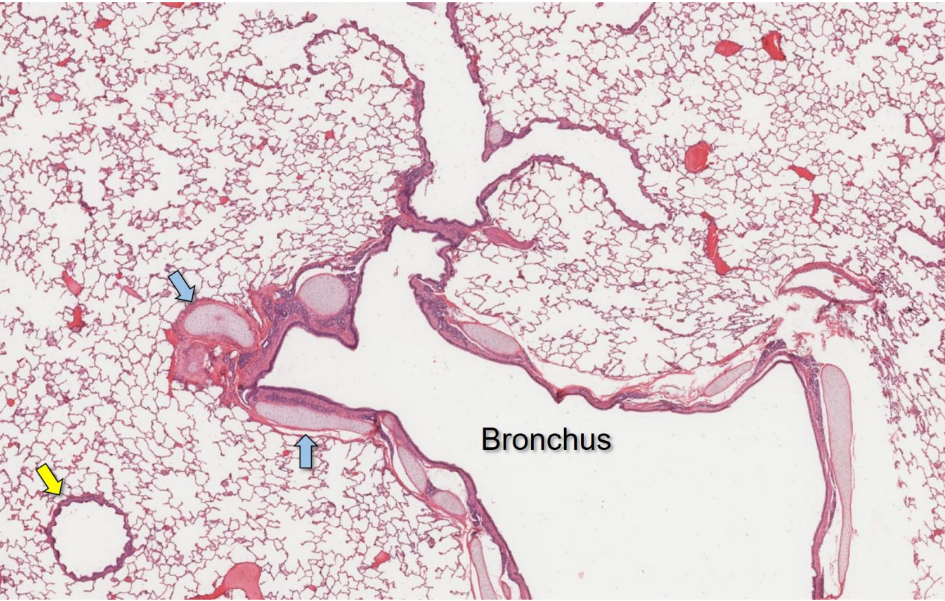
Name the structure indicated by the blue arrow.
Cartilage

Name the structure indicated by the yellow arrow.
Bronchiole
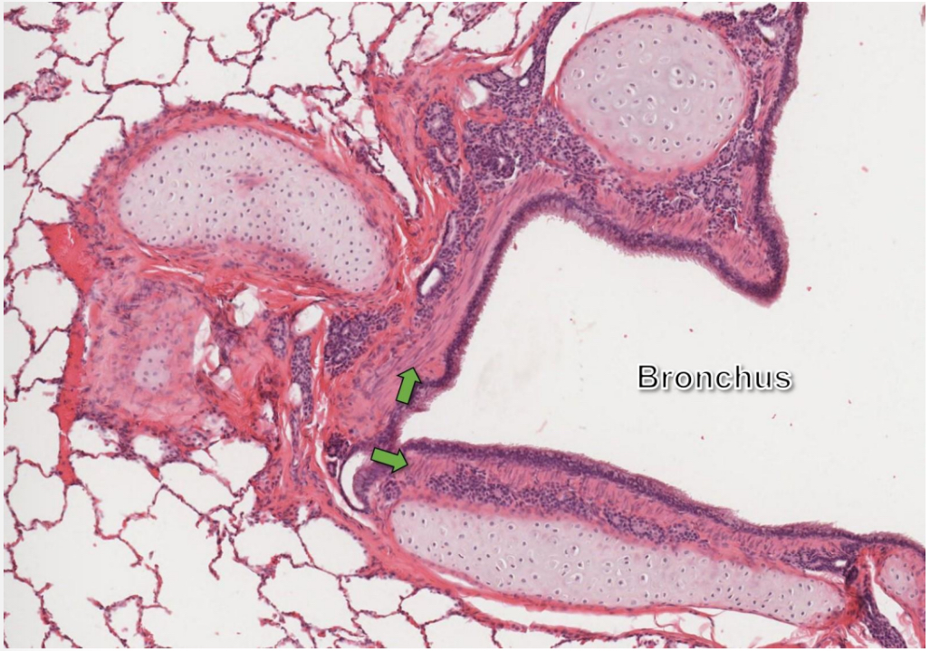
Name the structure indicated by the green arrow.
Smooth muscle

Name the structure indicated by the green arrow.
Alveolar space
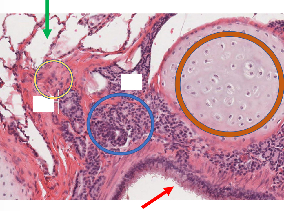
Name the structure indicated by the yellow circle.
Smooth muscle

Name the structure indicated by the blue circle.
Bronchial glands

Name the structure indicated by the orange circle.
Hyaline cartilage

Name the structure indicated by the red arrow.
Respiratory epithelium
Name the structure indicated by 2.
Cartilage

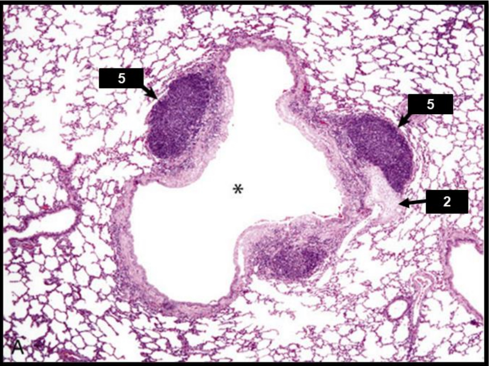
Name the structure indicated by 5.
BALT

Name the structure indicated by 1.
Respiratory bronchiole
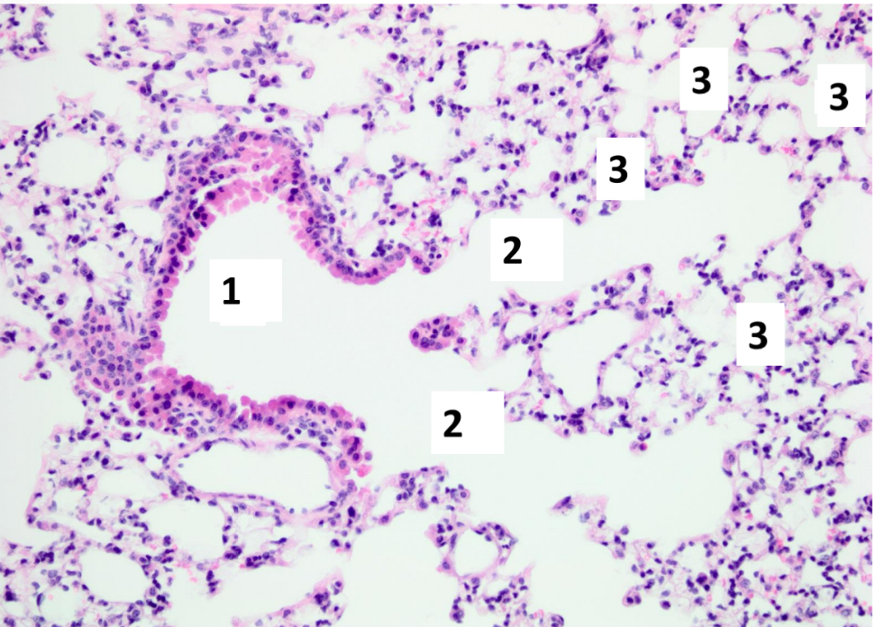
Name the structure indicated by 2.
Alveolar duct

Name the structure indicated by 3.
Alveolar spaces

Name the structure indicated by the red arrow.
Type II pneumocyte
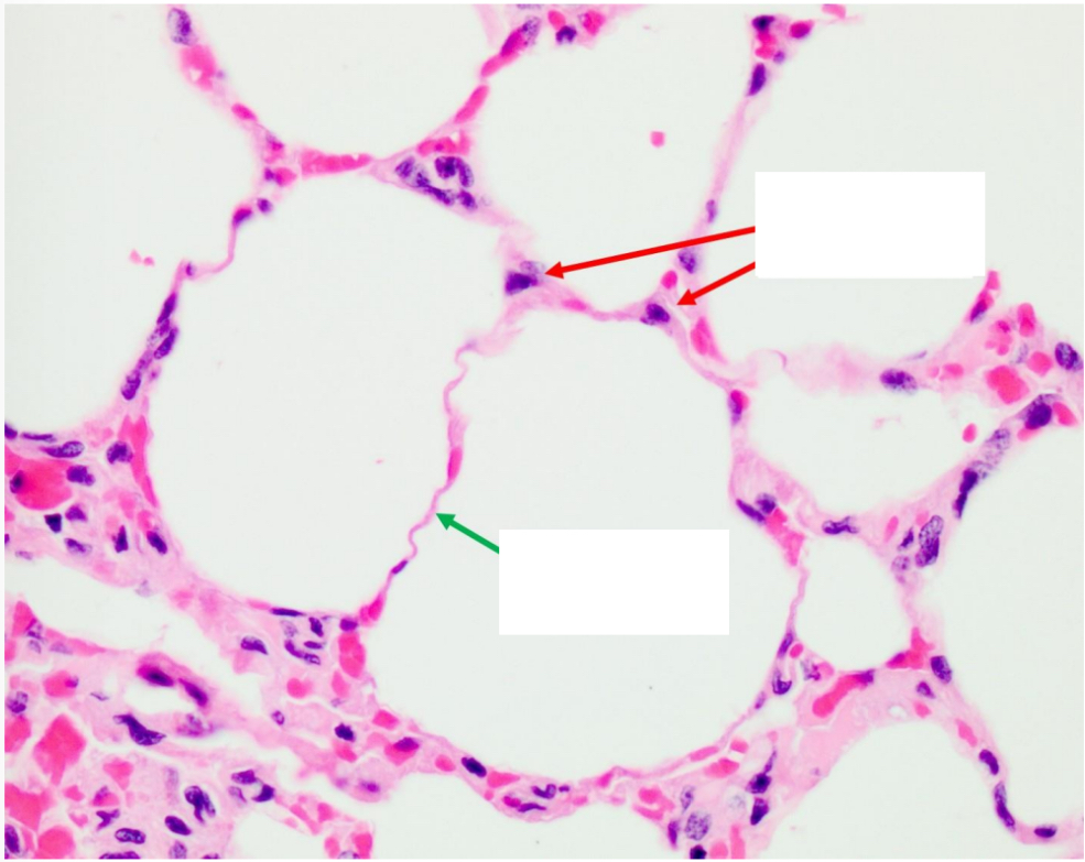
Name the structure indicated by the green arrow
Type I pneumocyte

Name the structure indicated by 1.
Pleura
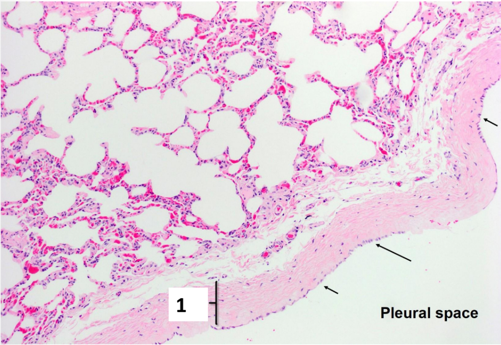
Name the structure indicated by the black arrow.
Mesothelium
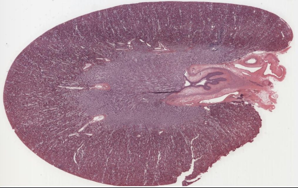
What organ is shown in the picture?
Kidney

Name the structure indicated by A.
Glomeruli

Name the structure indicated by B.
Proximal tubule
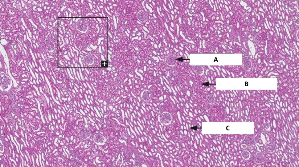
Name the structure indicated by C.
Distal tubule

Name the structure indicated by A.
Proximal tubule

Name the structure indicated by B.
Blood vessel
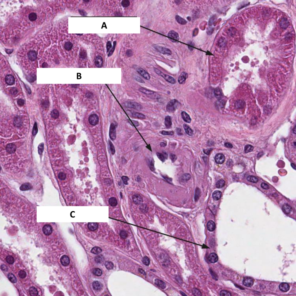
Name the structure indicated by C.
Distal tubule
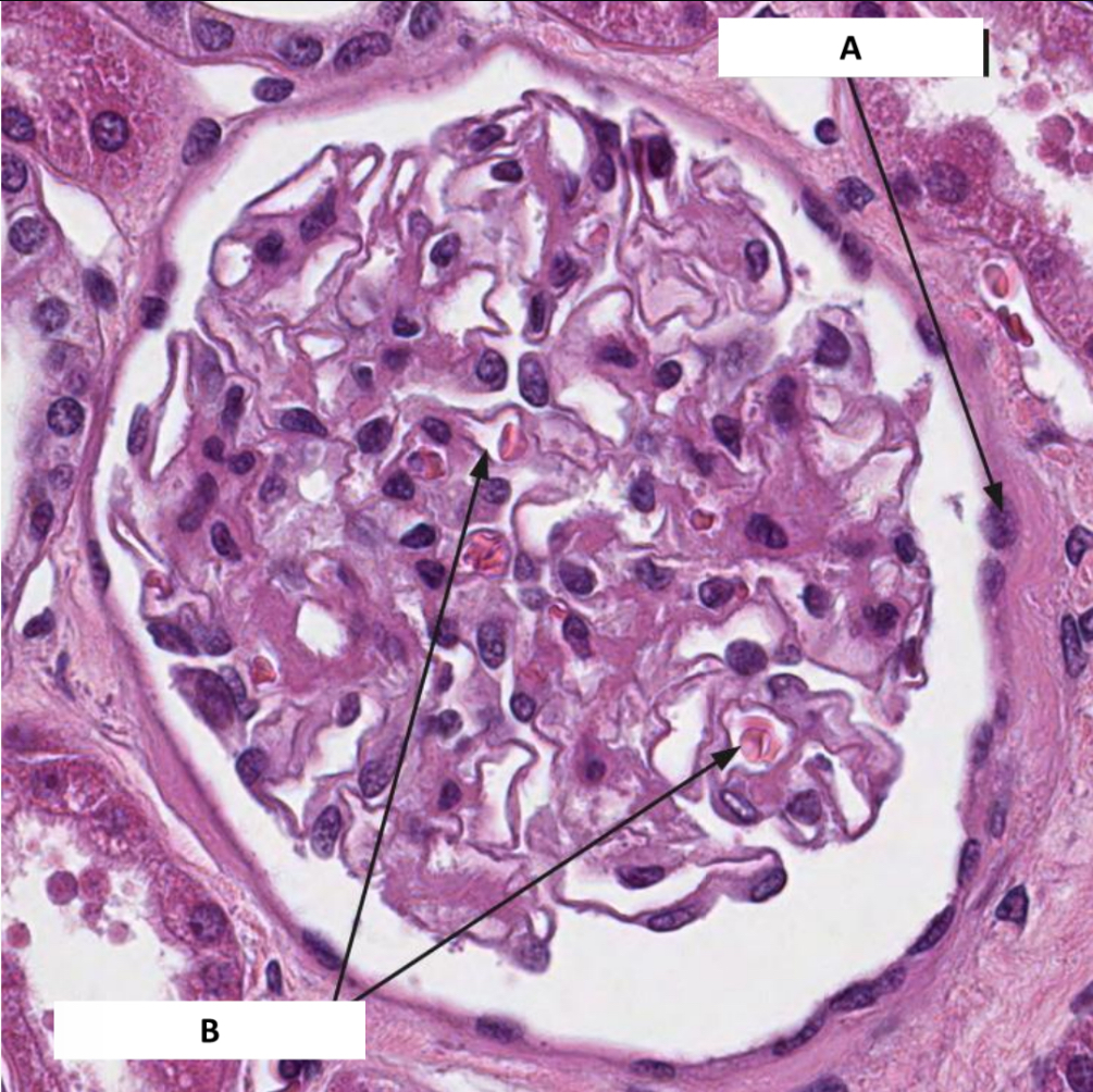
Name the structure indicated by A.
Bowman’s capsule

Name the structure indicated by B.
Capillary loops

Name the structure indicated by 1.
Parietal layer of capsule

Name the structure indicated by 2.
Capsular space
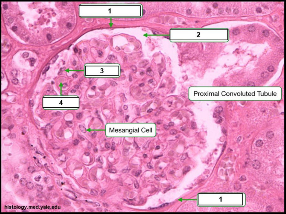
Name the structure indicated by 3.
Visceral layer of capsule (podocytes)

Name the structure indicated by 4.
Capillary

Name the structure indicated by A.
Bowman’s capsule

Name the structure indicated by B.
Glomerular capillary loop

Name the structure indicated by C.
Juxtaglomerular cells
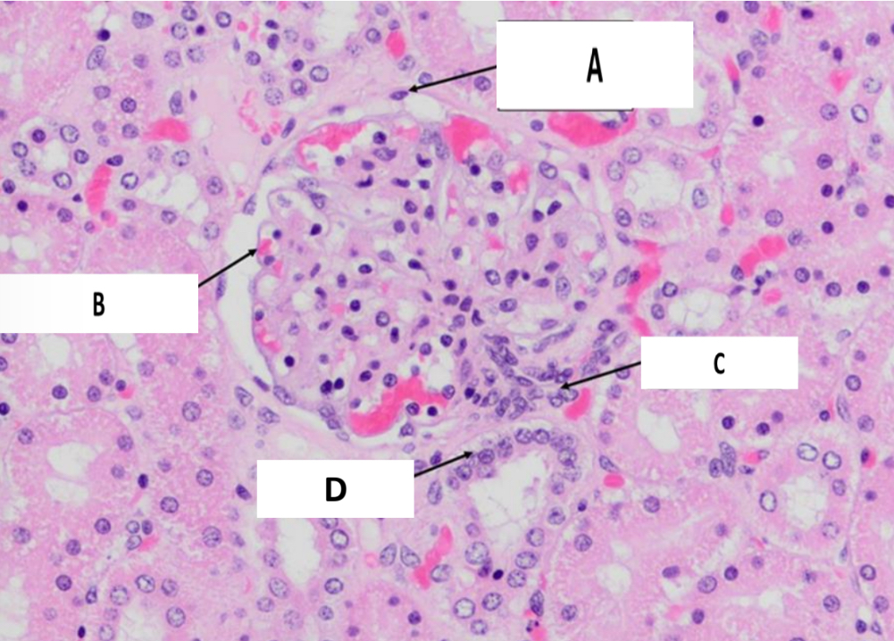
Name the structure indicated by D.
Macula densa
What organ is shown in the picture?
Urinary bladder

Name the structure indicated by A.
Mucosa
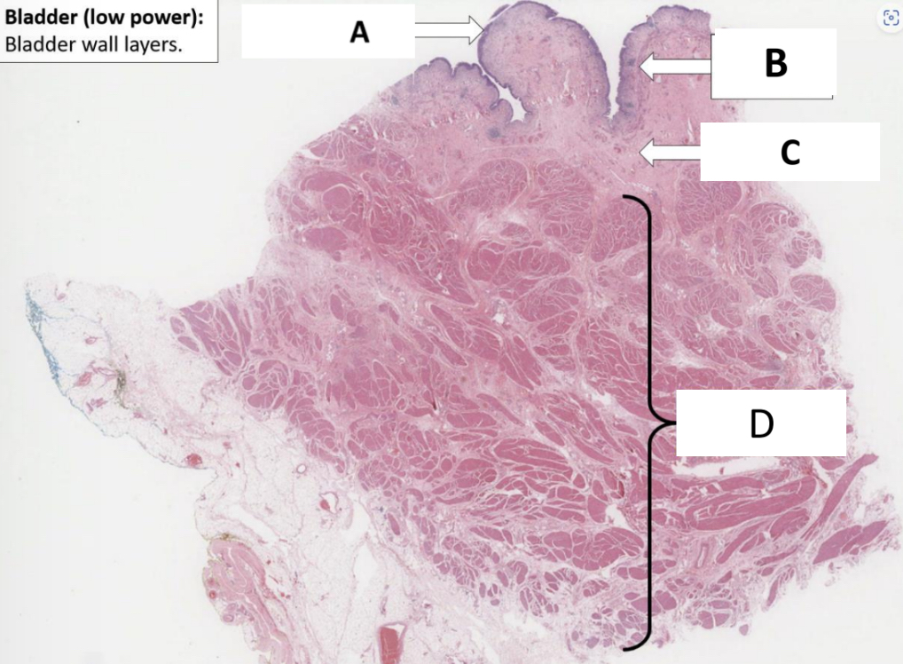
Name the structure indicated by B.
Muscularis mucosae

Name the structure indicated by C.
Lamina propria

Name the structure indicated by D.
Detrusor smooth muscle
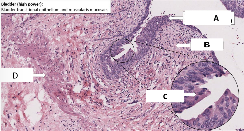
Name the structure indicated by A.
Urothelium

Name the structure indicated by B.
Lamina propria

Name the structure indicated by C.
Umbrella cell

Name the structure indicated by D.
Muscularis mucosa
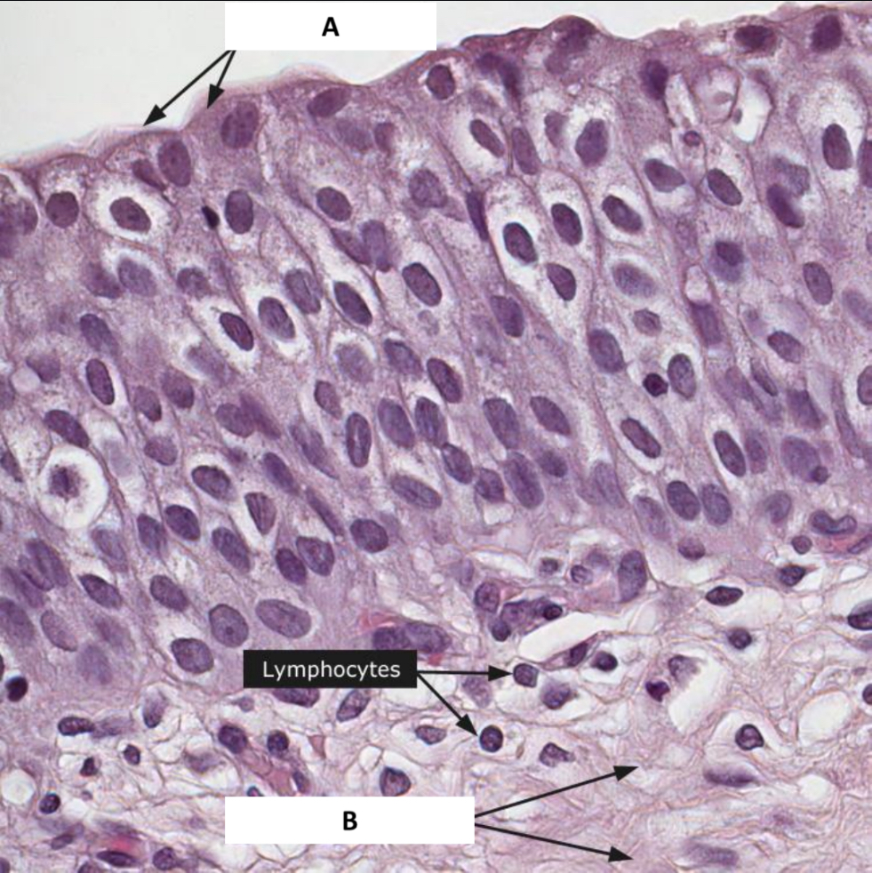
Name the structure indicated by A.
Umbrella cells

Name the structure indicated by B.
Connective tissue
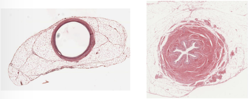
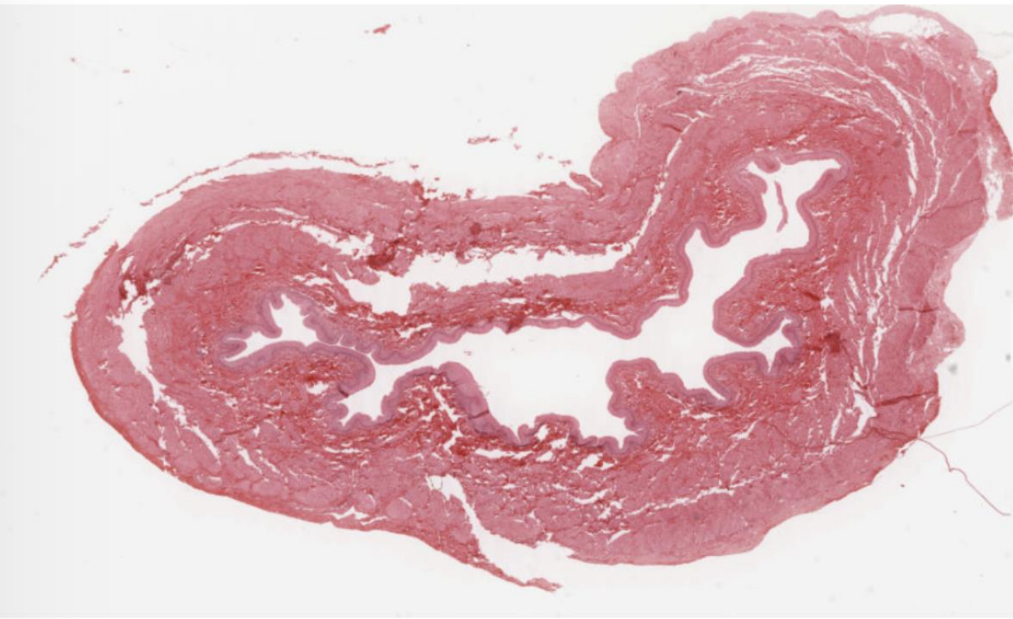
What organ is represented in the picture?
Ureter

Name the structure indicated by A.
Urothelium
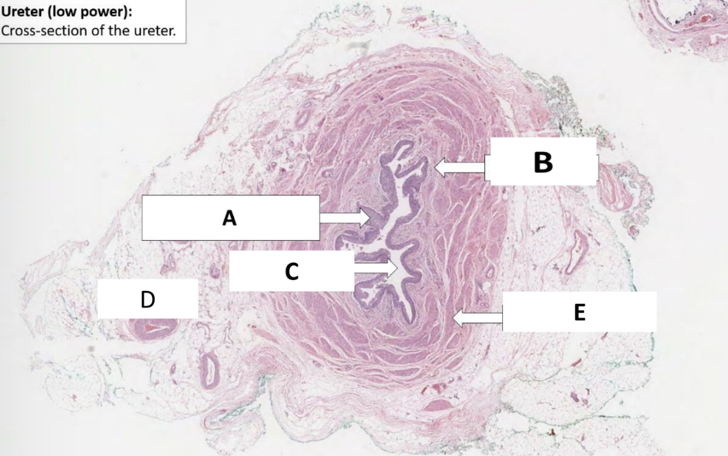
Name the structure indicated by B.
Lamina propria

Name the structure indicated by C.
Lumen

Name the structure indicated by D.
Adventitia

Name the structure indicated by E.
Muscularis propria

Name the structure indicated by A.
Urothelium

Name the structure indicated by B.
Lamina propria
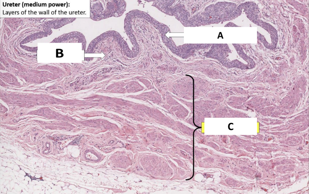
Name the structure indicated by C.
Muscularis propria
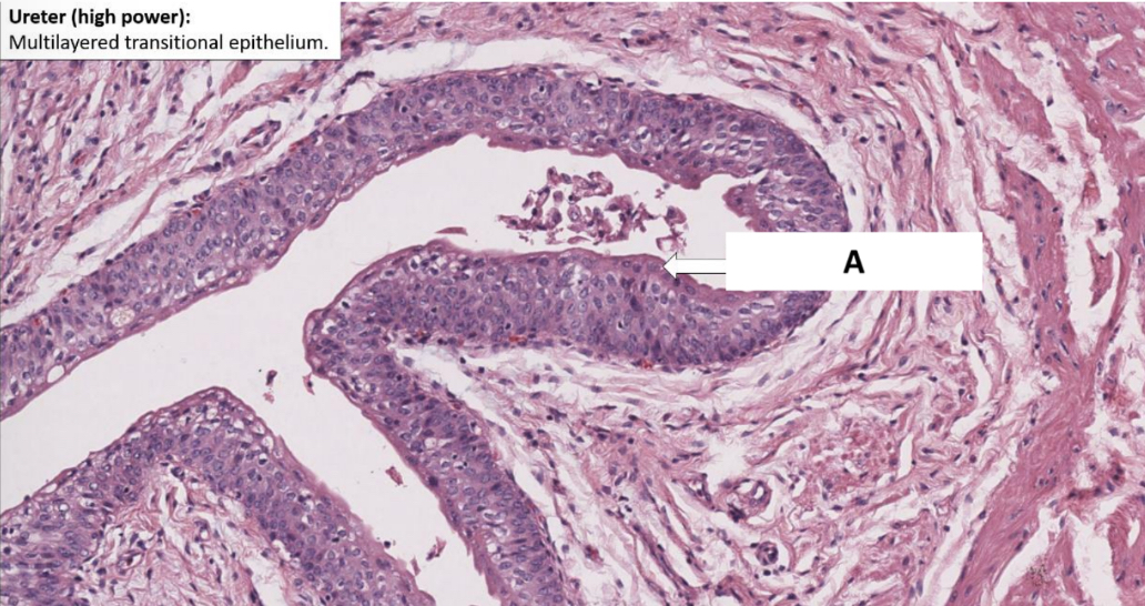
Name the structure indicated by A.
Umbrella cells

Name the structure indicated by A.
Urothelium

Name the structure indicated by B.
Lamina propria

Name the structure indicated by C.
Inner longitudinal muscle layer
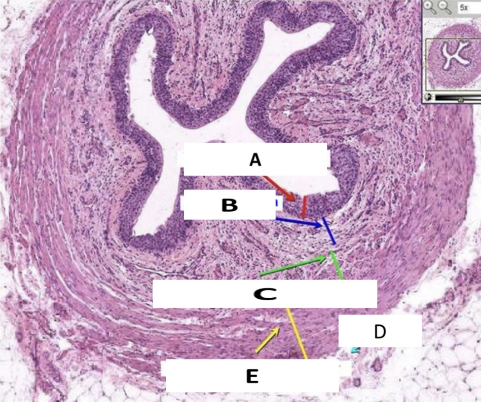
Name the structure indicated by D.
Adventitita

Name the structure indicated by E.
Outer circular muscle layer
Name the structure indicated by 1.
Lumen

Name the structure indicated by 2.
Urothelium