Anatomy Lower Limb, everything you will ever need to know about Week 1
1/108
Earn XP
Description and Tags
Movement of muscles and joints in lower limb
Name | Mastery | Learn | Test | Matching | Spaced | Call with Kai |
|---|
No analytics yet
Send a link to your students to track their progress
109 Terms
The hip joint is a.k.a a multiaxial joint, name all the movements it carries out (7):
Flexion, extension, abduction, adduction, internal rotation, external rotation, and circumduction.
Which muscle mainly extends the thigh assisting with lateral rotation if force is needed?
Gluteus maximus.
Describe the 2 movements that the gluteus medius and minimus perform:
Abduction and medial rotation of the thigh.
This muscle stabilises hip and knee joints and supports
gait cycle and climbing, as well as assists gluteus medius and minimus (abduction and medial rotation of the thigh):
Tensor fasciae latae.
The tensor fasciae latae is able to carry out climbing via a fibrous reinforcement on the lateral thigh, which stabilises the knee primarily, but also the hip joint, during locomotion and against gravity on knee flexion. What is this structure called?
Iliotibial band/tract.
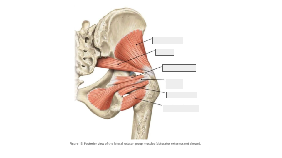
What are the 6 lateral rotators?
Piriformis, quadratus femoris, superior gemellus, obturator internus, obturator externus and inferior gemellus
All but one muscle in the lateral rotator group ABDUCTS the thigh when the hip is flexed, which muscle ADDUCTS the thigh when the hip is flexed instead?
Quadratus femoris
Which muscle in the posterior compartment of the thigh has 2 heads?
Biceps femoris
Which head of the 2 headed muscle in the posterior compartment of the thigh solely carries out extension of the thigh?
long head
Describe the movement of the biceps femoris, semitendinosus and semimembranosus:
Extension of hip, flexion of knee
This muscle exists in the posterior compartment, it has 2 portions, together they adduct the hip:
adductor magnus
The Ischiocondylar portion of the A.M. does what?
Extension of the hip
The Pubofemoral portion of the A.M. does what?
Flexion of the hip
The sacrum and coccyx, together with the hip bone make up the pelvic girdle (also known as the pelvis). What does the sacrum and coccyx articulate with?
L5 and L/R hip bones
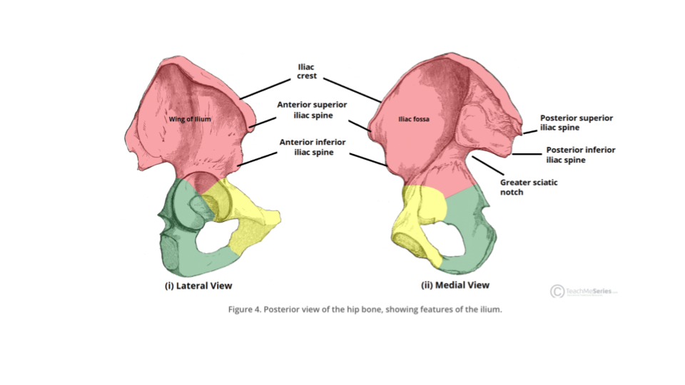
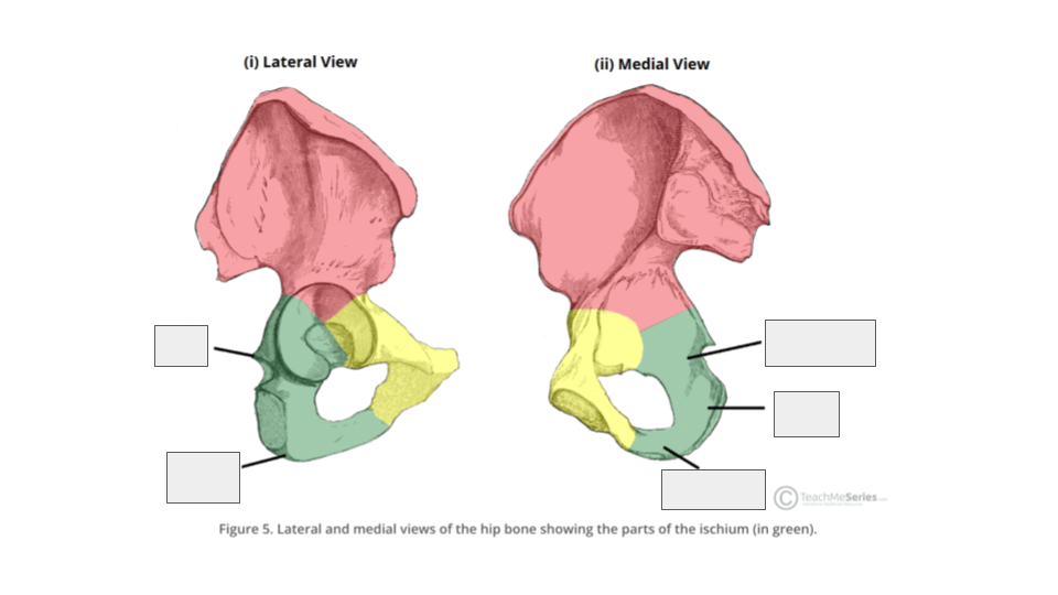
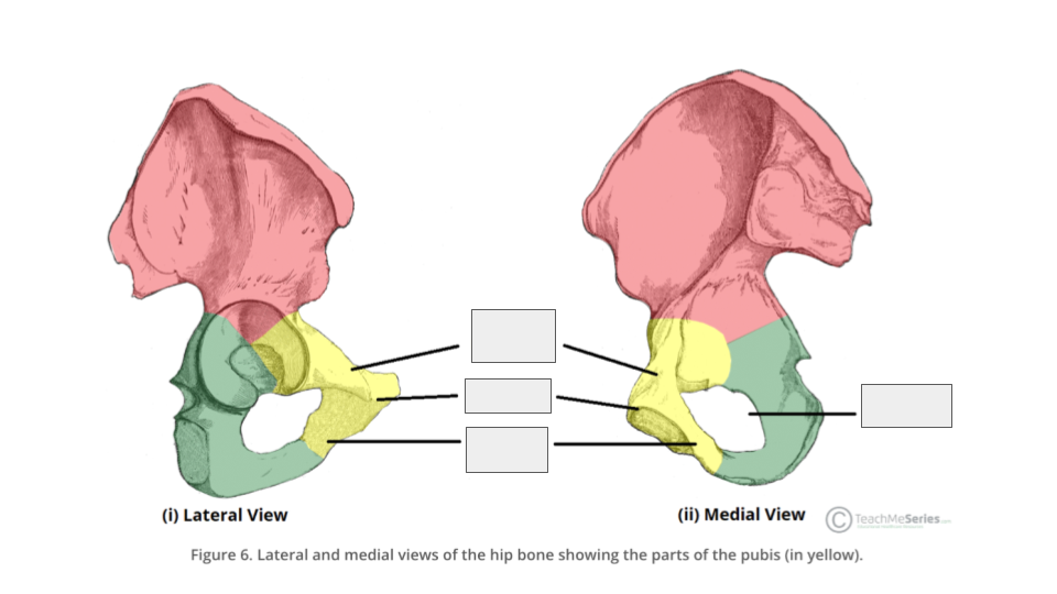
The hip bone is composed of what 3 bones?
ilium, pubis and ischium
where do the 3 bones of the hip bone fuse together?
the acetabulum
what are the 3 joints in the pelvis?
sacroiliac joint, pubic symphysis and hip joint
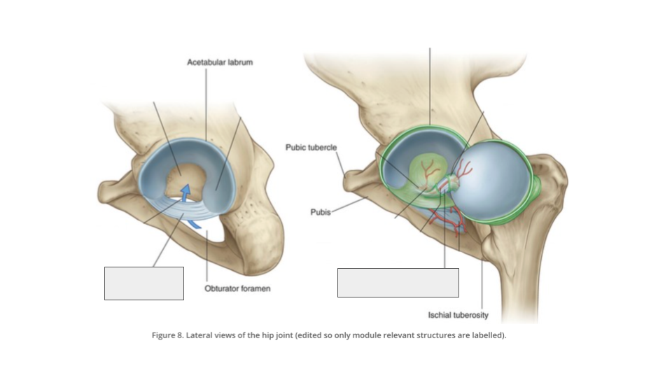
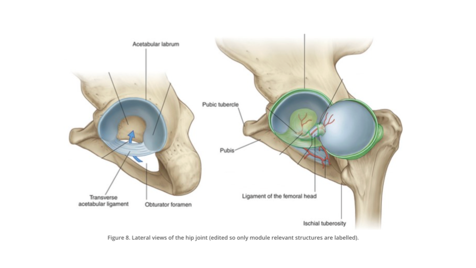
describe what the labrum is:
A ring of cartilage that surrounds the brim of the acetabulum. Stabilises and lubricates the hip joint and absorbs shock.
describe the location of the hip joint:
between the hip bone (acetabulum and labrum) and the femur (femoral head)
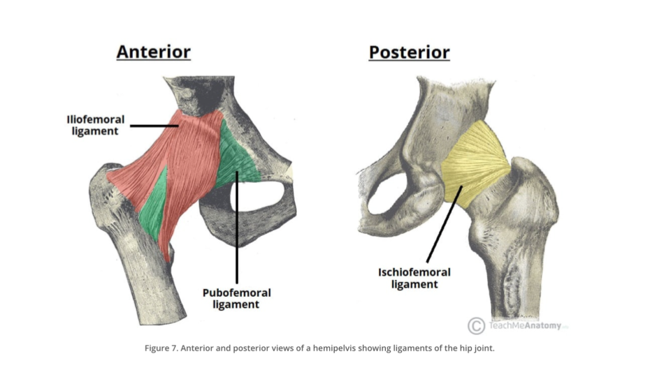
what are the 2 types of ligaments that surround the hip joint?
capsular and intracapsular
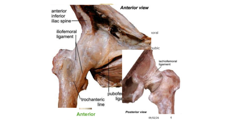
name the 3 types of capsular ligaments surrounding the hip joint:
iliofemoral ligament, pubofemoral ligament and ischiofemoral ligament
name the 2 types of intracapsular ligaments surrounding the hip joint:
transverse acetabular ligament, ligament of the head of the femur (LHF, ligamentum capitis femoris, ligamentum teres)
which nerve (on the anterior aspect) provides innervation to the anterior compartment of the thigh?
femoral nerve
which nerve (on the inferior aspect) provides innervation to the medial compartment of the thigh?
obturator nerve
which nerve (to the superior aspect) provides innervation to the gluteus medius and minimus muscles?
superior gluteal nerve
Which nerve (on the posterior aspect) provides innervation to the quadratus femoris and inferior gemellus muscles?
nerve to quadratus femoris
what provides blood supply to the hip joint?
anastomosis of the deep femoral artery, obturator artery and superior and inferior gluteal artery
describe the location of the the gluteal region:
posterior to the pelvic girdle, inferior to the iliac crest and superior to the gluteal fold
what are the 2 groups of muscles in the gluteal region?
gluteal group and lateral rotator group
what muscles are in the gluteal group?
gluteus maximus, gluteus medius, gluteus minimus and tensor fascia latae
What nerve supplies innervation to the gluteus maximus?
inferior gluteal nerve
what nerve provides innervation to the tensor fascia latae?
superior gluteal nerve
define fascia lata:
a deep band or sheet of dense, fibrous connective tissue that surrounds and supports the thigh
what provides innervation to the piriformis muscle?
nerve to piriformis (:p)
what provides innervation to the quadratus femoris?
nerve to quadratus femoris (:p)
what provides innervation to the obturator internus?
nerve to obturator internus (:p)
what provides innervation to the superior gemellus?
nerve to obturator internus (:<)
what provides innervation to the inferior gemellus?
nerve to quadratus femoris (:<)
the longest, widest single nerve in the body that goes from the anterior lumbar and sacral nerve fibres from the spinal cord → nerve roots unite into a single nerve anterior to the piriformis muscle → exits the pelvis between the piriformis and the superior gemellus into the gluteal region → travels down into the posterior thigh → …. is?
the sciatic nerve
what is the bone in the thigh?
femur
what are the 4 muscles in the posterior compartment of the thigh?
biceps femoris, semitendinosus, semimembranosus and adductor magnus
The nerve in the posterior thigh comes from the gluteal region → between adductor magnus and biceps femoris, lateral to semimembranosus/semitendinosus → divides at the apex of the popliteal fossa (behind the knee) is?
the sciatic nerve
This nerve divides to form the tibial and common fibular nerve:
sciatic nerve
fill in the missing terms:
fill in the blanks:

fill in the blanks:
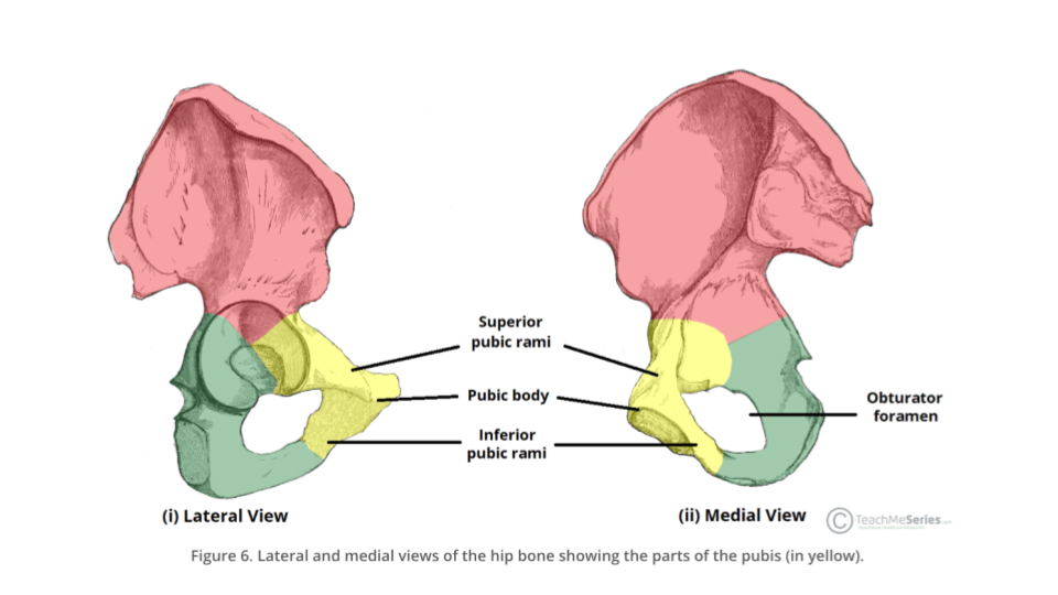
describe the pubic symphysis:
articulation between the left and right hip bones, on the anterior side of the pelvis
describe the sacroiliac joint:
articulation between the left and right hip bones with the sacrum, on the posterior side of the pelvis
At the hip joint, how much of the femoral head inserts and sits within the acetabulum?
2/3
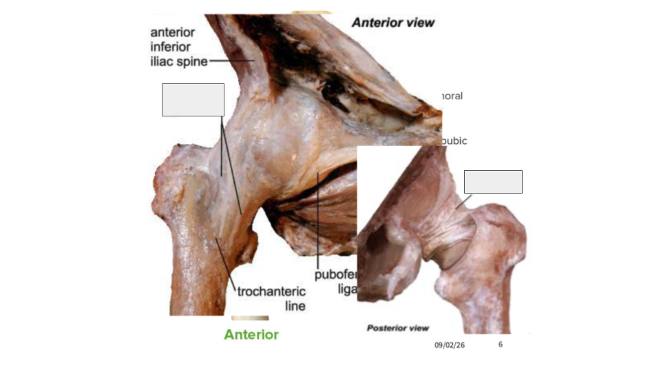
describe the shape of the iliofemoral ligament:
thick Y shape
the iliofmeoral joint blends in with the?
joint capsule
the iliofemoral ligament goes from the ________ inferior iliac spine and the ____________ rim to the intertrochanteric line of the femur:
anterior, acetabular
the strongest ligament in the BODY is:
iliofemoral
the iliofemoral joint prevents ______________ of the hip joint when standing:
hyperextension
what shape are the pubofemoral and ischiofemoral ligaments?
band shape
what aspect is the pubofemoral ligament on the hip joint?
anteroinferior
what aspect is the ischiofemoral ligament on the hip joint?
posteroinferior
the pubofemoral ligament goes from the ____________ ramus and pubic bone to the lower aspect of the ______________ line of the femur, blending with the joint capsule and the iliofemoral ligament
iliopubic, intertrochanteric
the pubofemoral joint prevents:
excessive abduction of the hip joint by tightening during extension and abduction movements.
the ischiofemoral ligament goes from the ______ below the acetabulum to the ______ trochanter of the ______.
ischium, greater, femur
the ischiofemoral ligament limits:
internal rotation of the hip
fill in the blanks:
fill in the blanks:
the transverse acetabular ligament is a strong flat ligament which completes the _________ aspect of the acetabular rim by bridging the acetabular ______, effectively converting the notch into a foramen and completing the circular rim of the acetabulum. By doing so, it supports the ___________ labrum and deepens the hip socket, increasing joint _______ and ensuring the femoral head is securely contained.
inferior, notch, acetabular, stability
the foramen created beneath the ligament serves as a protected passageway for what kind of vessels, as well as nerves, to enter the joint cavity and reach the head of the femur?
nutrient
what shape is the ligament of the head of femur?
flattened triangular band
the ligament of the head of the femur connects the femoral head to the acetabulum and which ligament?
transverse acetabular ligament
In infants and children, the primary role of the LHF is to serve as a protected conduit for the acetabular branch of the obturator artery, this branch provides critical blood supply to the developing head of the femur. What happens to this branch as adults?
In adults, this artery usually becomes narrow or obliterated, and the ligament functions instead as a secondary stabiliser for the hip joint.
fill in the blanks:
What muscle am I describing:
From the ilium between the inferior and anterior gluteal lines to the femur. Covered by the gluteus medius. Its main action is to abduct and medially rotate the thigh. It also helps stabilise the pelvis during locomotion. It is innervated by the superior gluteal nerve.
gluteus minimus
What muscle am I describing?
From between the posterior gluteal line and the iliac crest to the femur. Partially covered by the gluteus maximus. Its main action is to abduct and medially rotate the thigh. It also helps stabilise the pelvis during locomotion (same as the gluteus minimus). It is innervated by the superior gluteal nerve.
gluteus medius
What muscle am I describing:
From the posterior gluteal line and the sacrum to the femur. It is the main extensor of the thigh. Its large size is one of the most characteristic features of the human muscular system and allows us to maintain an erect posture. It is innervated by the inferior gluteal nerve.
gluteus maximus
What muscle am I describing?
From the iliac crest to the iliotibial band/tract. Its main action is to tense the iliotibial band, stabilising the hip and knee joints. This supports locomotion and weight-bearing. It also assists the gluteus medius and minimus in abduction and medial rotation of the thigh. It is innervated by the superior gluteal nerve.
tensor fascia latae
describe the shape of the piriformis muscle:
flat and pyramidally-shaped
describe the shape of the superior and inferior gemellus:
small and tubular
describe the shape of the quadratus femoris
flat and quadrilateral
fill in the blanks

why is abduction of the flexed thigh important in the action of locomotion?
because it shifts the body weight to the opposite side when the foot is being lifted, which prevents falling
Lateral rotators oppose what kind of movement by the gluteus medius and minimus muscles?
medial rotation
the muscles in the posterior compartment of the thigh are commonly known as the?
hamstrings
what is the general function of the posterior compartment of the knee?
extend the hip and flex the knee
all the muscles in the posterior compartment of the thigh have a common origin where?
on the ischial tuberosity
all the muscles of the posterior compartment of the thigh are innervated by?
the sciatic nerve
all muscles in the posterior compartment receive blood blood from which artery which is a branch of the femoral artery?
deep femoral artery
Both heads of the biceps femoris attach to the fibula on the leg; however, which head comes from the ischial tuberosity?
the long head
as the ____ head crosses the hip joint, only that portion acts on that joint, both heads act on the _____ joint.
long, knee
the semitendinosus is ___________ to the semimembranosus
superficial
the semitendinosus has a ________, more tubular tendon than the semimembranosus
longer
where does the semitendinosus insert on the tibia?
on the medial side at the pes anserinus
the semimembranosus is the most ______ of the 3 hamstrings
medial
the semimembranosus has a flat tendon and the muscle belly is ________ than semitendinosus
bigger
where does the semimembranosus insert on the tibia?
medial side
both the semitendinosus and semimembranosus receives additional blood supply on it’s proximal end by which artery?
inferior gluteal artery
define the pes anserinus:
the conjoined tendons of semitendinosus, sartorius, and gracilis muscles
what is the insertion of the adductor magnus?
distal femur
where does the sciatic nerve arise from
the lumbosacral plexus
The common iliac arteries bifurcate just below ___ into the internal and external iliac arteries. The ________ iliac artery supplies the pelvic cavity and pelvis. The main branches are the superior and inferior _________ arteries and the _______ artery.
L4, internal, gluteal, obturator
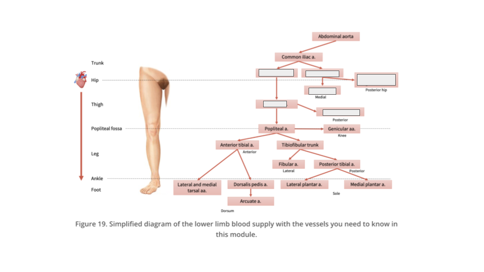
fill in the blanks:
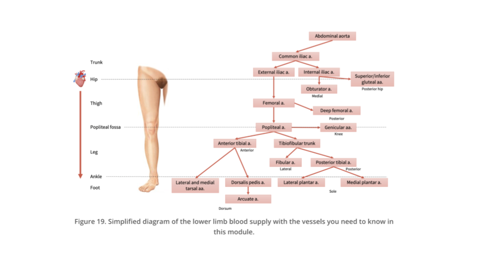
The ________ iliac artery continues downwards and after passing under the inguinal ligament into the lower limb, it changes name into _________ artery. The ________ artery travels into the anterior thigh, supplying the anterior compartment muscles. At the proximal thigh, the femoral artery gives a branch, the _____ femoral artery, which travels posteriorly and then medially to supply the muscles in the posterior and medial compartments.
external, femoral, femoral, deep
Where does the venous drainage of the lower limbs ultimately drain into?
inferior vena cava