Anatomy Practical 2
1/122
There's no tags or description
Looks like no tags are added yet.
Name | Mastery | Learn | Test | Matching | Spaced | Call with Kai |
|---|
No analytics yet
Send a link to your students to track their progress
123 Terms
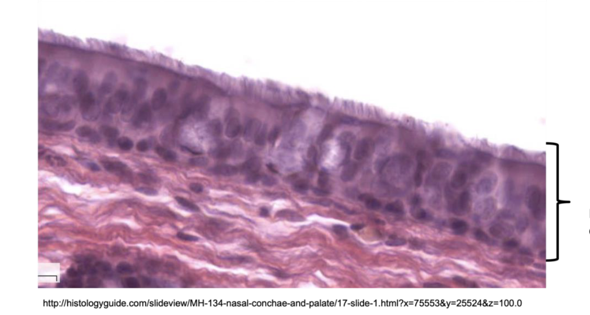
Pseudo stratified ciliated columnar
found in trachea, primary and secondary bronchi
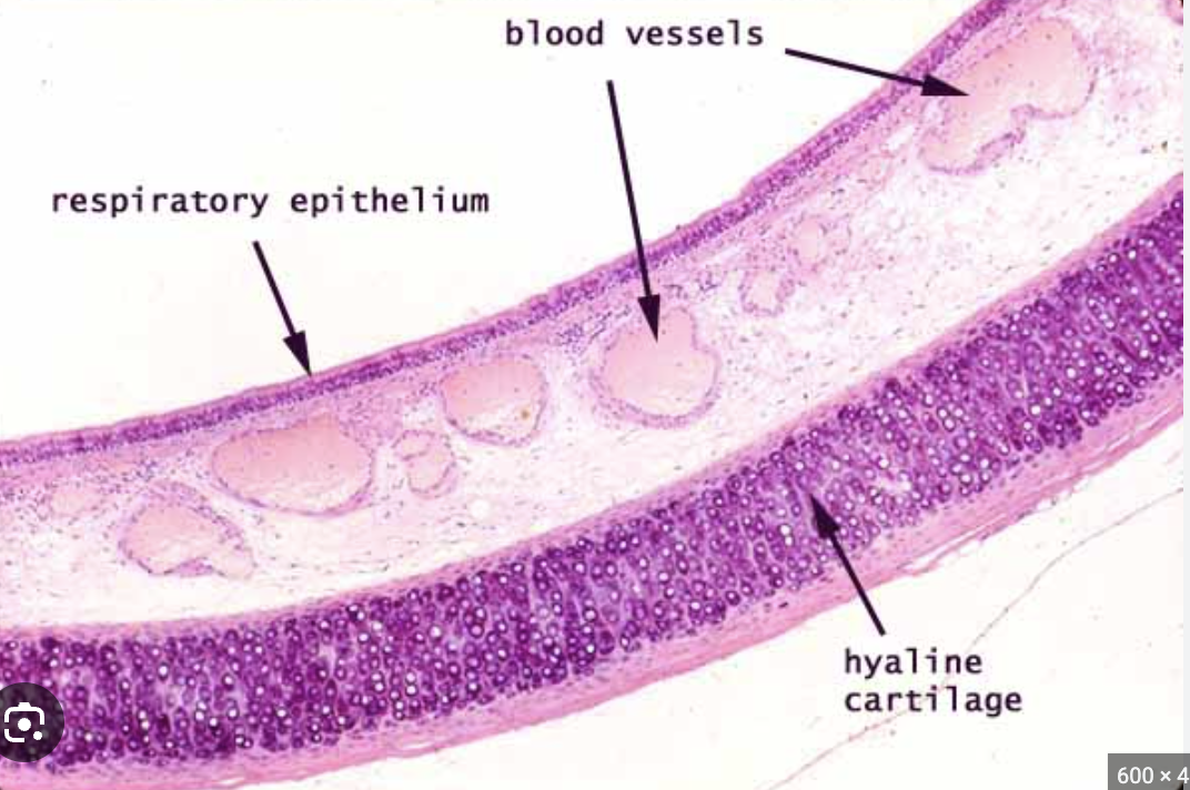
Hyaline cartilage
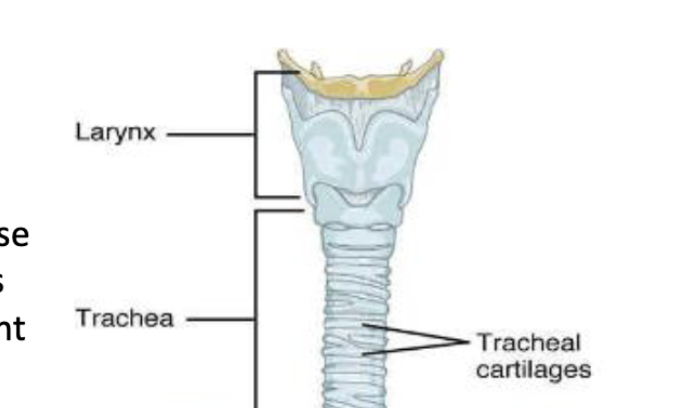
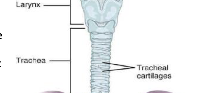
forms 16 to 20 C-shaped rings that provide rigid structural support to the trachea, ensuring the airway remains open
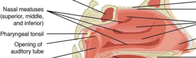
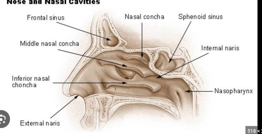
Conchae; superior, middle, inferior
increase the surface area of the nasal cavity and to disrupt the flow of air as it
enters into the nose, causing air to bounce along the epithelium, where it is cleaned and
warmed
superior, inferior, and middle meatus
conserve water and prevent dehydration of the nasal epithelium by trapping water during
exhalation.
external and internal nares
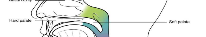
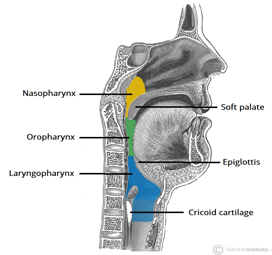
hard and soft palate
The hard palate at the anterior region of the nasal cavity is composed of bone, while the soft palate, at the posterior portion of the nasal cavity, consists of muscle tissue
uvula
move like a pendulum during swallowing, swinging upward to close off the
nasopharynx to prevent ingested materials from entering the nasal cavity
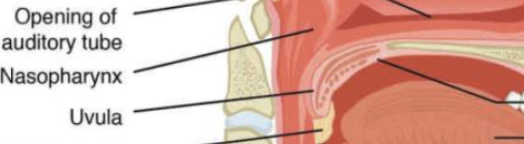
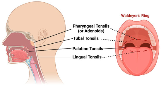
pharyngeal tonsil
the immune system’s first line of defense against ingested or inhaled foreign pathogens, trapping bacteria and viruses entering through the mouth and nose

palatine tonsil
the immune system’s first line of defense against ingested or inhaled foreign pathogens, trapping bacteria and viruses entering through the mouth and nose
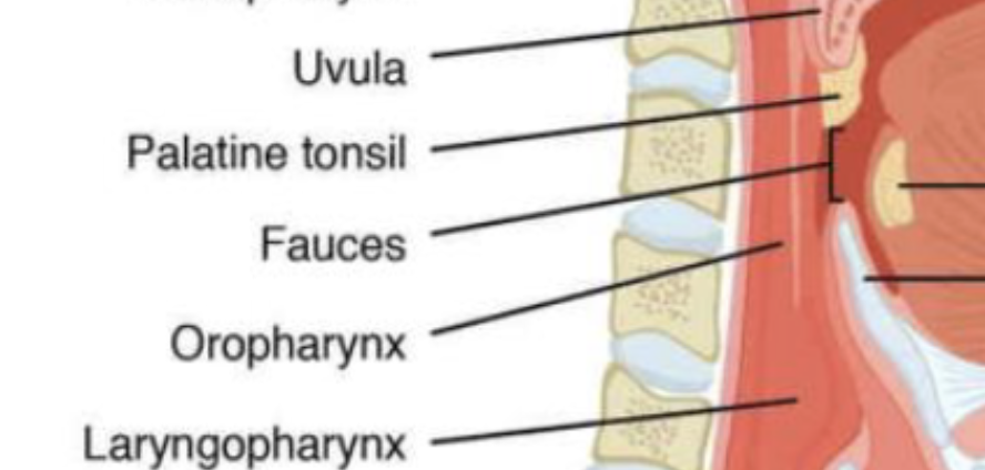
lingual tonsil
the immune system’s first line of defense against ingested or inhaled foreign pathogens, trapping bacteria and viruses entering through the mouth and nose
nasopharynx, oropharynx, laryopharynx
serves as an airway
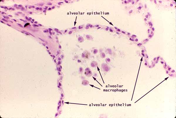
simple squamous epithelium
found in Respiratory bronchioles
epiglottis
composed of elastic cartilage, is a plate-like structure that
closes the opening of the larynx (called the glottis) during swallowing
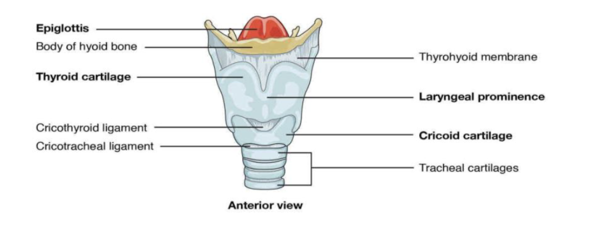
larynx
commonly known as the “voice box” because it is an important organ for sound
production in humans
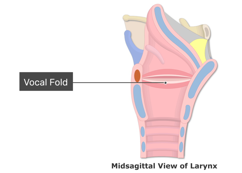
vestibular and vocal cords of the larynx
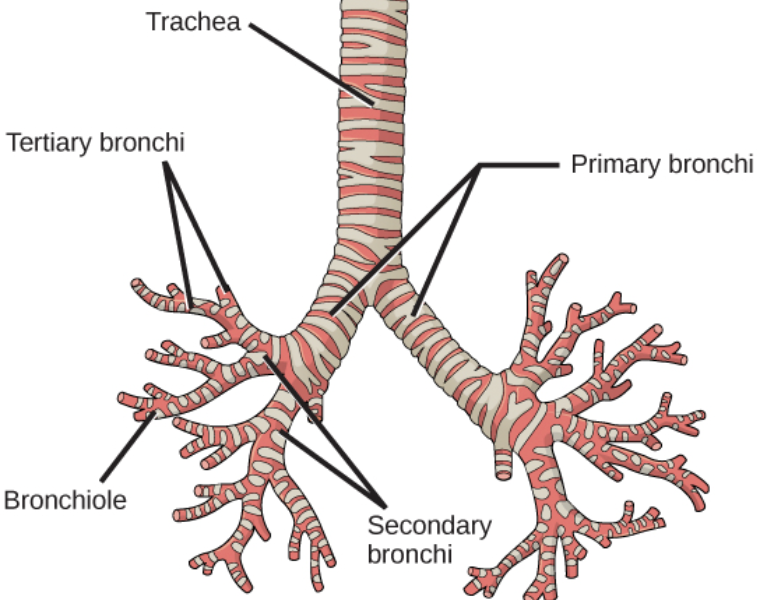
trachea
commonly known as the “windpipe” because it extends and carries air from the larynx toward the lungs
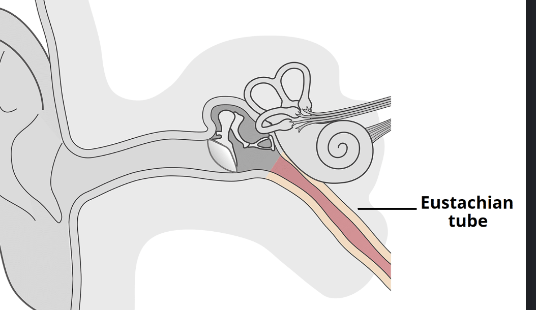
pharyngotympatic tube
connect the nasopharynx to each middle ear cavity
Thyroid, arytenoid, cricoid cartilages

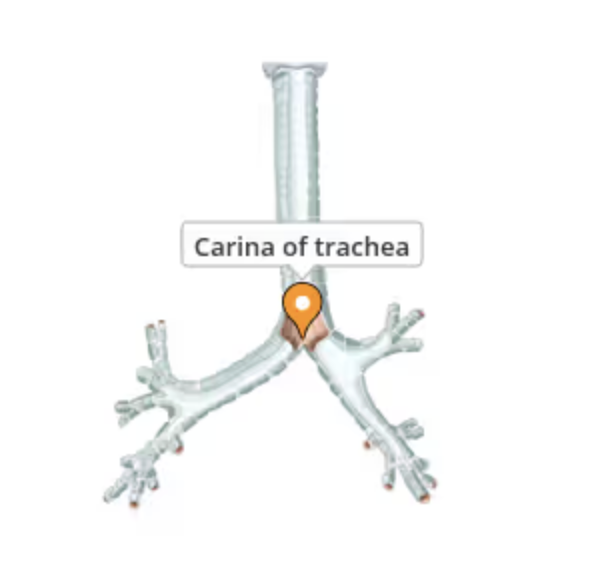
trachea; carina
triggers a strong cough reflex if foreign objects touch it and helps divide air towards the lungs
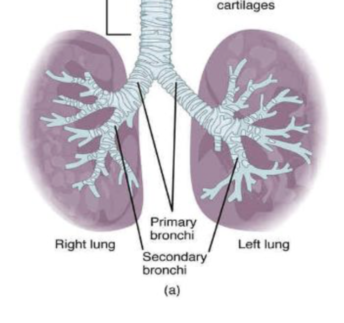
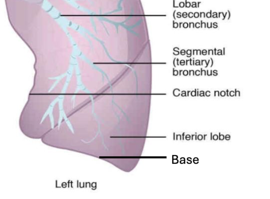
primary bronchi
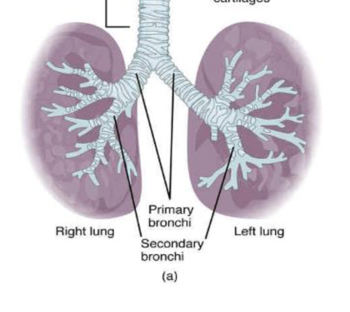
secondary bronchi
tertiary broncho
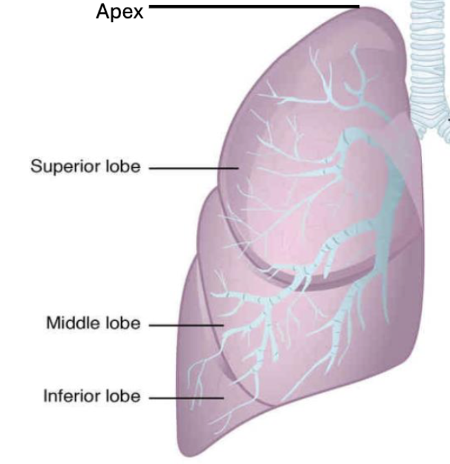
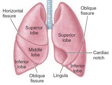
superior, middle, inferior lobe of lung
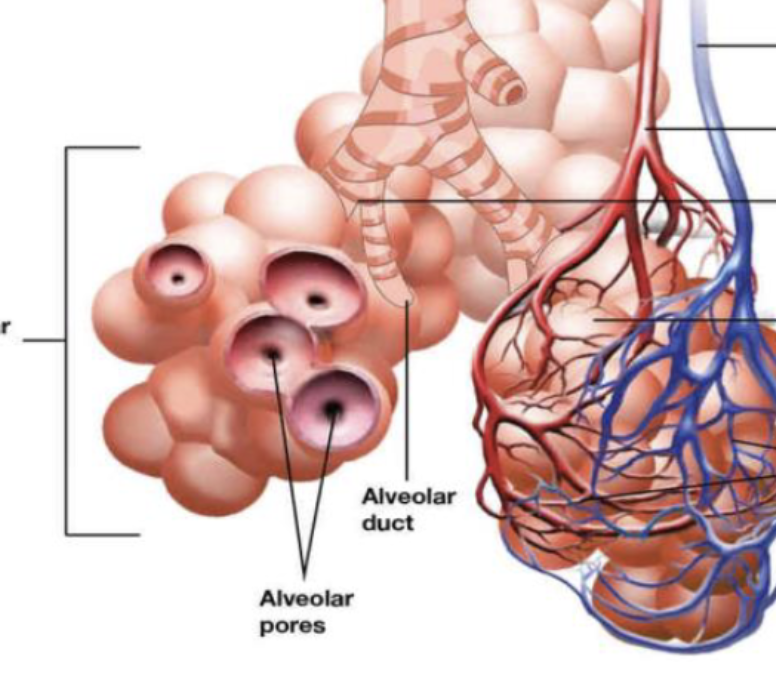
alveolar duct
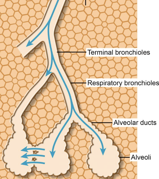
terminal bronchiole
small respiratory tubes with smooth muscle in their walls, no cartilage, and an inner lining of respiratory epithelium
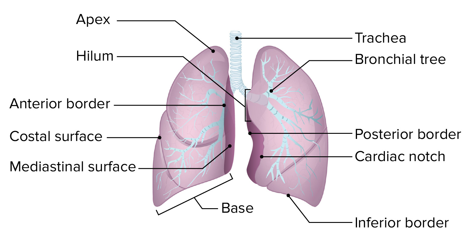
cardiac notch
an indentation on the surface of the left lung that allows space for the heart
horizontal and oblique fissures
separate the lobes of the lungs
respiratory bronchiole
the smallest type of bronchiole, which then leads to an alveolar duct
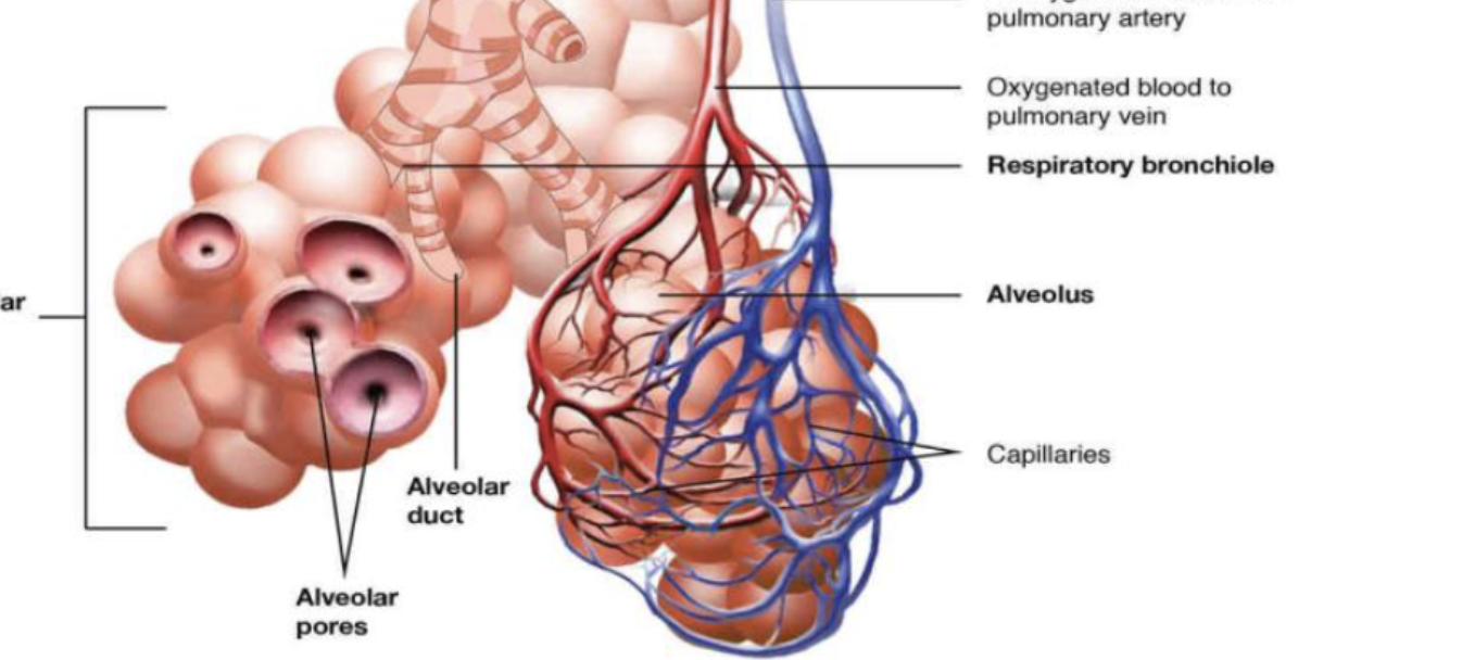
alveolar sac

alveoli
the primary site for gas exchange
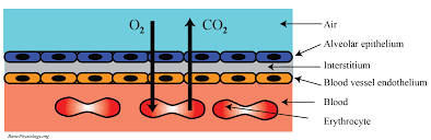
respiratory membrane
allows for gas exchange
hilus, apex, and base of lungs
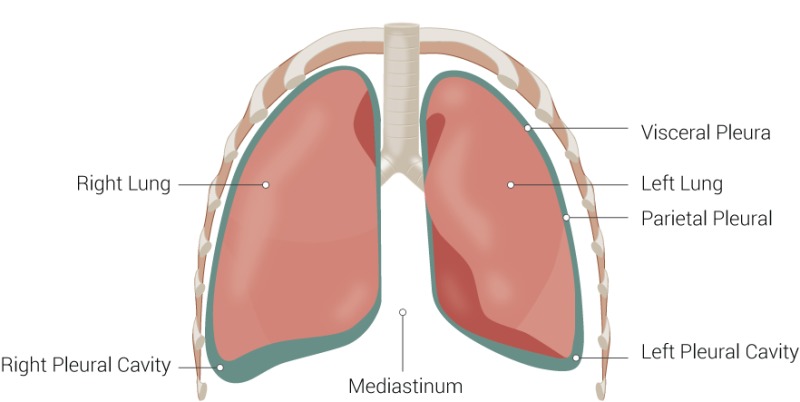
visceral and parietal pleural cavity
create a frictionless, fluid-filled pleural cavity that allows for smooth lung movement
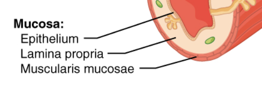
muscosa: lamina propria, epithelium, and muscularis mucosa
epithelium that is in contact with the contents in the lumen; underlying connective tissue called the lamina propria; and a thin layer of smooth muscle called the muscularis mucosae
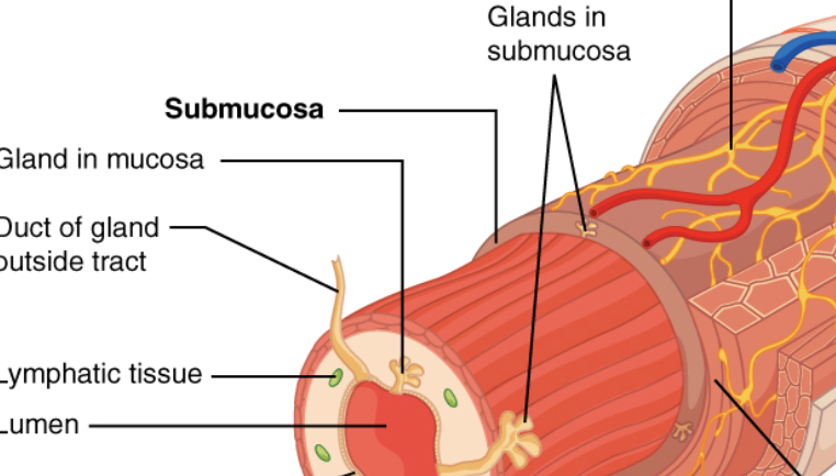
submucosa, muscularis externa, serosa/adventitia
the four layers from the lower esophagus to the anal canal
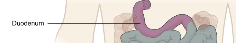
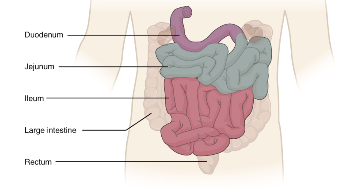
duodenum
layers of small intestine are the duodenum closest to the stomach
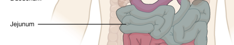
Jejunum
middle region of the small intestine
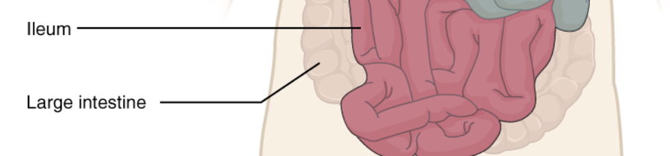
ileum
the longest and most distal region of the small intestine
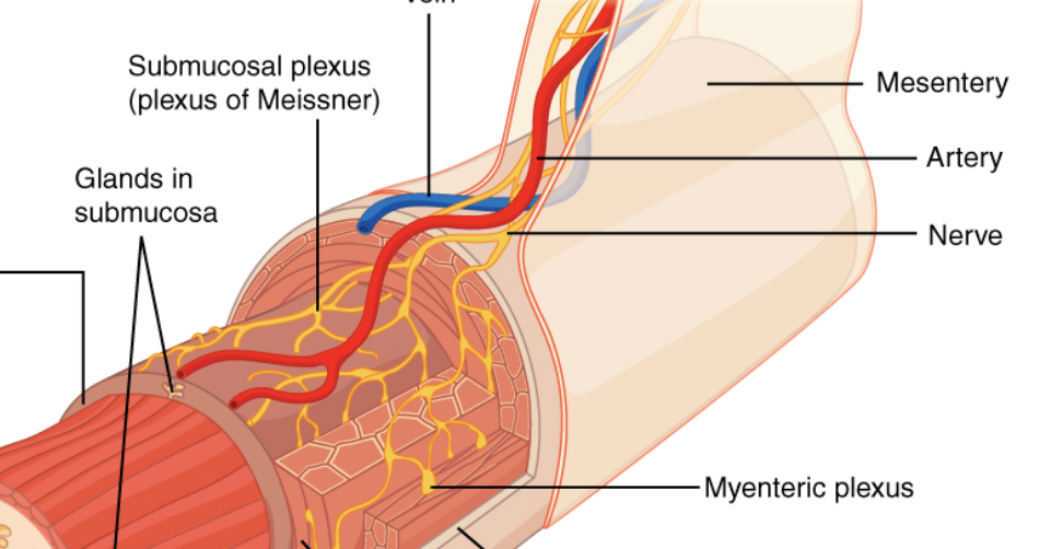
myenteric and submucosal plexus
myenteric plexus (located between muscle layers) regulates motility/peristalsis, while the submucosal plexus (located in the inner submucosal layer) regulates glandular secretion, absorption, and local blood flow
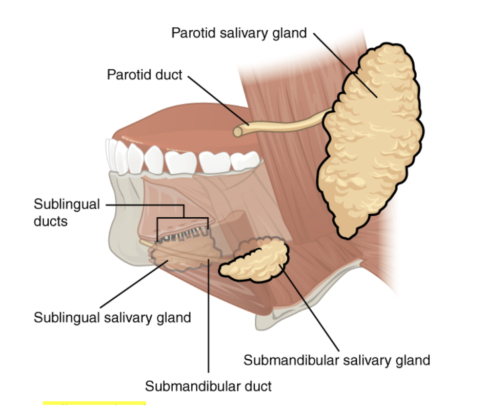
parotid salivary gland
found outside the mouth and superficial to masseter on both side
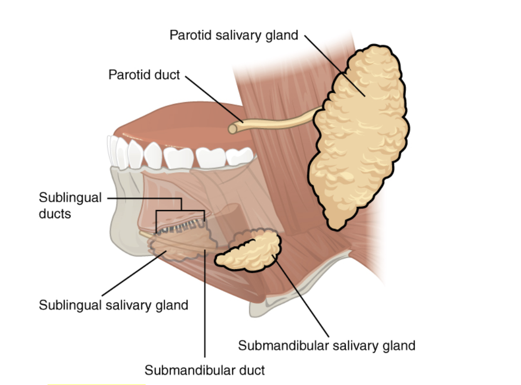
submandibular glands,
located just below the jaw
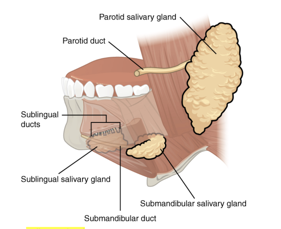
sublingual glands
located under the tongue
tonsils: lingual, palatine, and pharyngeal
act as the first line of defense against ingested or inhaled pathogen
fundus of stomach
stores undigested food and gases
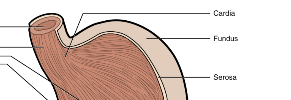
greater curvature
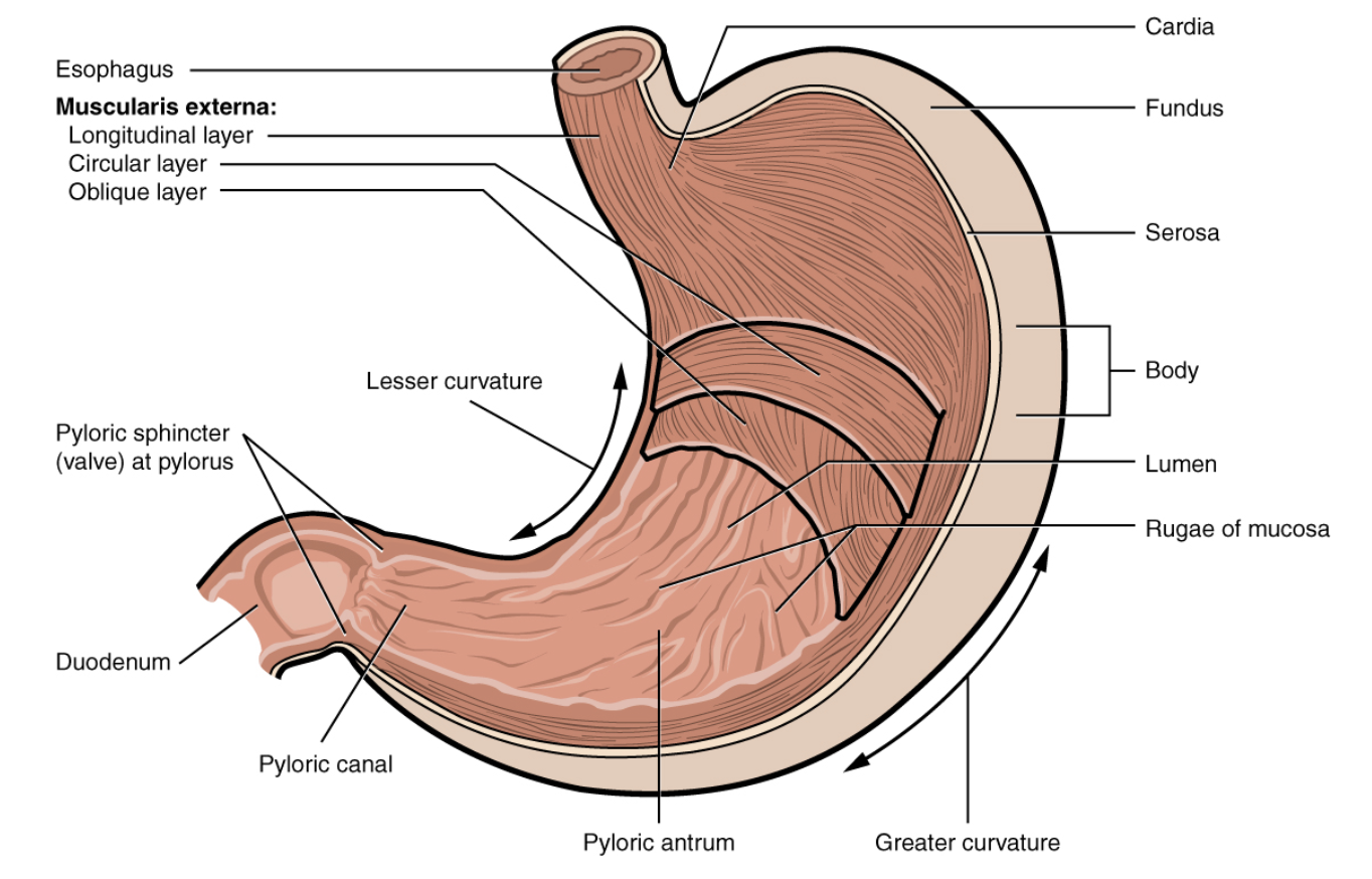
lesser curvature
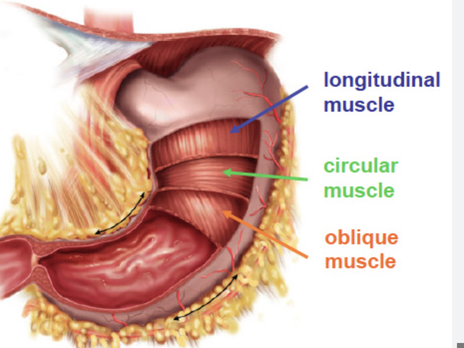
layers of smooth muscle in the stomach
facilitates the mechanical digestion, churning, and mixing of food with gastric juices to form chyme
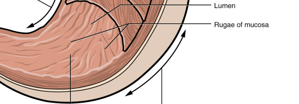
rugae
allow for stomach expansion
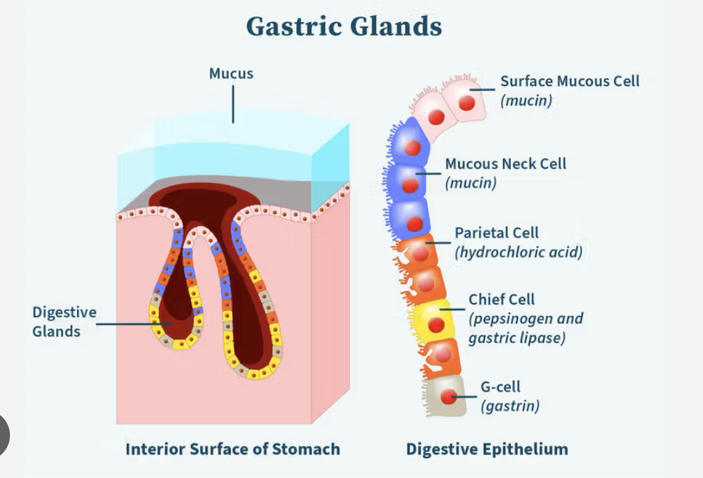
gastric pits of the stomach
small openings in the stomach lining that lead to tubular glands producing gastric juices essential for digestion
regions of the small intestine
duodenum, jejunum, and ileum
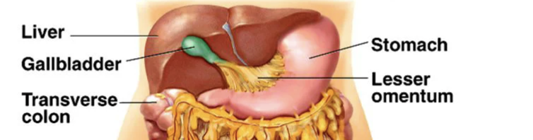
greater omenta
covers the small intestine to protect it against damage
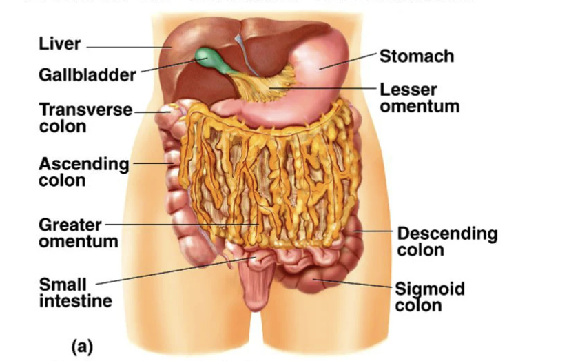
lesser omenta
small openings in the stomach lining that lead to tubular glands producing gastric juices essential for digestion
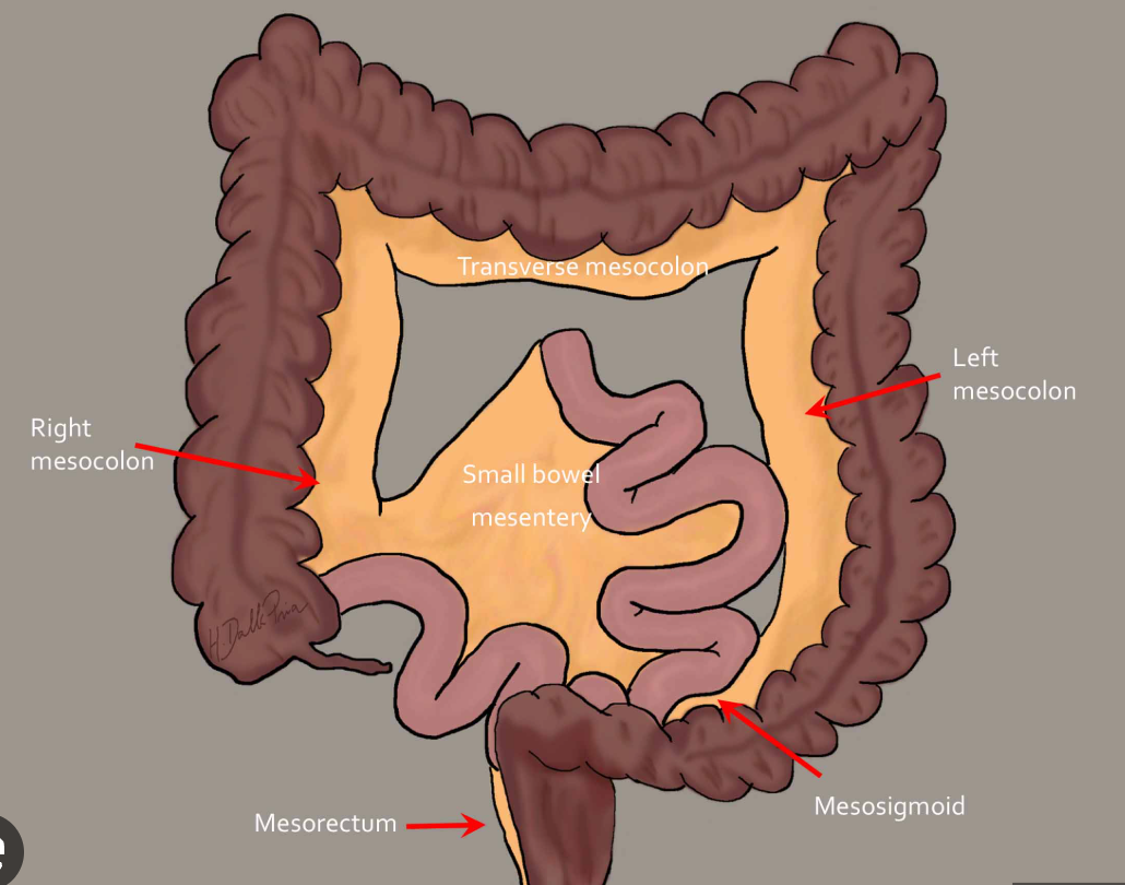
mesocolon
specialized, double-layered fold of peritoneum that attaches the colon (large intestine) to the posterior abdominal wall, anchoring it while providing mobility and supporting blood vessels, nerves, and lymphatics
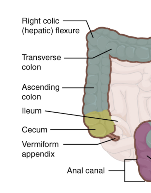
Ileocecal valve
cecum and vermiform appendix
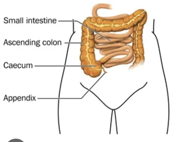
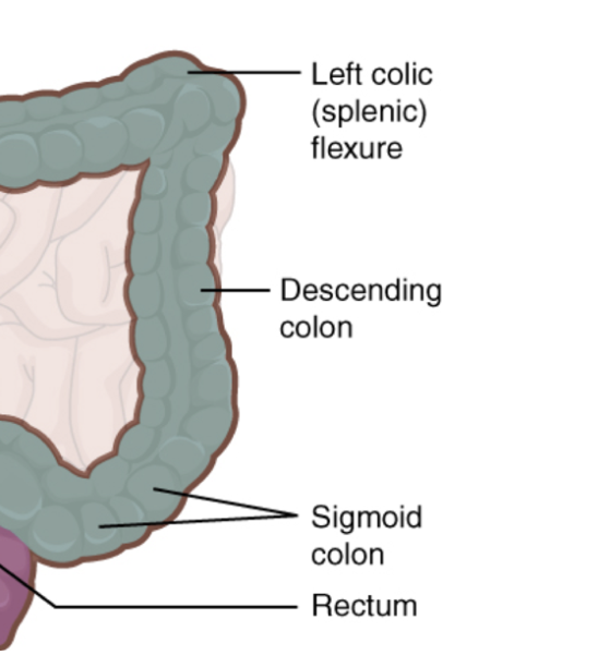
ascending colon
descending colon
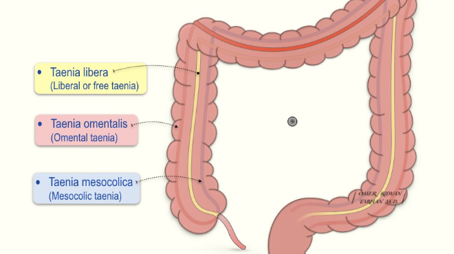
taenia coli and epiploic appendages
Epiploic appendages are small, fat-filled, serosa-covered pouches that project from the colon's surface, mostly bordering the taeniae libera and omentalis
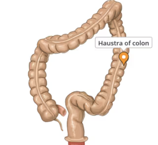
haustrum
small, sac-like pouch of the large intestine (colon) created by sacculation, giving the colon its characteristic segmented appearance
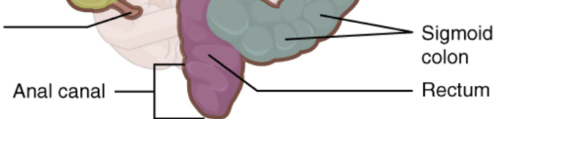
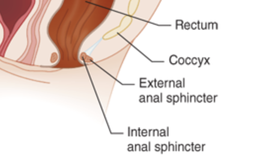
rectum and anal canal
located at the bottom of the large intestine
acinar cells
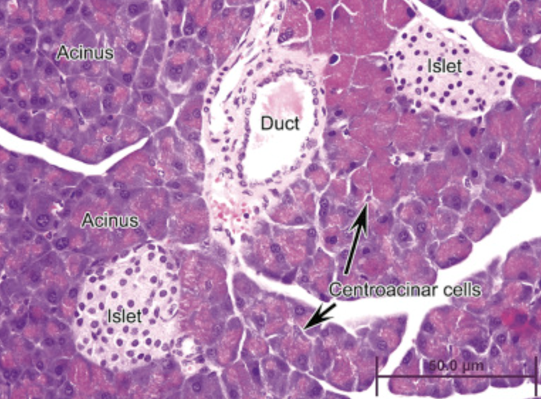
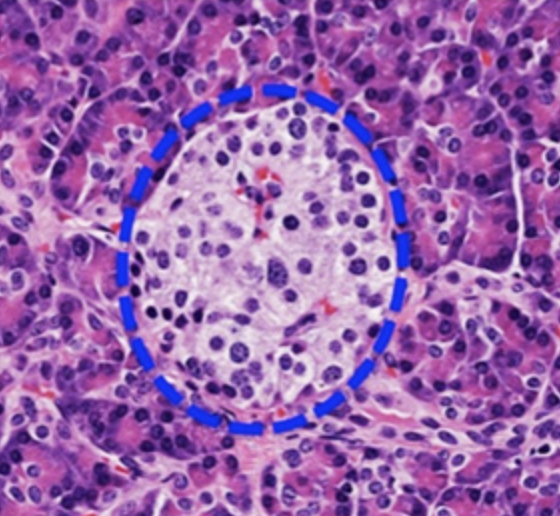
islets of langerhans
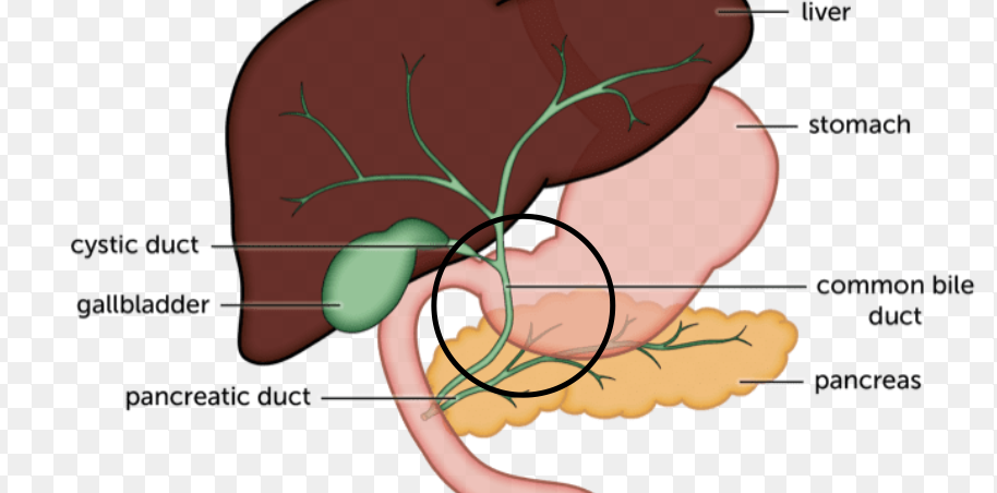
hepatopancreatic ampulla
a small, spherical reservoir in the duodenal wall where the common bile duct and pancreatic duct join
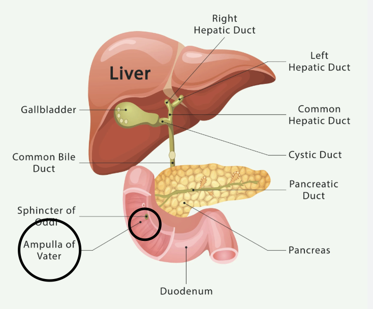
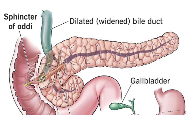
sphincter of oddi
muscular valve surrounding the exit of the bile and pancreatic ducts into the small intestine, controlling digestive fluid flow
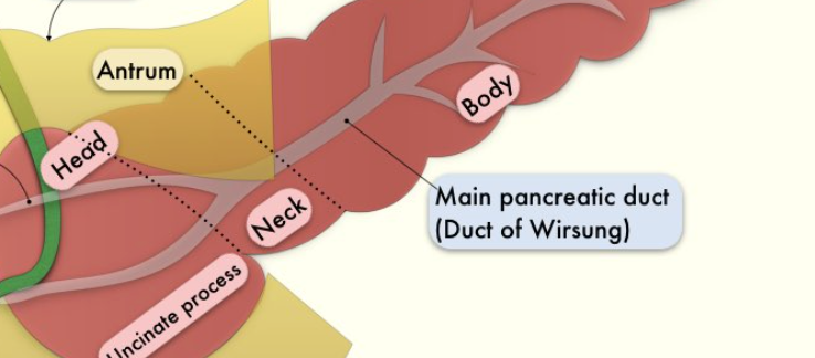
pancreatic duct
a crucial tube running through the pancreas that transports digestive enzymes to the small intestine
external anal sphincter
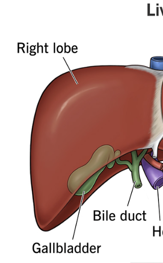
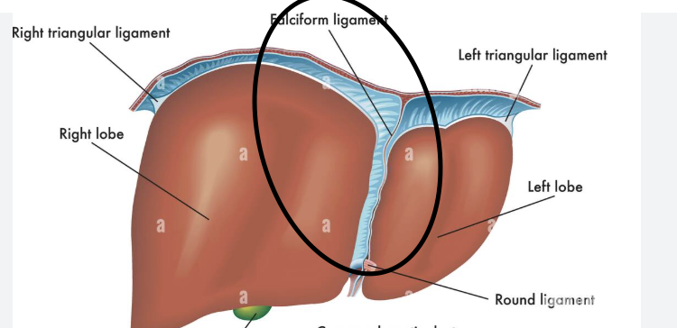
right lobe of liver
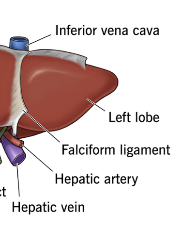
left lobe of liver
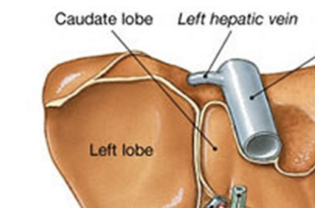
caudate lobe
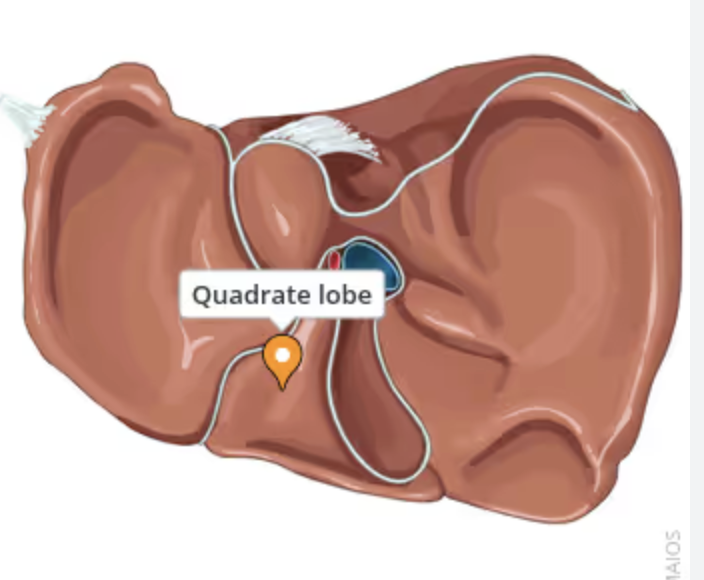
quadrate lobe
falciform ligament
thin, sickle-shaped fold of peritoneum that attaches the liver to the anterior abdominal wall and diaphragm, separating the left and right lobes
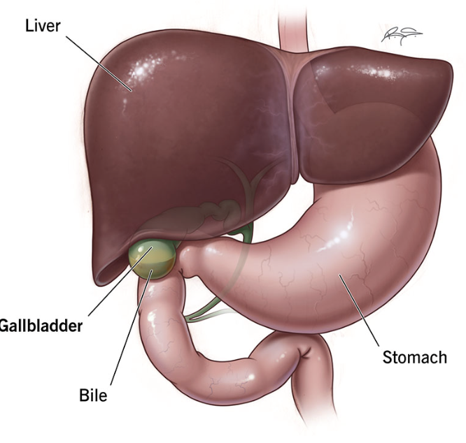
gall bladder
stores bile to help digest fat
cystic duct
transporting bile into the gallbladder for storage and, upon hormonal signaling, transporting concentrated bile out toward the small intestine to aid in digestion
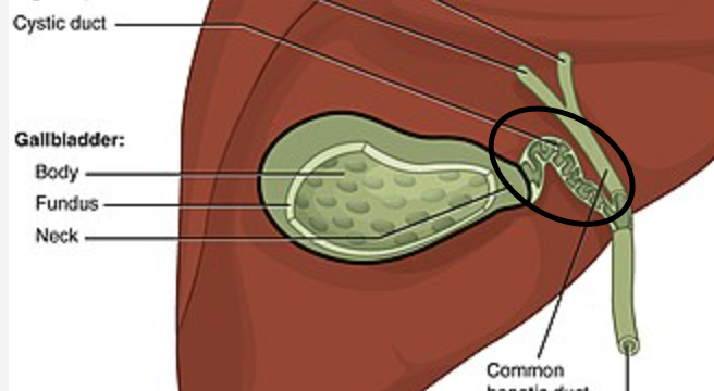
bile duct
carries bile between organs
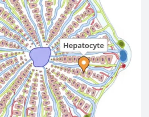
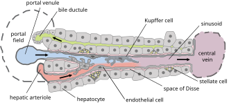
liver lobules
rough six-sided arrangement of hepatocytes (lever cells), blood vessels and bile
canaliculi
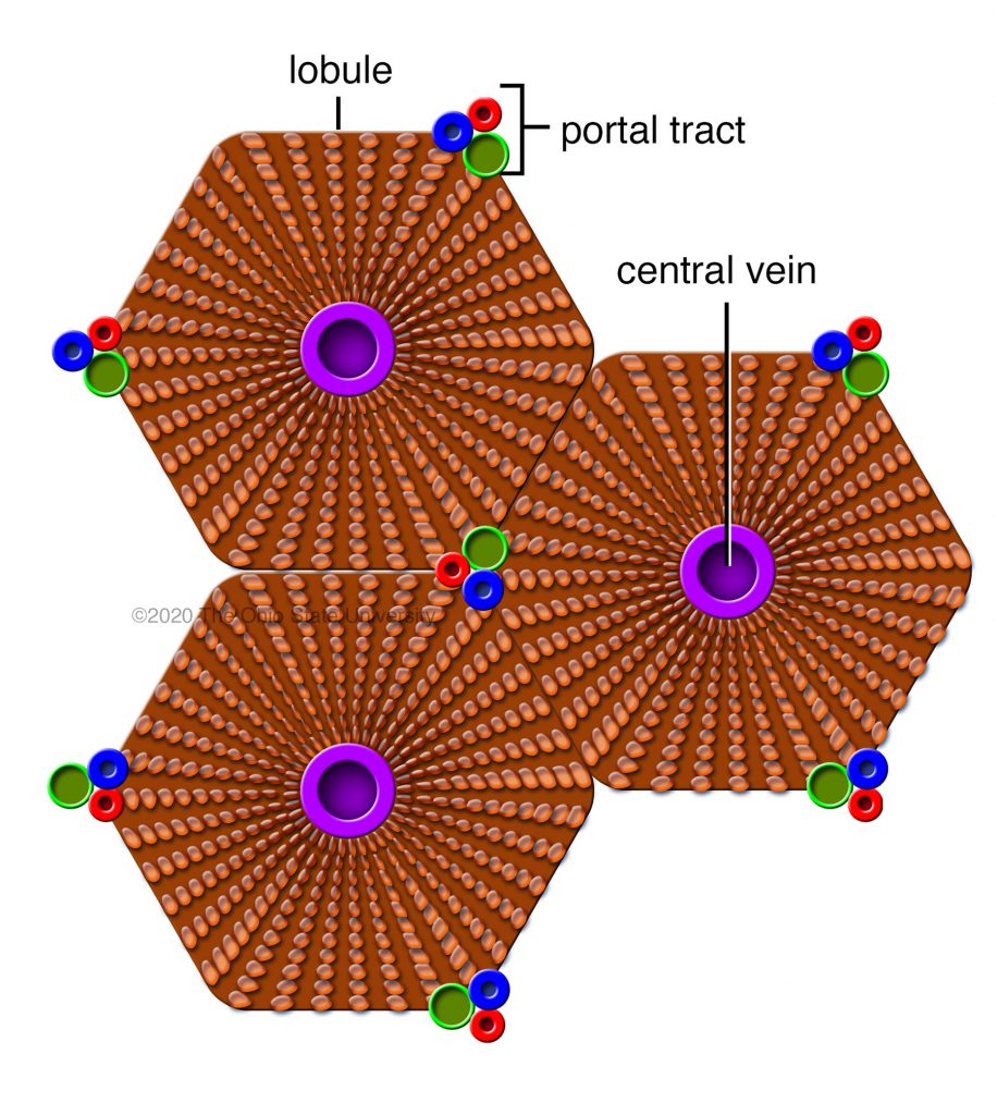
portal triad
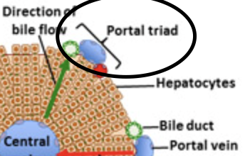
central vein
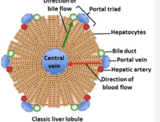
hepatocytes
kupffer cells
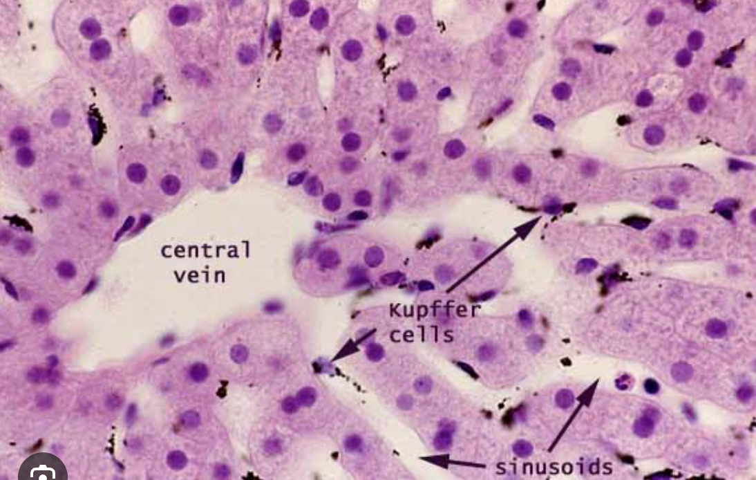
sinusoids
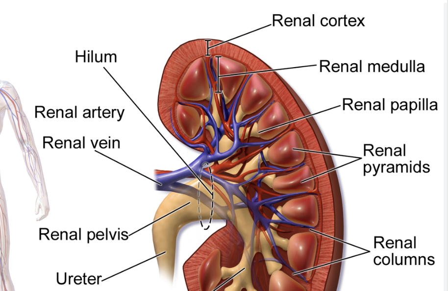
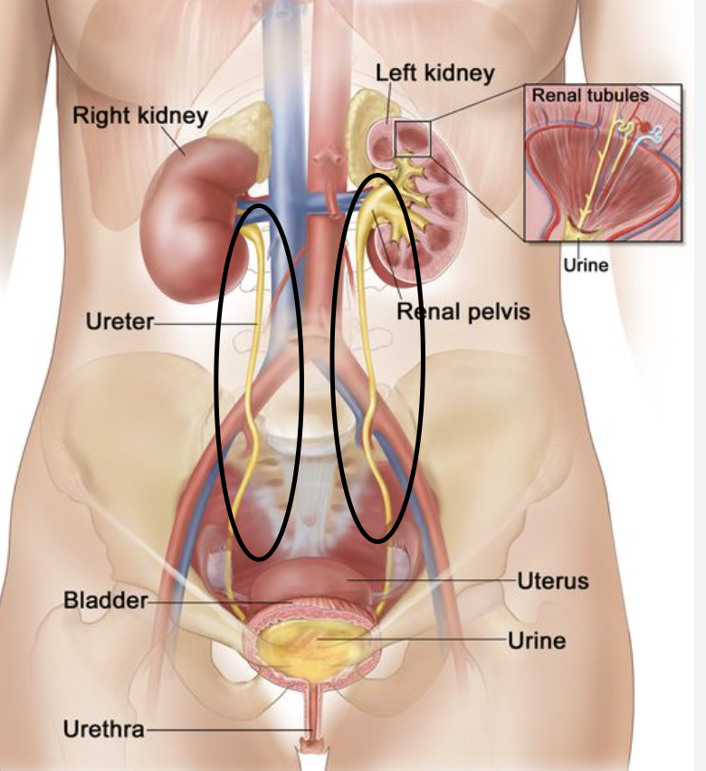
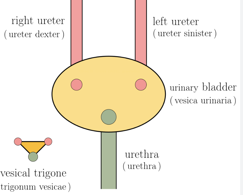
ureter
transport urine from the renal pelvis of each kidney to the urinary bladder
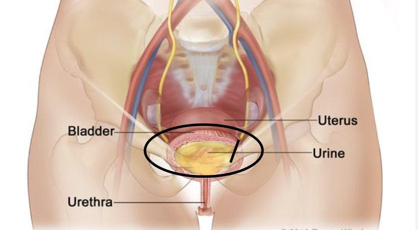
urinary bladder
stores urine before excretion
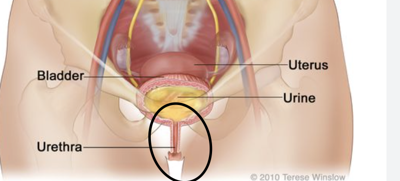
Urethra
tube connecting the bladder to the exterior of the body, functioning primarily to expel urine
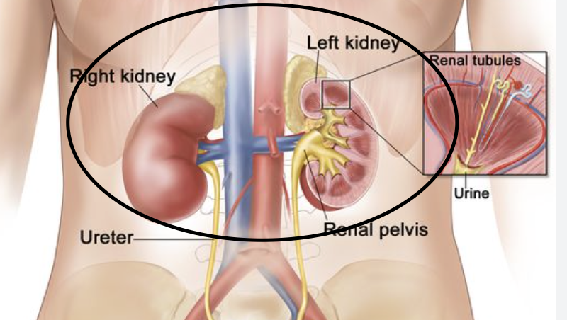
kidneys
filtering waste, balancing body fluids, regulating blood pressure, and producing hormones.
detrusor muscle
enables urination by contracting to expel urine and relaxing to store it
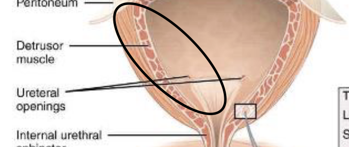
ureteral openings
the external exit points for urine to leave the body, allowing for waste elimination from the bladder
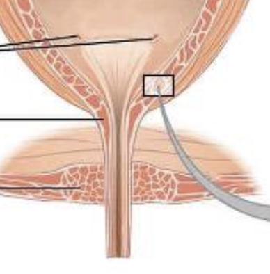
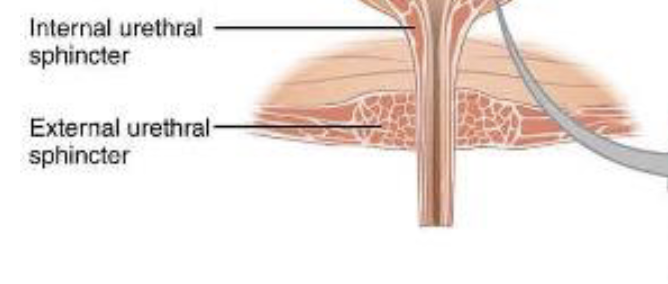
internal and external urethral sphincters
control urine outflow (micturition) and maintain continence - internal is involuntary while external is voluntary and allows s to hold our urine in
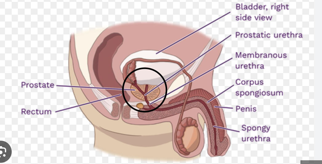
membranous urethra
helps control urination in men
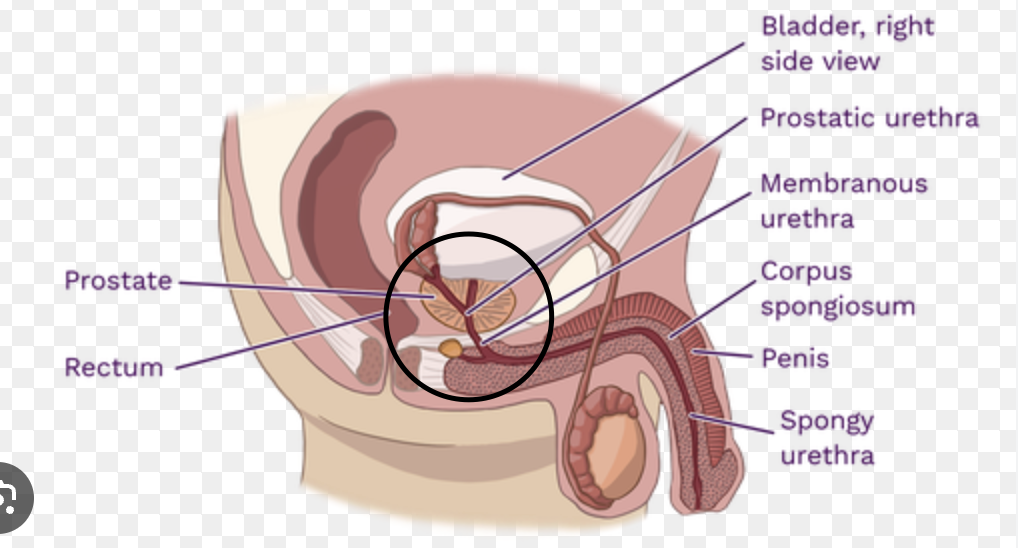
spongy urethra
Serves as the final common pathway for both urination and ejaculation
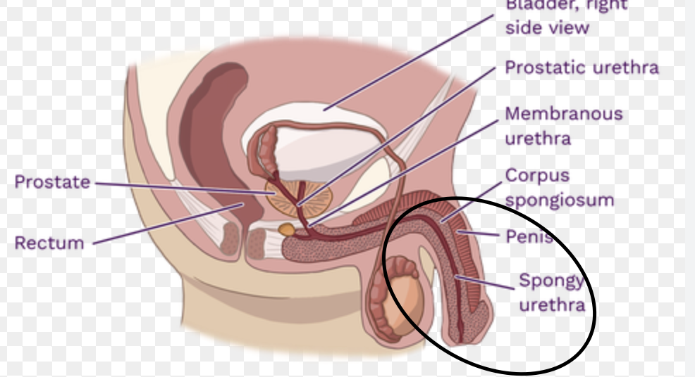
prostate and prostatic urethra
the 3–4 cm, widest segment of the male urethra that passes through the center of the prostate gland, extending from the bladder neck to the membranous urethra. It is crucial for conducting urine and semen, containing openings for ejaculatory ducts, and is surrounded by the prostate's glandular tissue

bulbourethral gland
two exocrine glands that secrete pre-ejaculation for lubrication the urethra and protects sperm
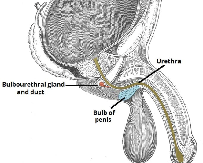
crus and bulb of penis
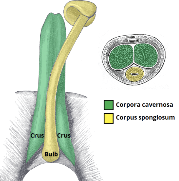
erectile tissue of penis
consists of three expandable, sponge-like columns—two corpora cavernosa and one corpus spongiosum—that fill with blood to produce an erection
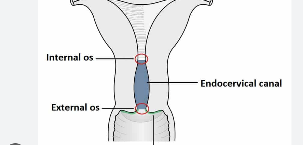
external and internal orifice
two openings of the uterine cervix. The internal os connects the cervical canal to the uterus, while the external os connects the cervix to the vagina
trigone
smooth, triangular, and highly sensitive region at the base of the bladder, bounded by two ureteral orifices and the internal urethral meatus
seminal vesicle
a pair of accessory glands in the male reproductive system that produce approximately 70-80% of the fluid in semen
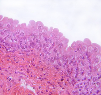
transitional epithelium in bladder
a specialized, stratified tissue lining the urinary bladder, ureters, and renal pelvis, allowing for significant distension
cortex of kidney
this region is essential for blood filtration, producing urine, and regulating blood pressure
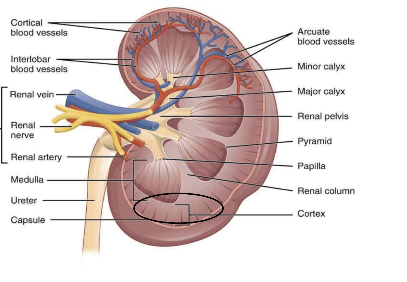
medulla
functions primarily to concentrate urine through a countercurrent multiplier system involving the loops of Henle and collecting ducts, which reabsorb water and electrolytes
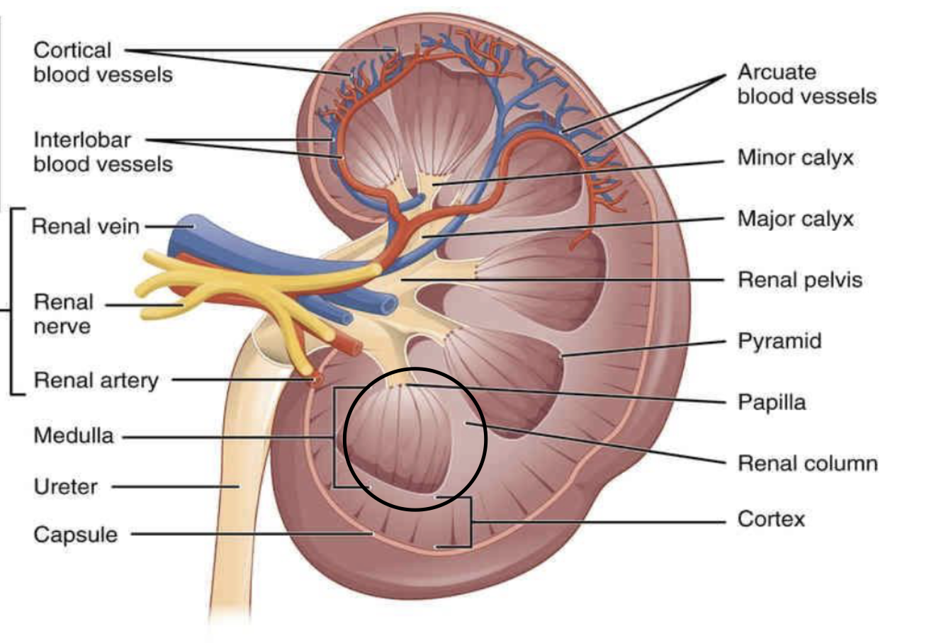
hilum
acts as the main gateway for structures entering and leaving the kidney, including the renal artery, vein, lymphatics, nerves, and the renal pelvis