BIOS 3754 Practical 3: Everything Else Besides Quiz 3
5.0(1)
Studied by 13 peopleCard Sorting
1/168
Last updated 8:06 AM on 12/1/22
Name | Mastery | Learn | Test | Matching | Spaced | Call with Kai |
|---|
No analytics yet
Send a link to your students to track their progress
169 Terms
1
New cards
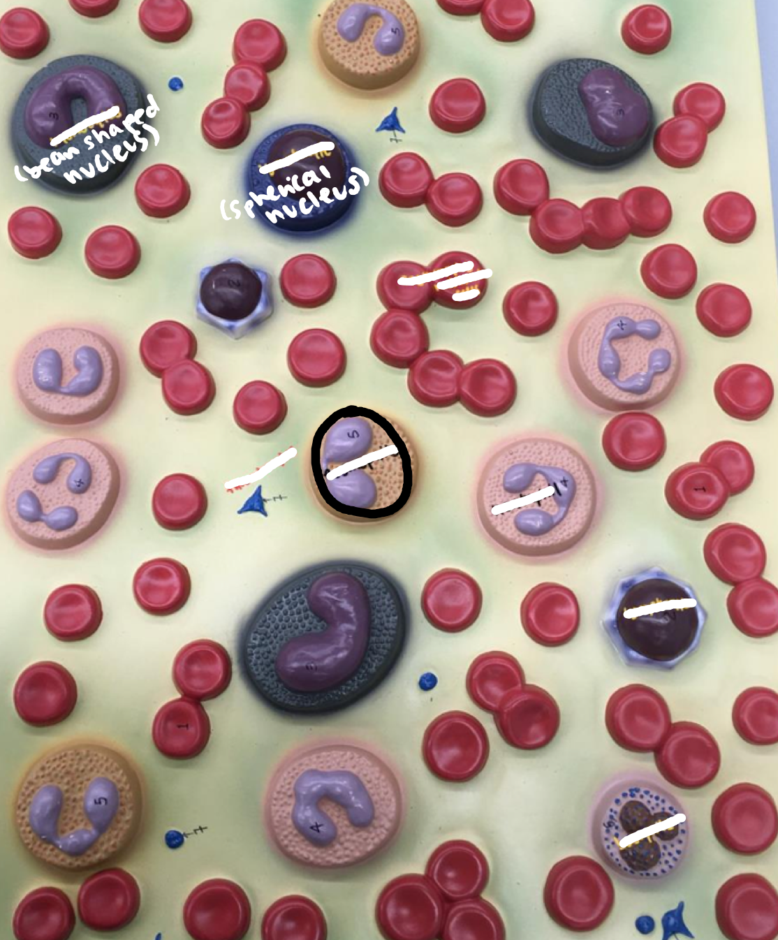
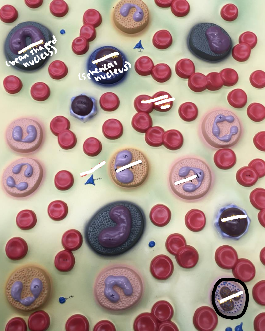
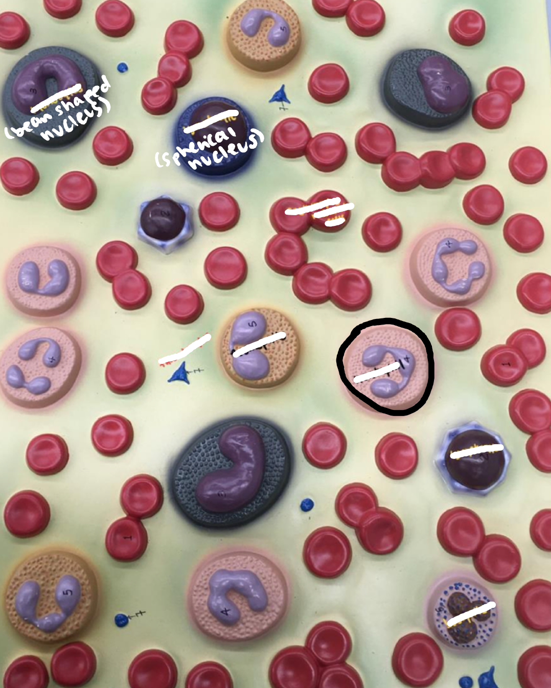
Erythrocytes
Red blood cells
2
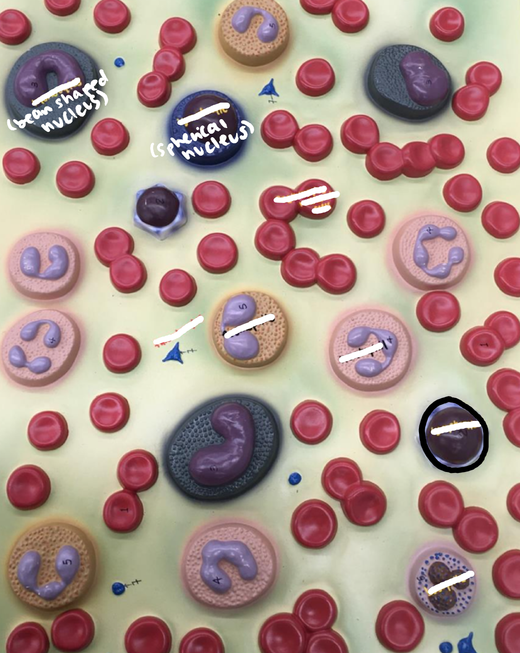
New cards
Leukocytes
3
New cards
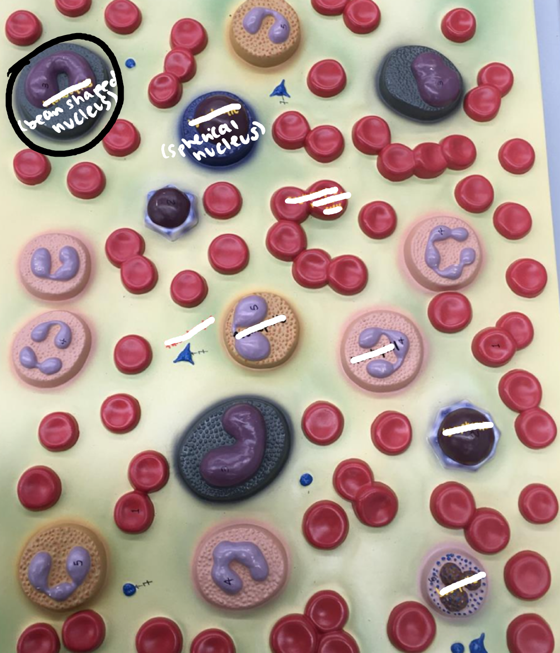
Granulocytes:
- Eosinophils
- Eosinophils
Beige circle with 2 purple dots
4
New cards
Granulocytes:
- Basophils
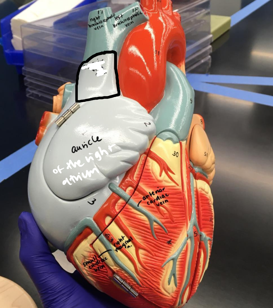
- Basophils
Purple circle with 3 purple dots
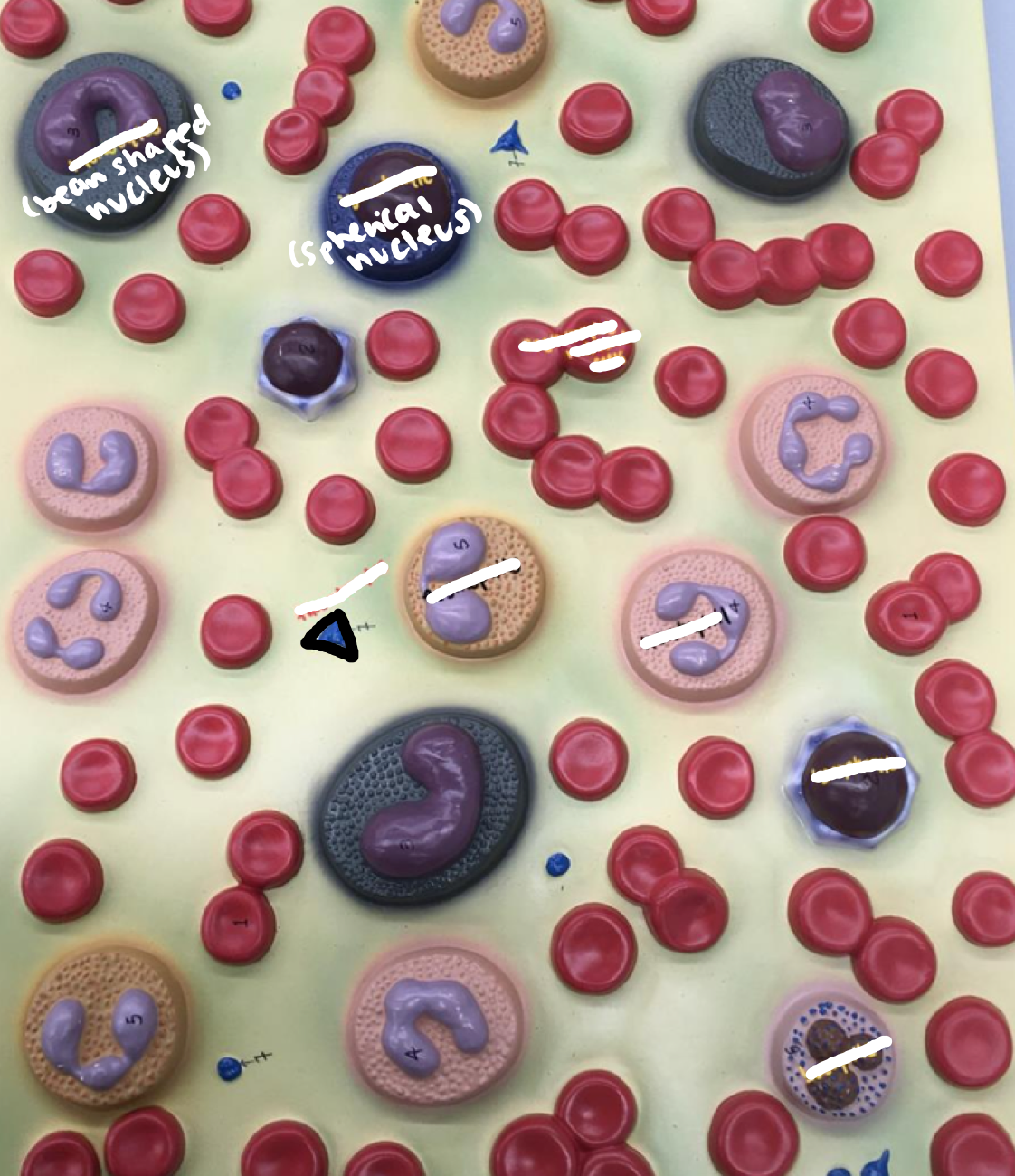
5
New cards
Granulocytes:
- Neutrophils
- Neutrophils
Neutral pink toned circle with purple dots
6
New cards
Agranulocytes:
- Monocytes
- Monocytes
Dark circles with the bean shaped nucleus
7
New cards
Agranulocytes:
- Lymphocytes
- Lymphocytes
Dark purple dot with the spherical nucleus
8
New cards
Thrombocytes (platelets)
Blue independent spots
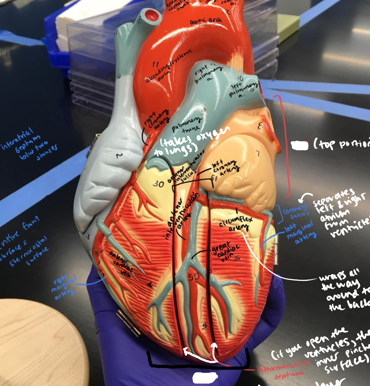
9
New cards
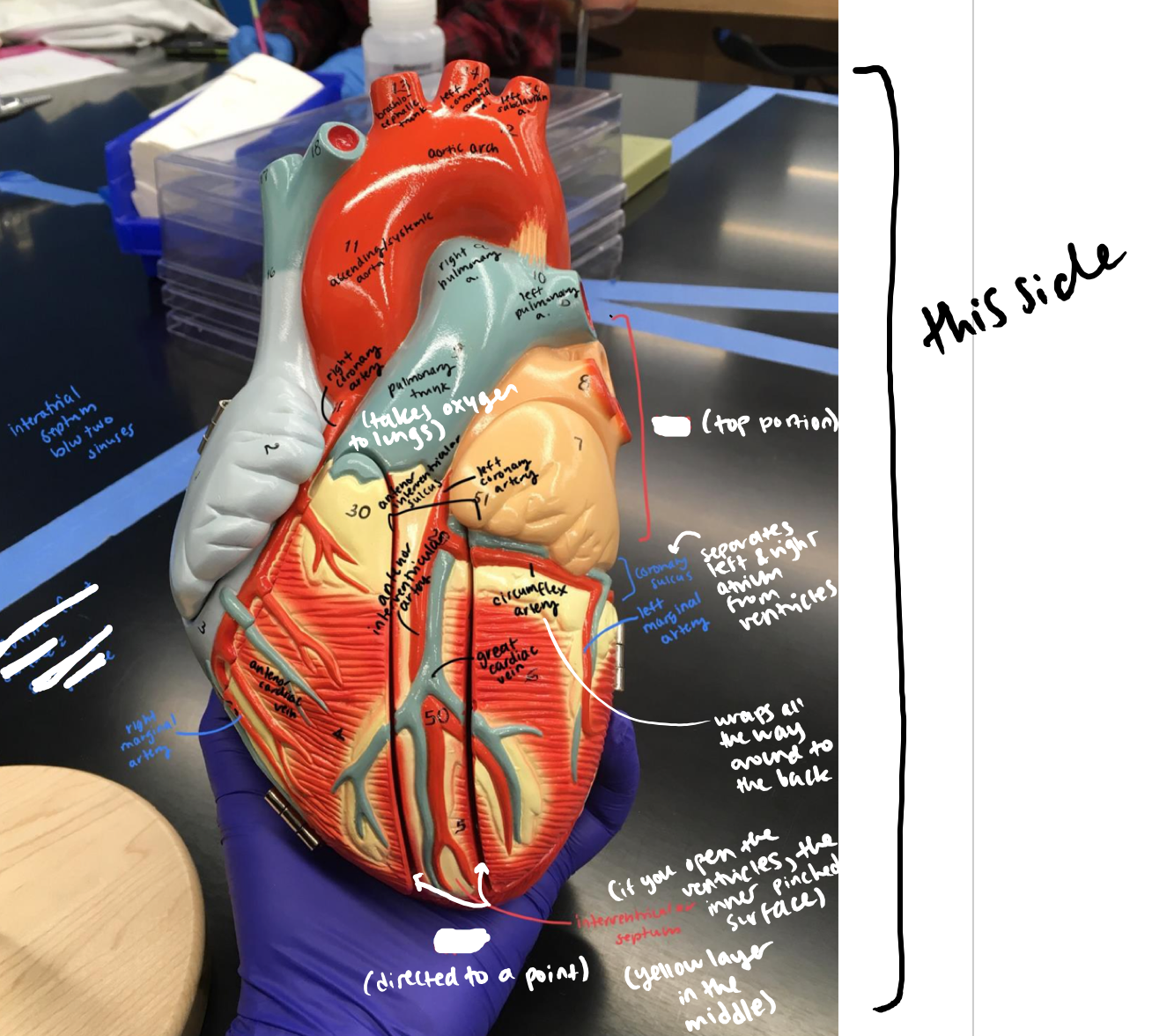
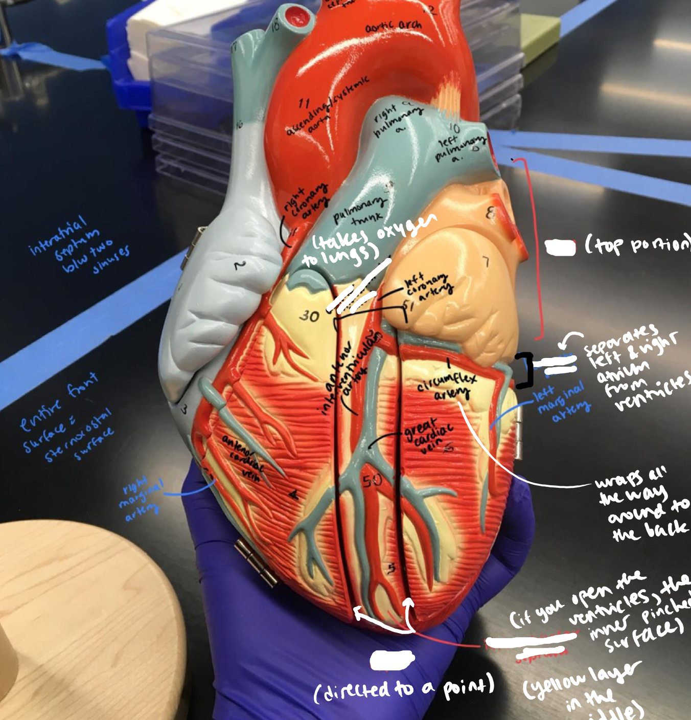
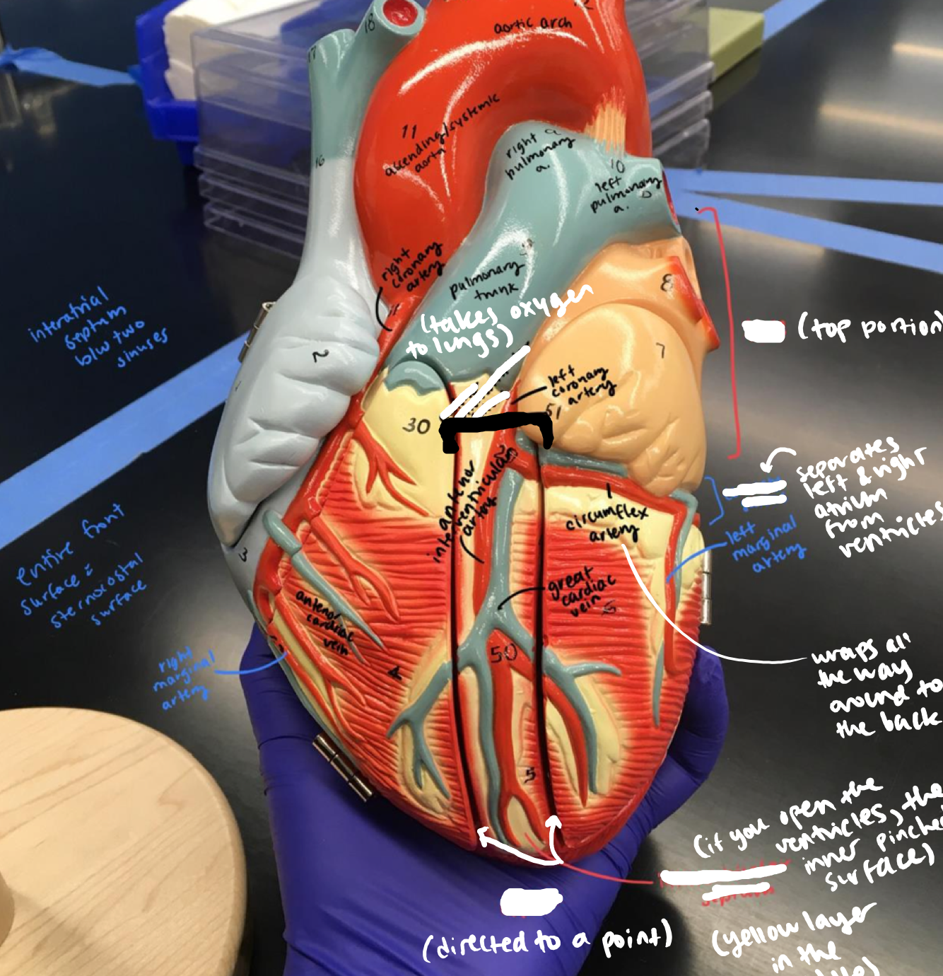
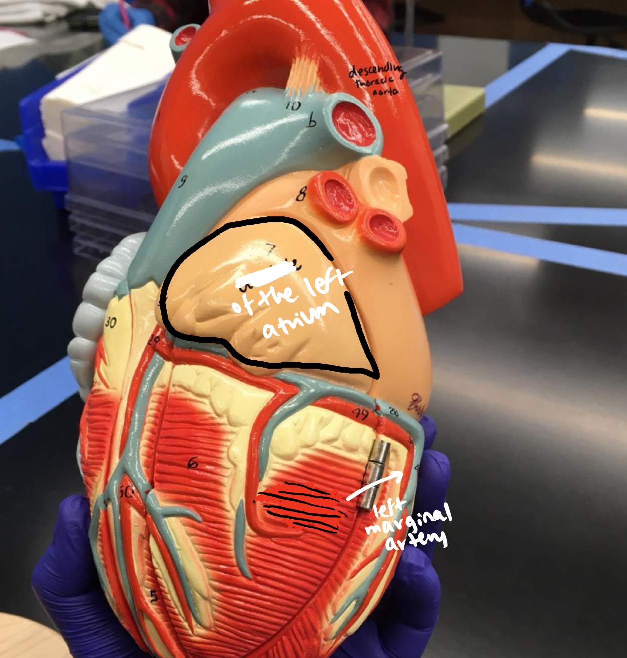
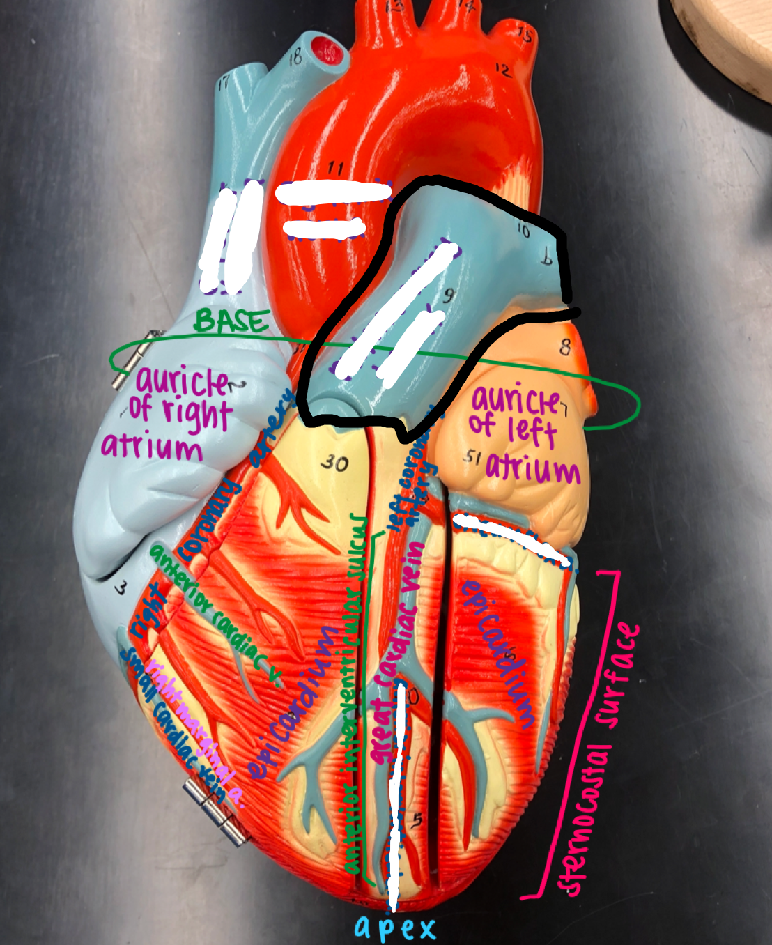
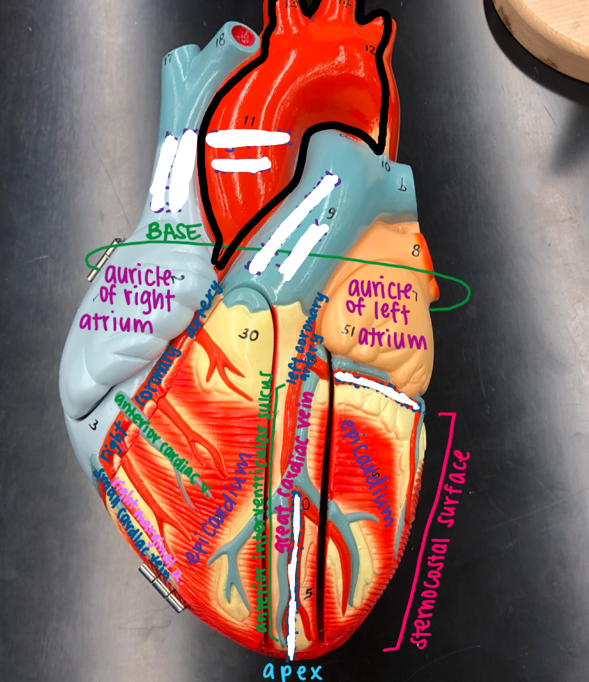
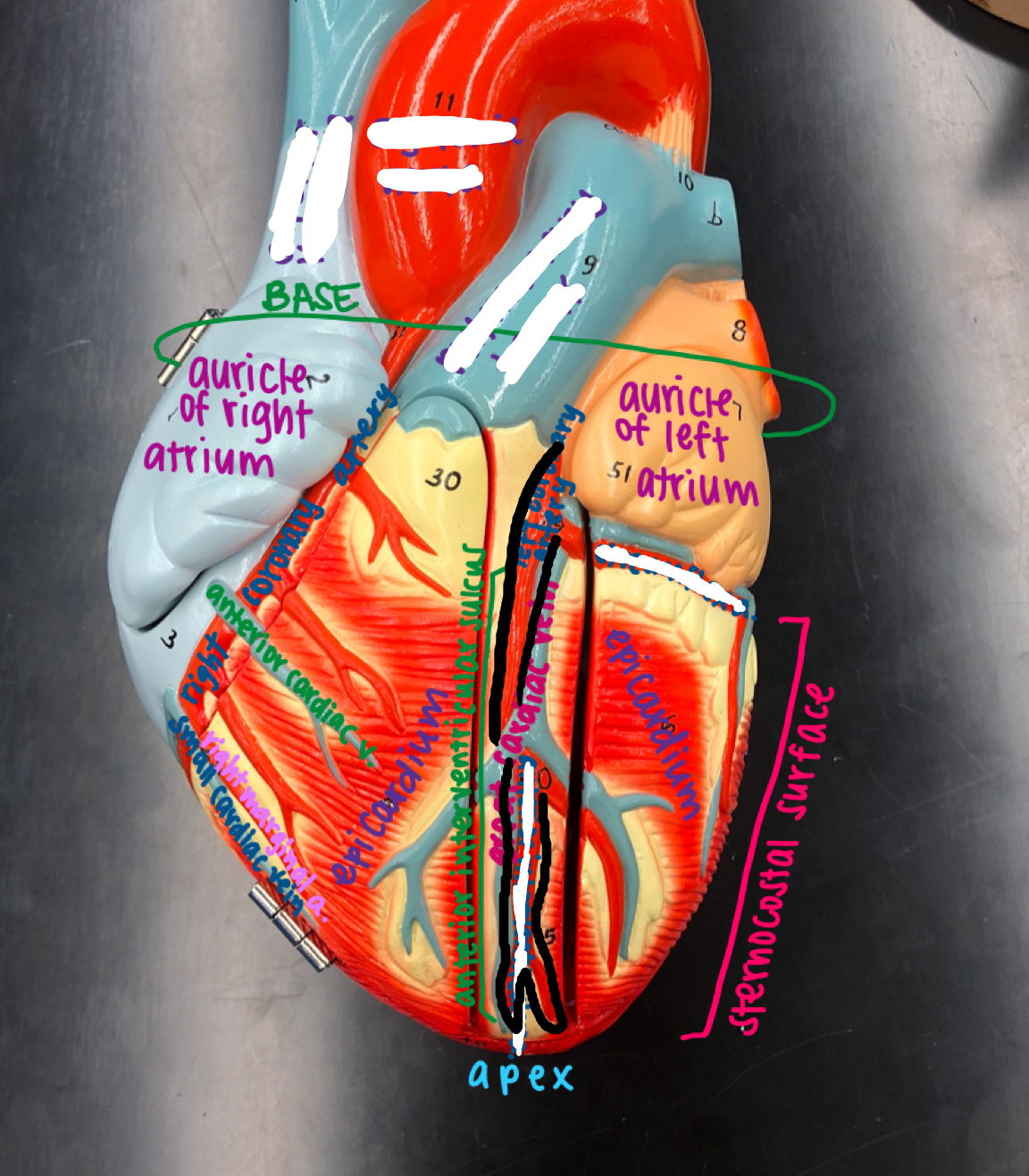
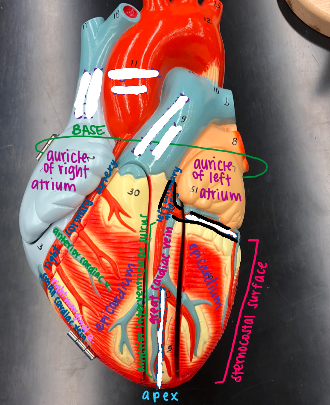
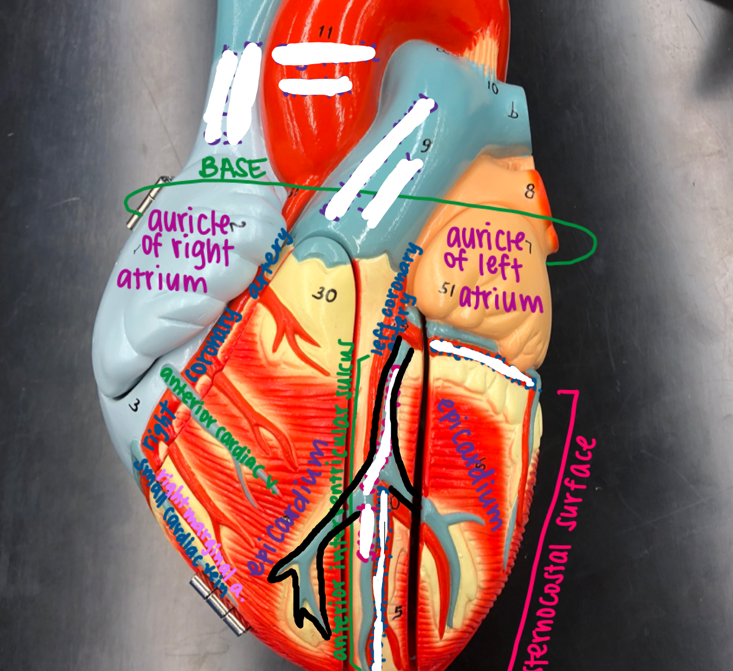
Regions of the heart
- Base
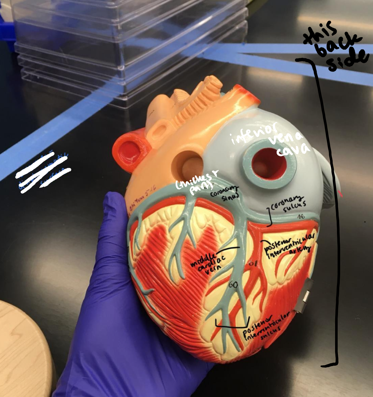
- Base
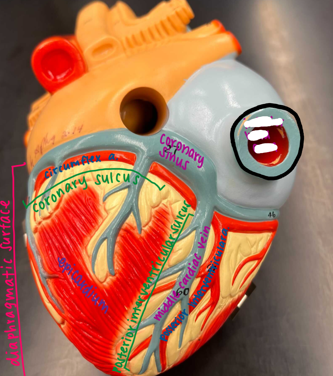
Top portion of the heart
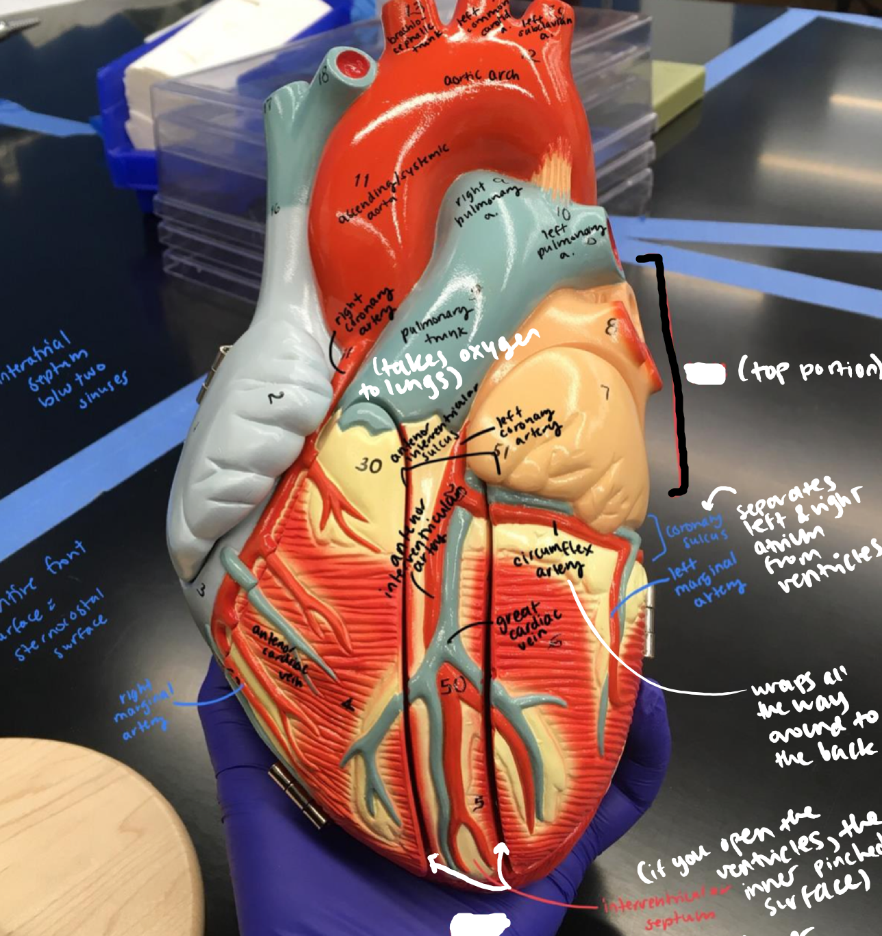
10
New cards
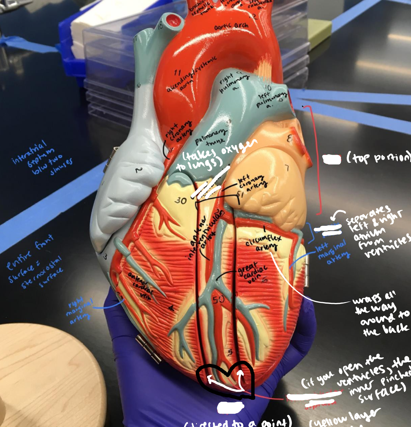
Regions of the heart
- Apex
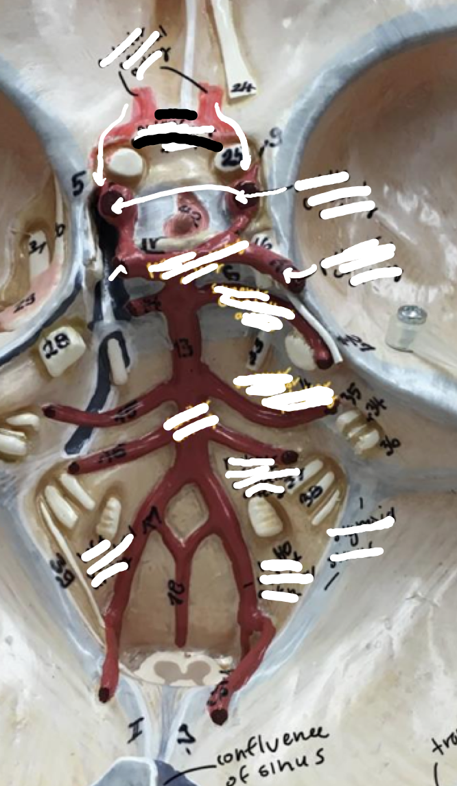
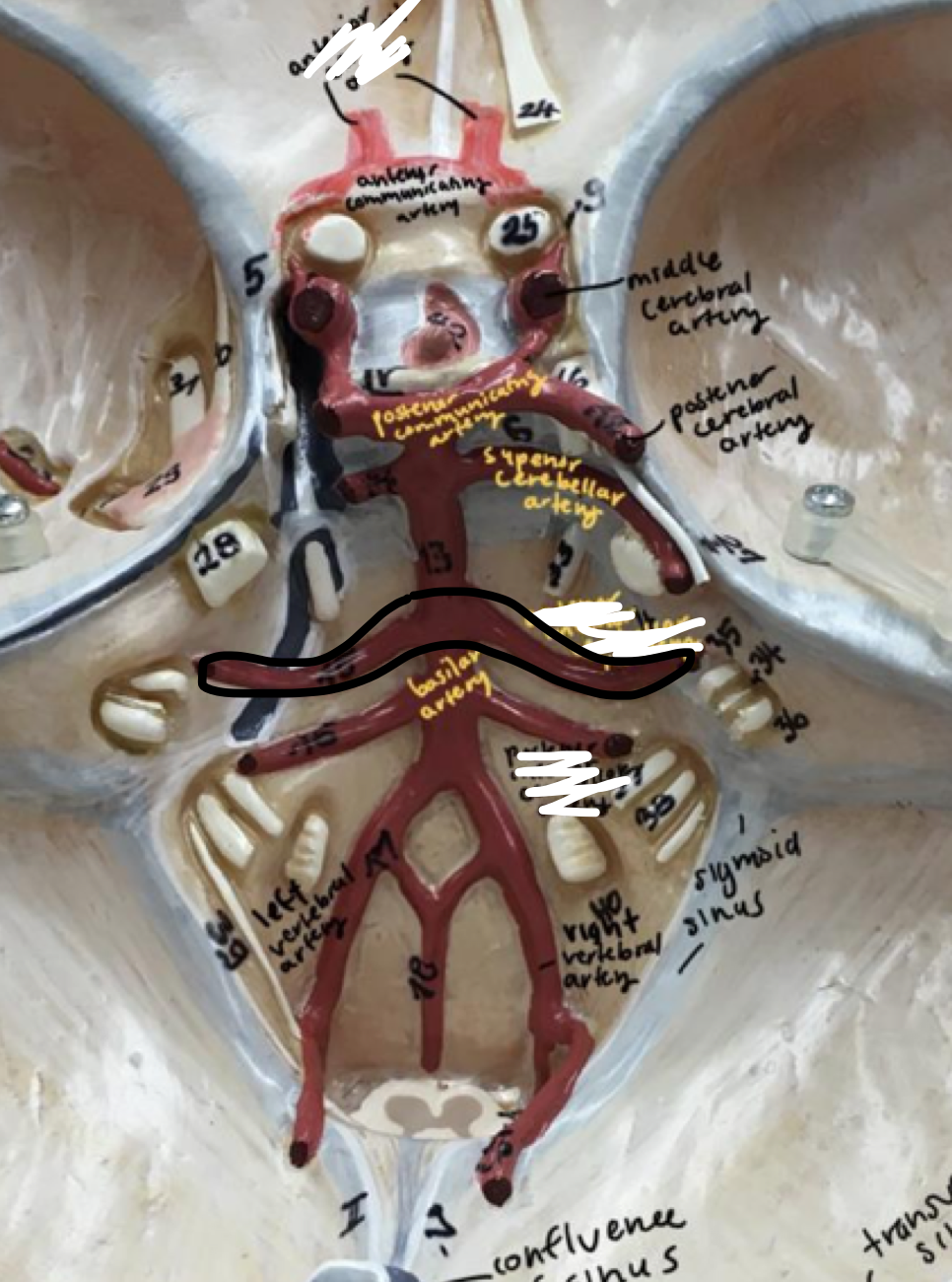
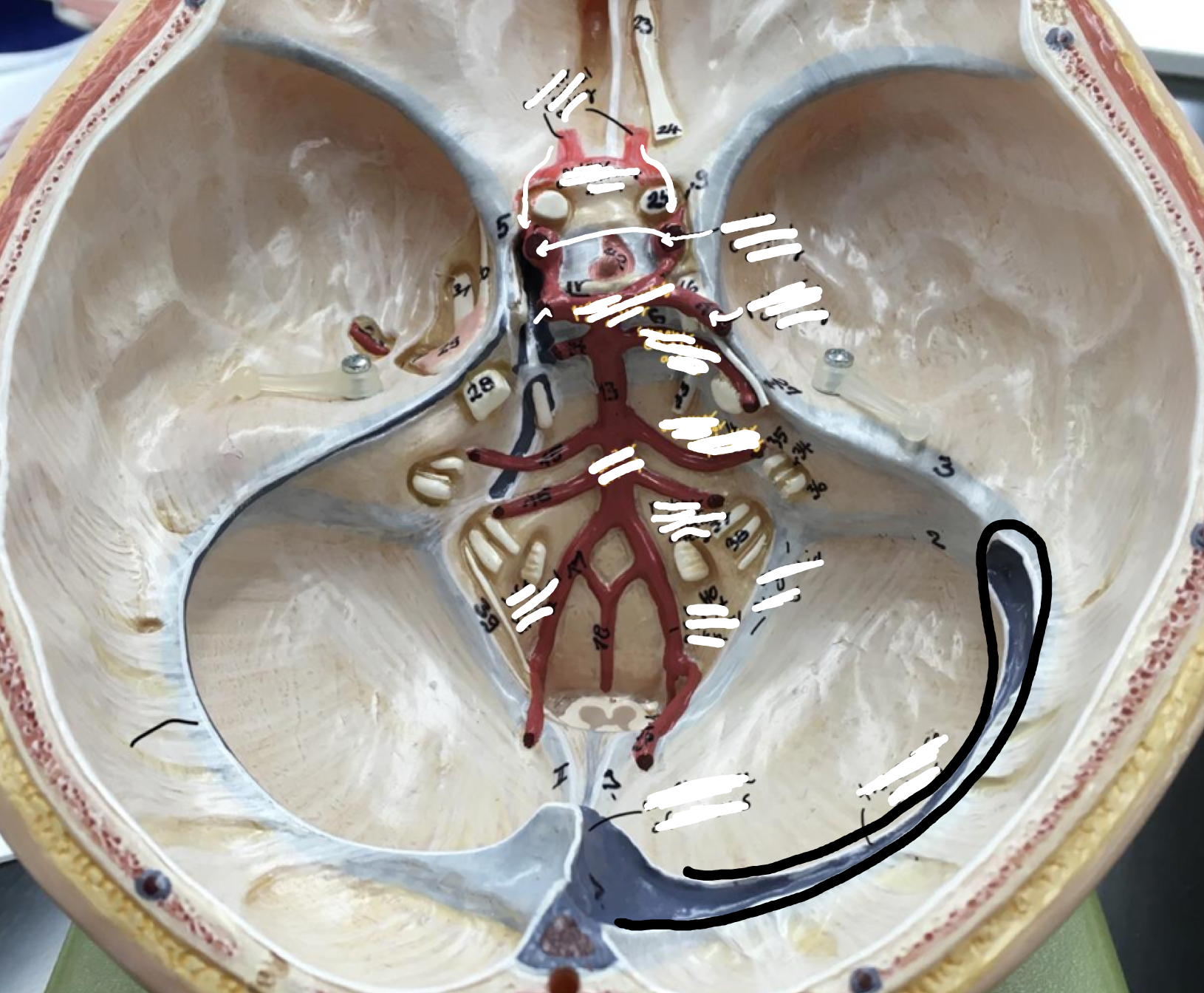
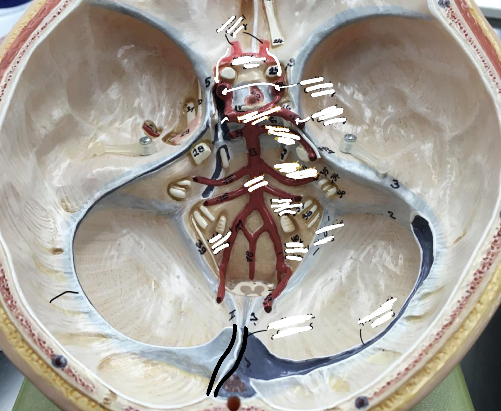
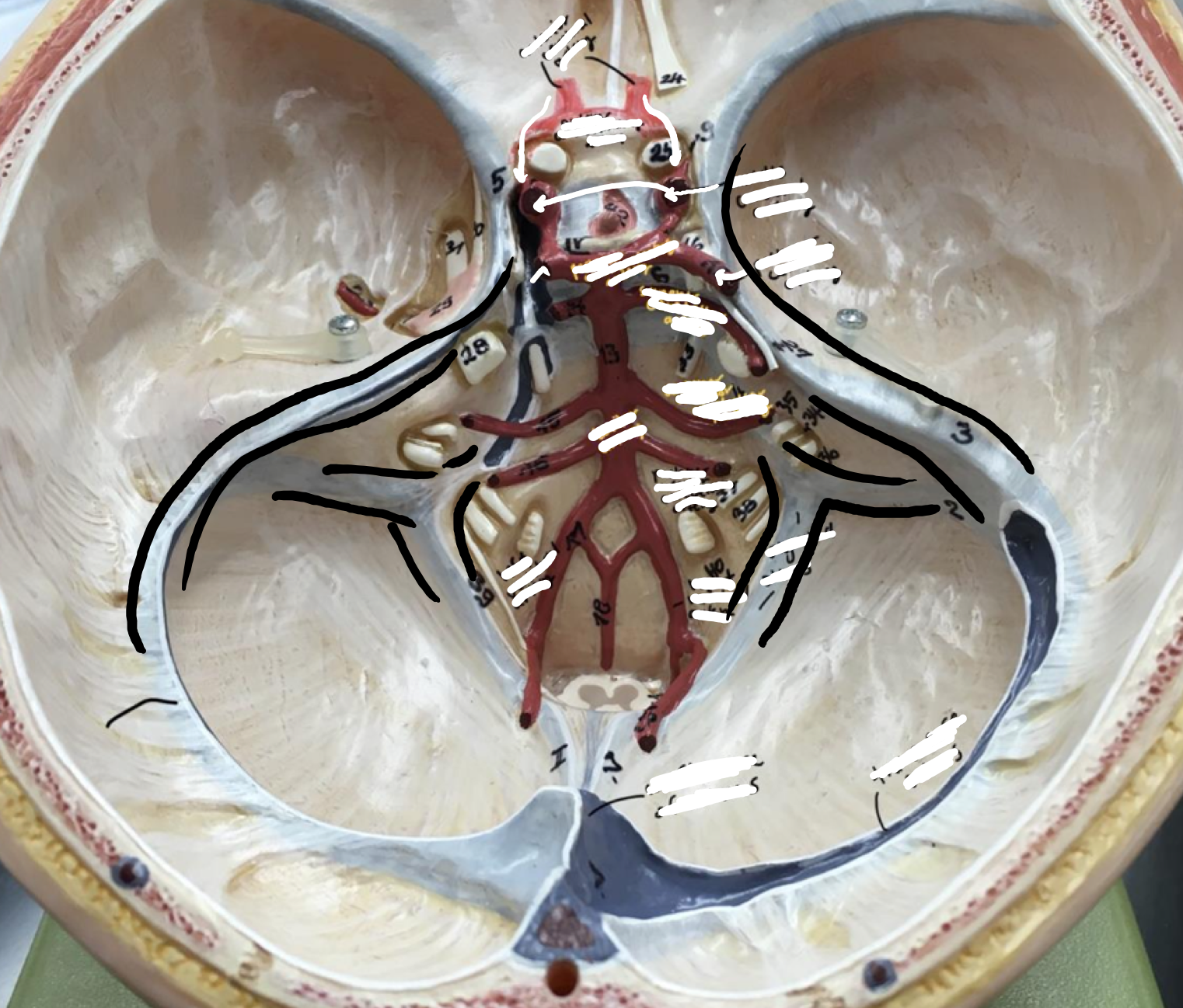
- Apex
Part of the heart directed to a point
11
New cards
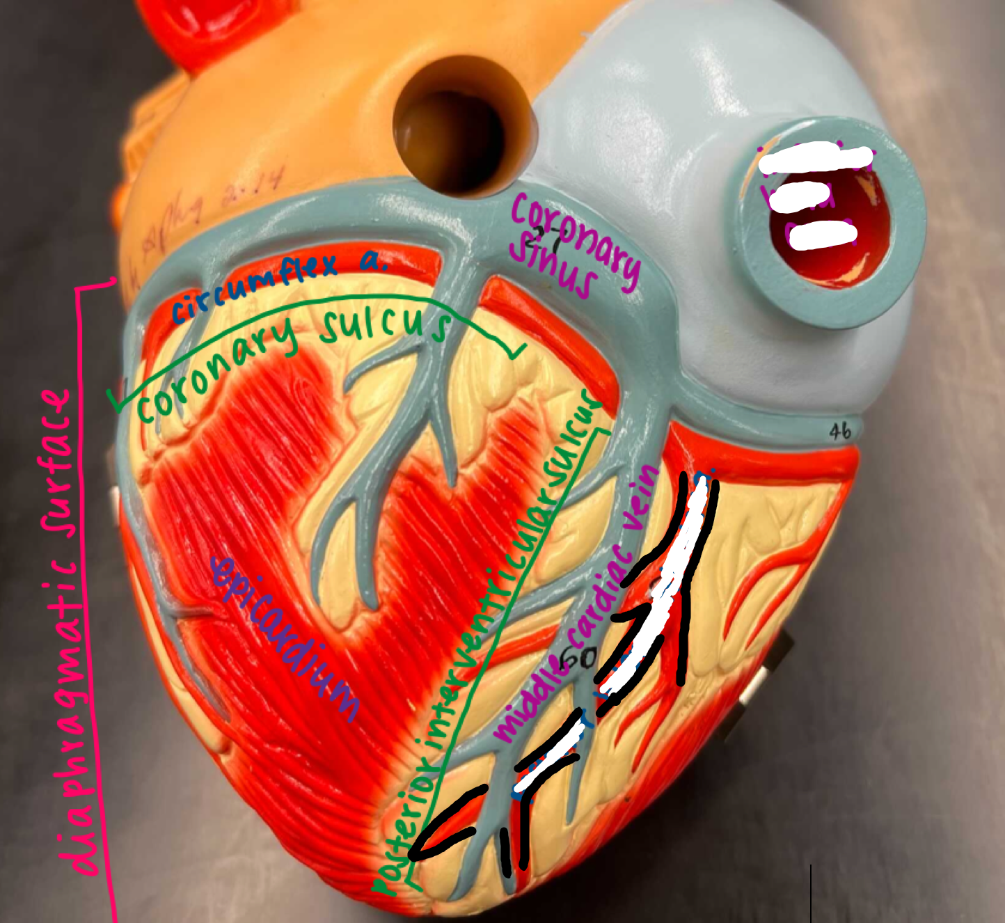
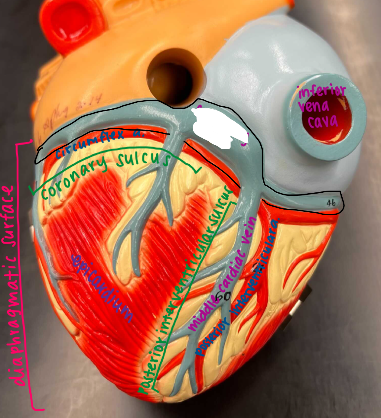
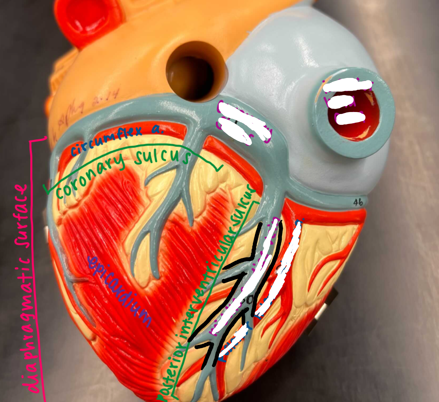
Regions of the heart
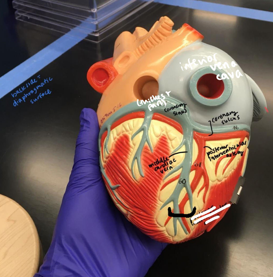
- Diaphragmatic surface
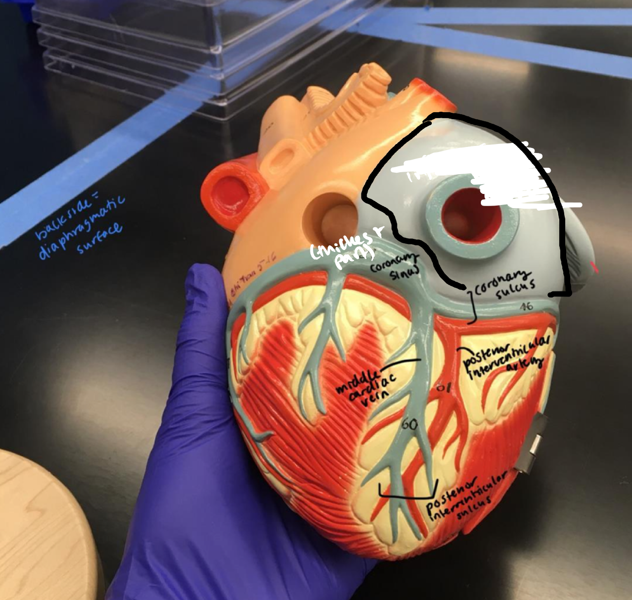
- Diaphragmatic surface
The entire back side of the heart
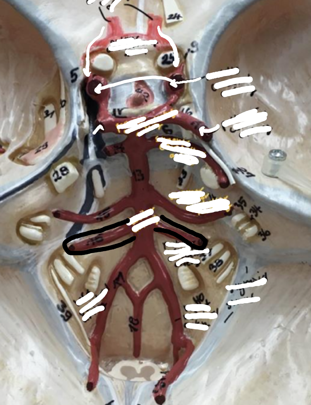
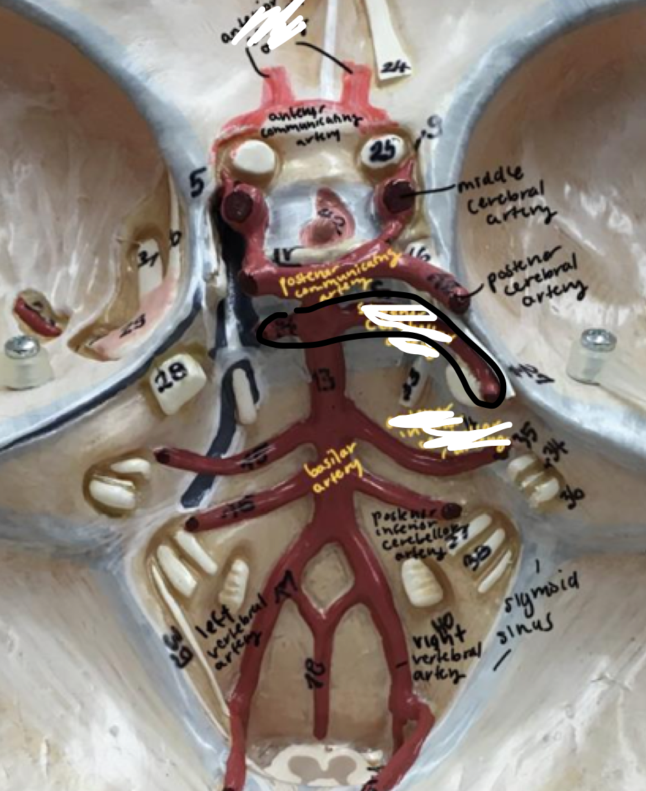
12
New cards
Regions of the heart
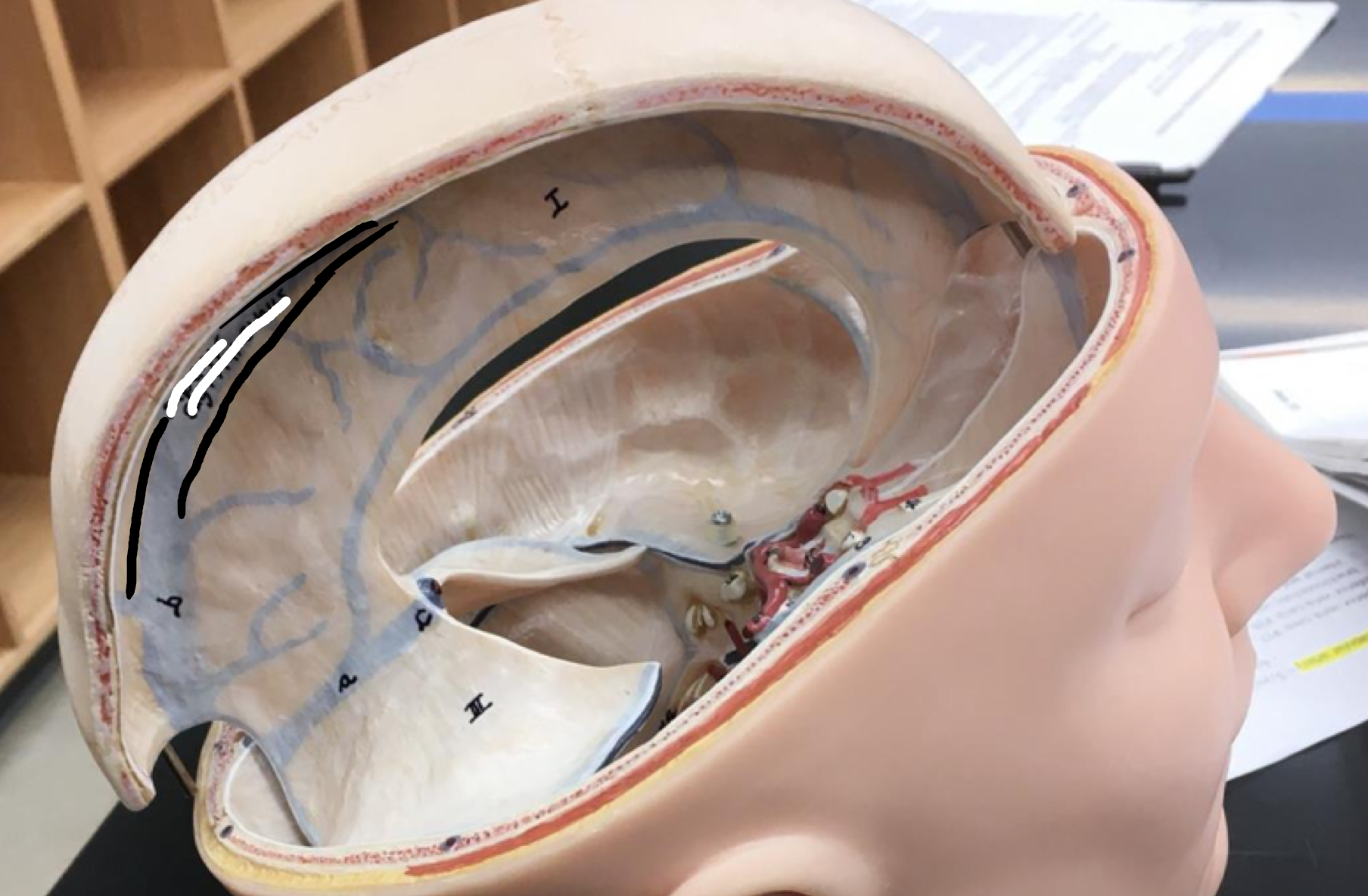
- Sternocostal surface
- Sternocostal surface
The entire front side of the heart
13
New cards
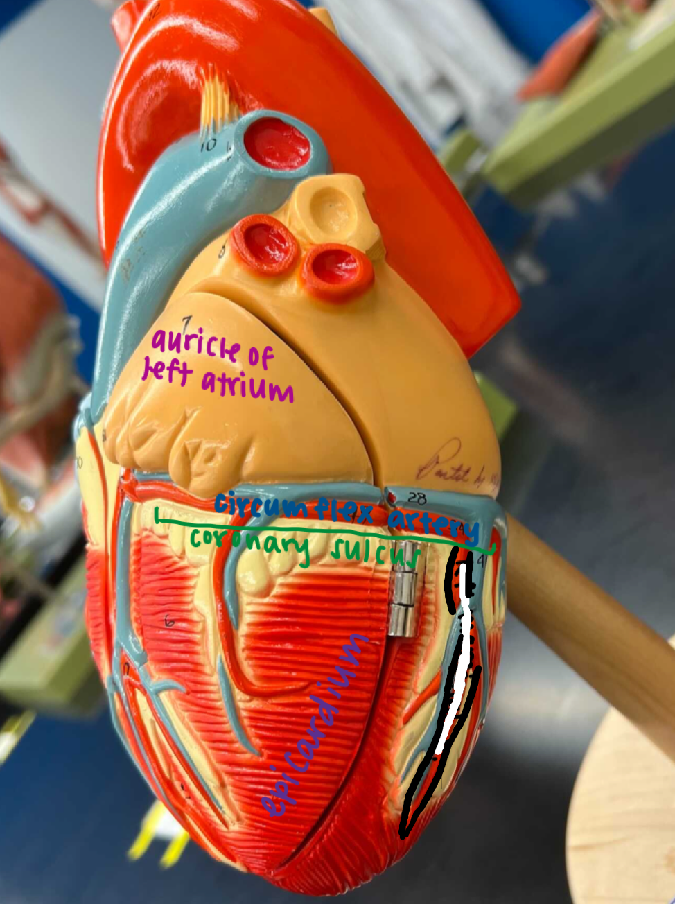
Regions of the heart
- Coronary sulcus
- Coronary sulcus
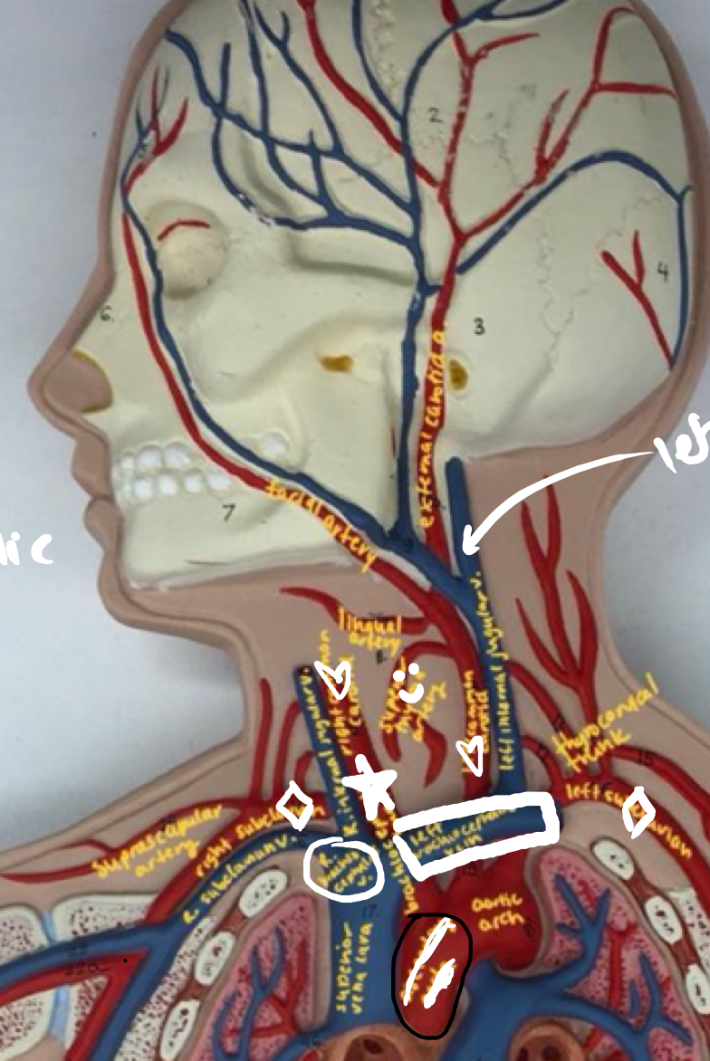
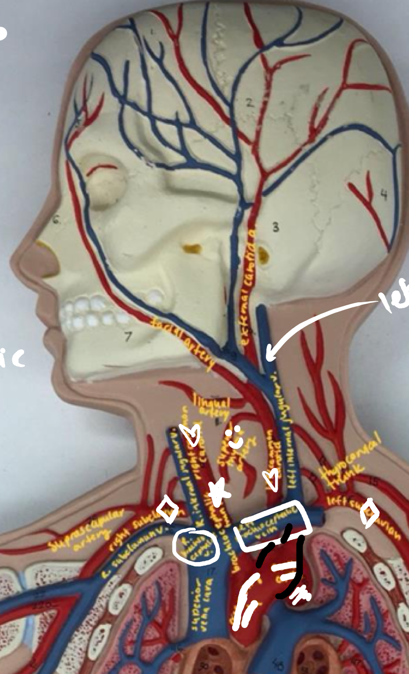
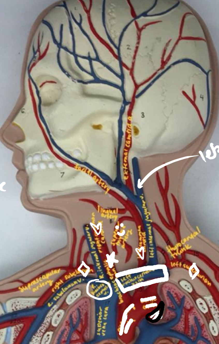
Separates left and right atrium from ventricles
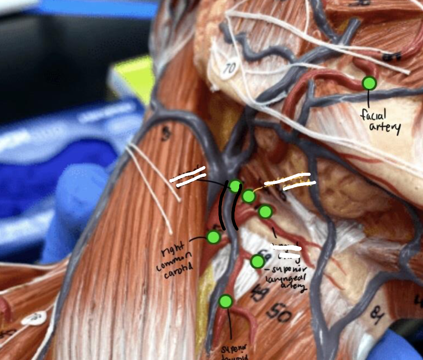
14
New cards
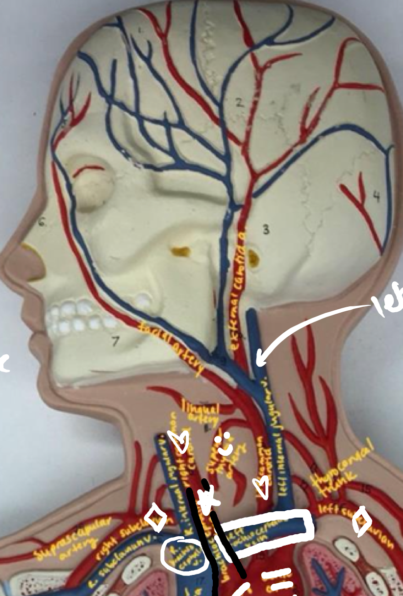
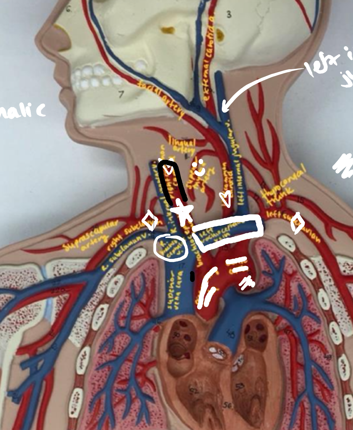
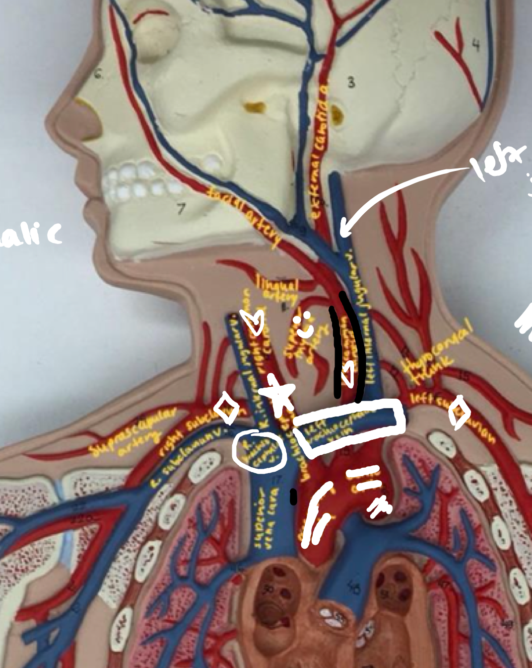
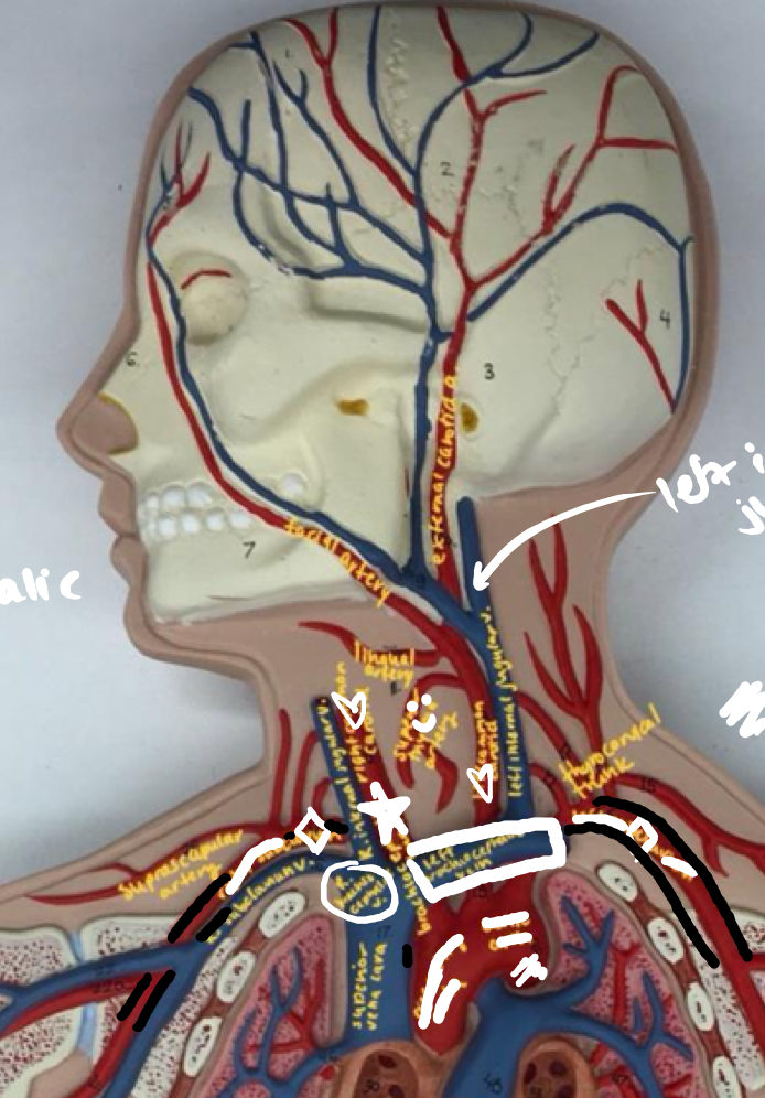
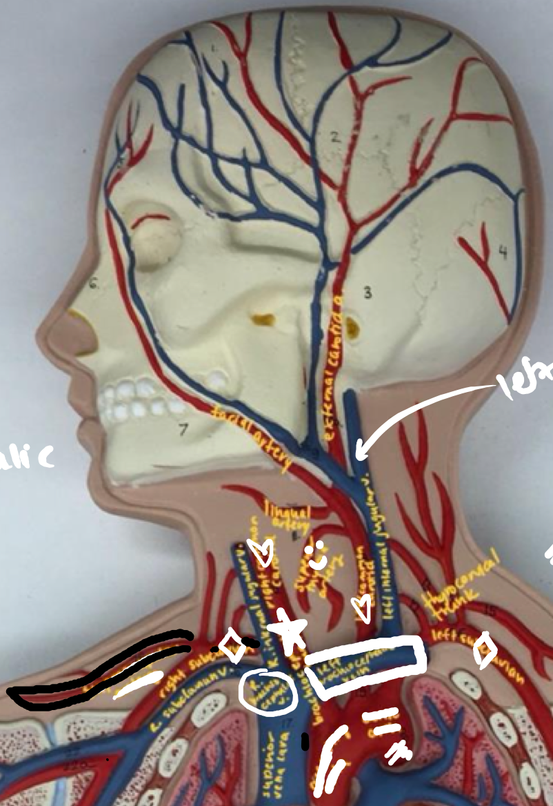
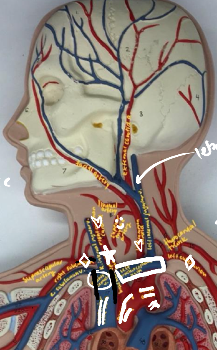
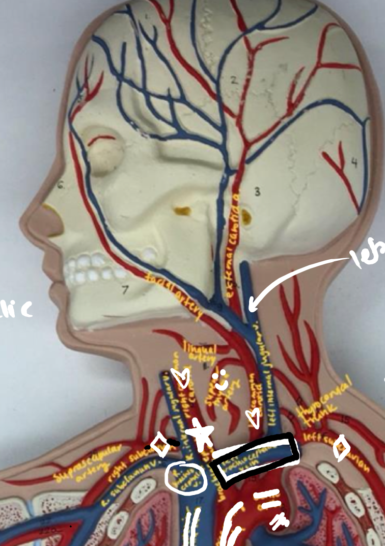
Regions of the heart
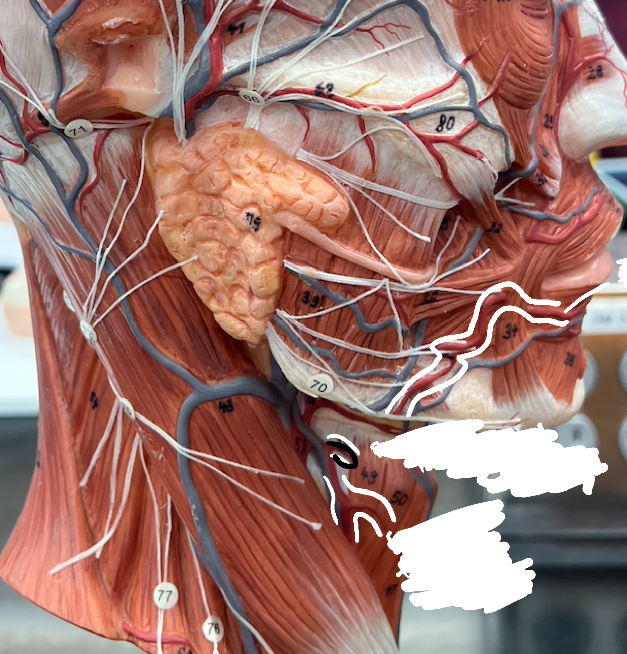
- Anterior interventricular sulcus
- Anterior interventricular sulcus
On the front of the heart, the section encapsulating the 2 wires
15
New cards
Regions of the heart
- Posterior interventricular sulcus
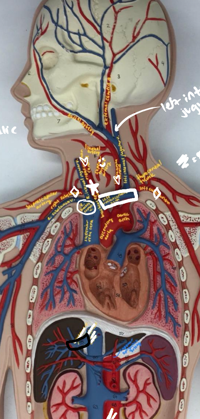
- Posterior interventricular sulcus
On the back of the heart, the section encapsulating the 2 wires
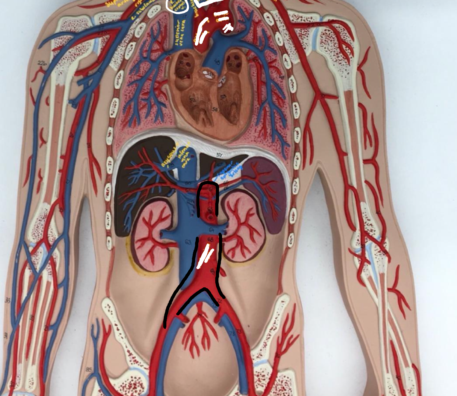
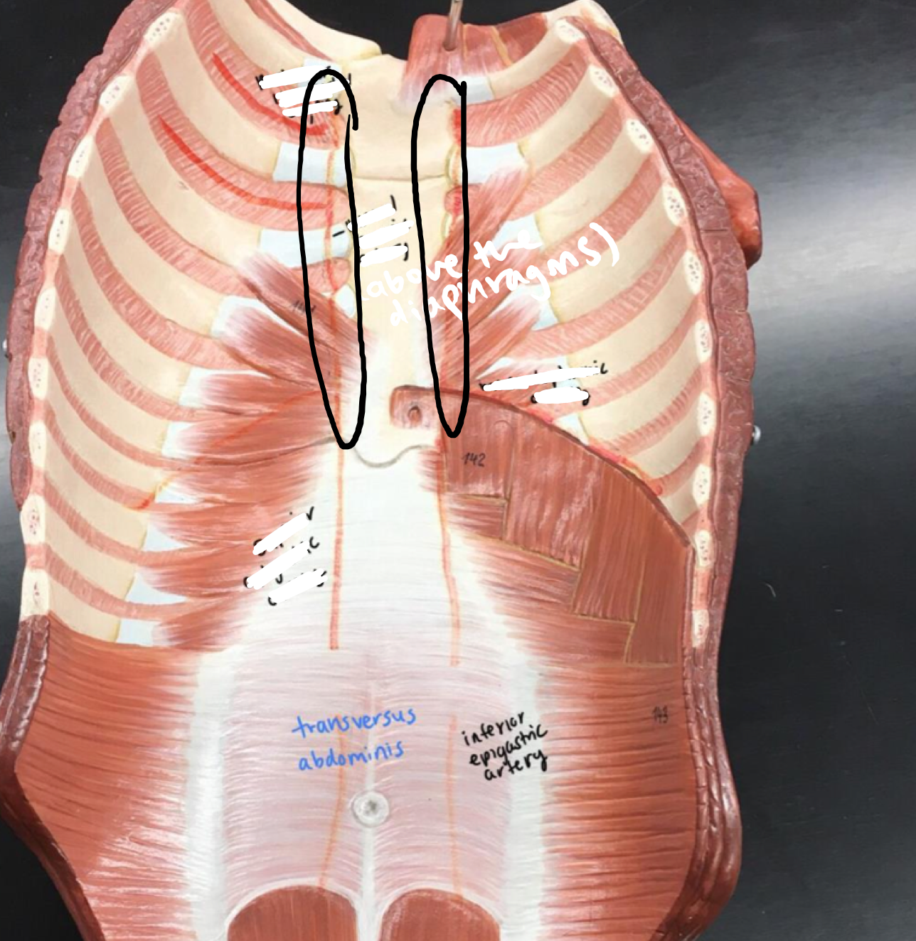
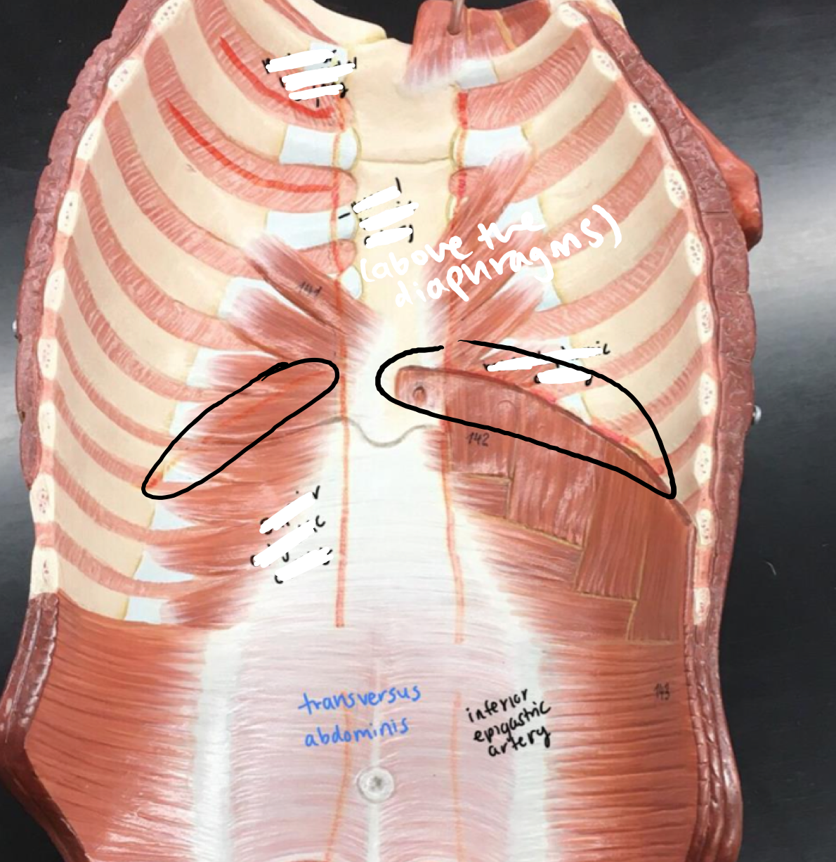
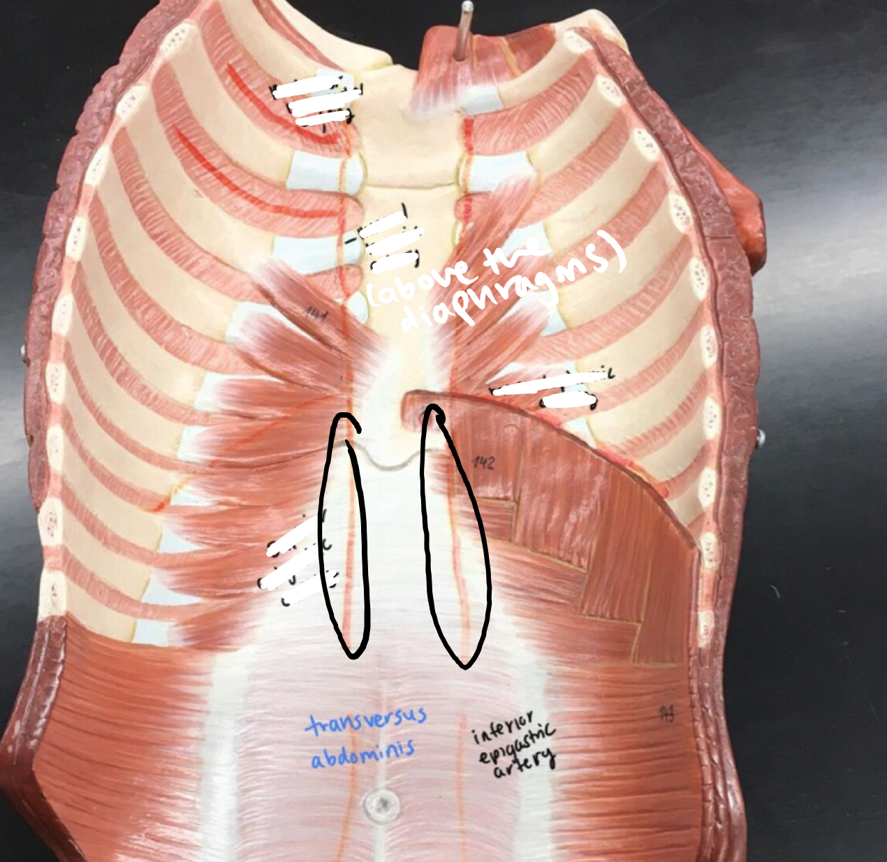
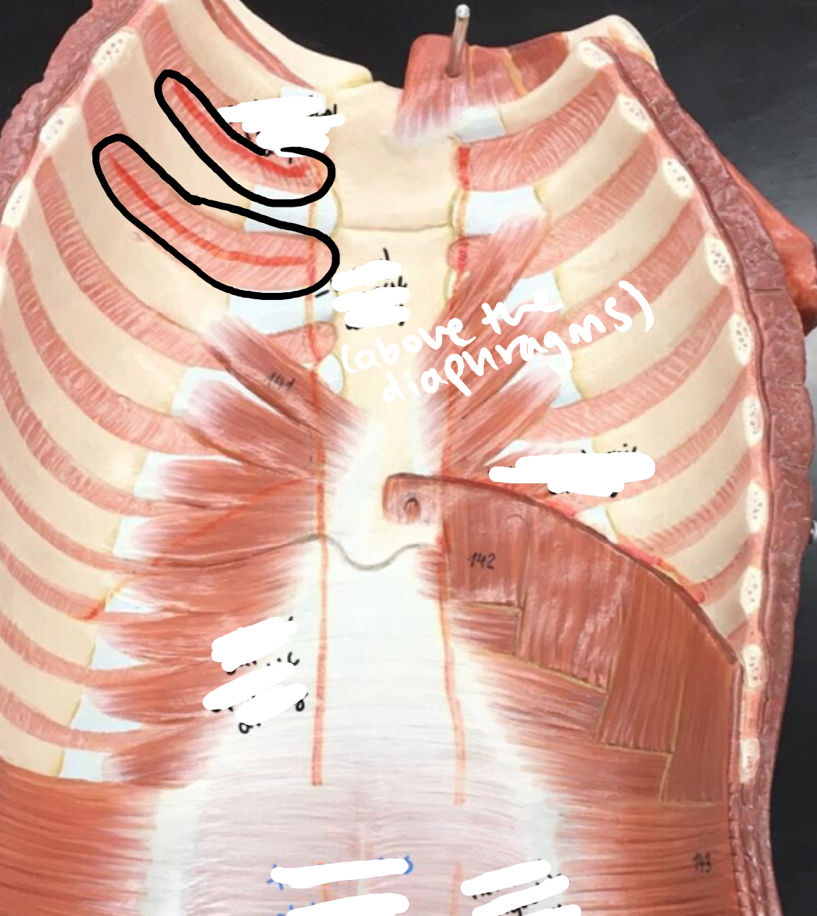
16
New cards
Regions of the heart
- Interventricular septum
- Interventricular septum
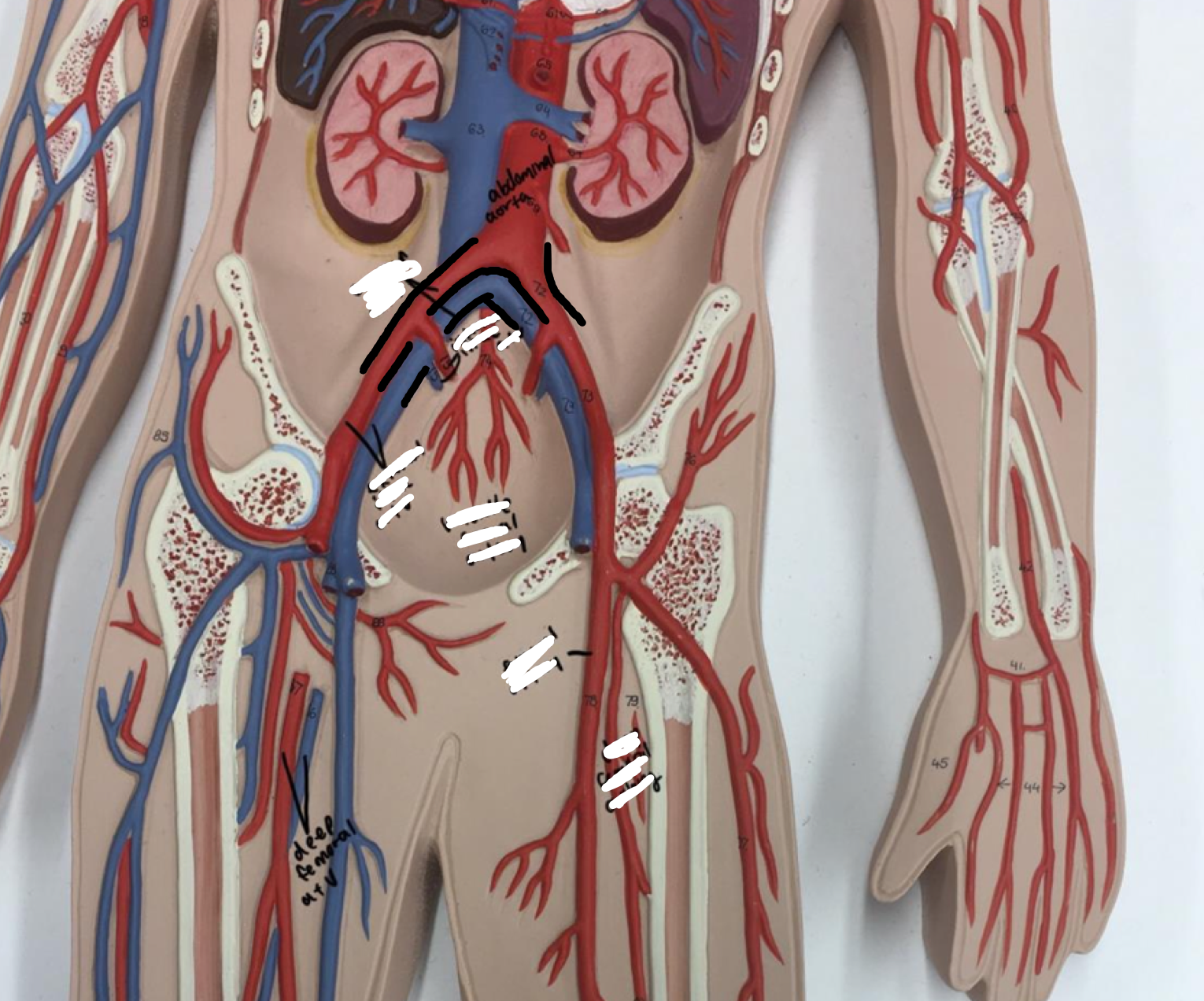
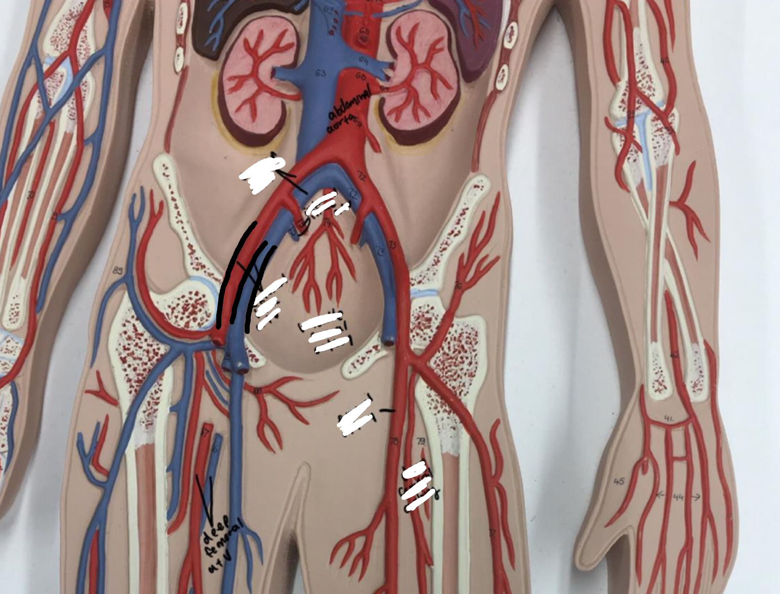
If you open the ventricles, the inner pinched surface
17
New cards
Regions of the heart
- Interatrial septum
- Interatrial septum
18
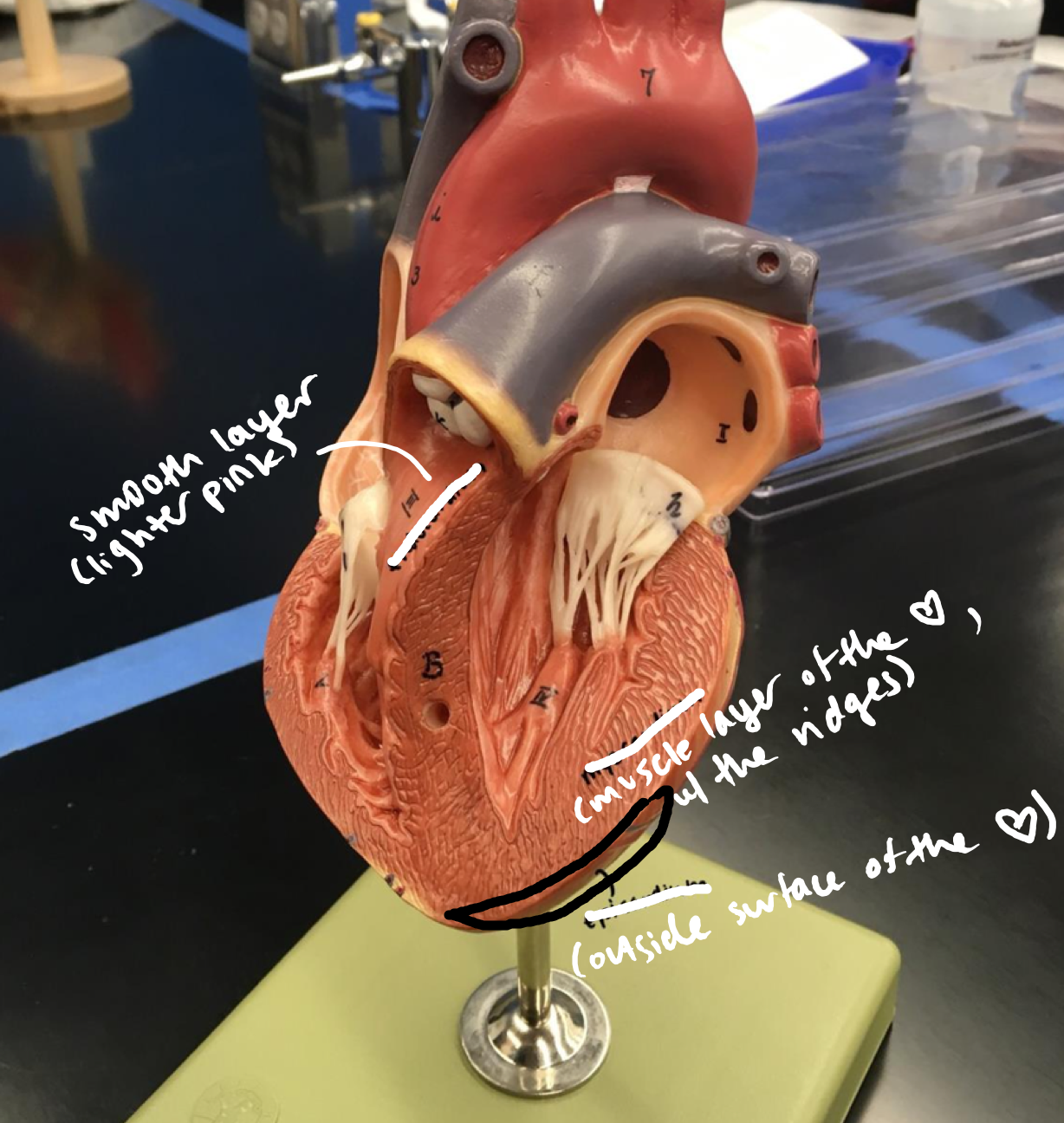
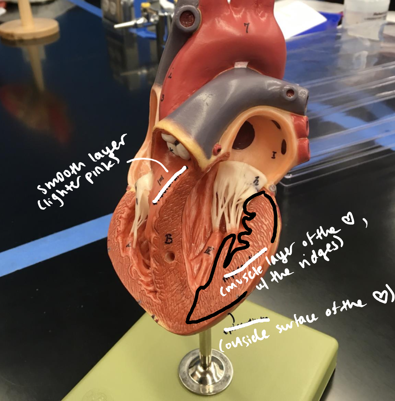
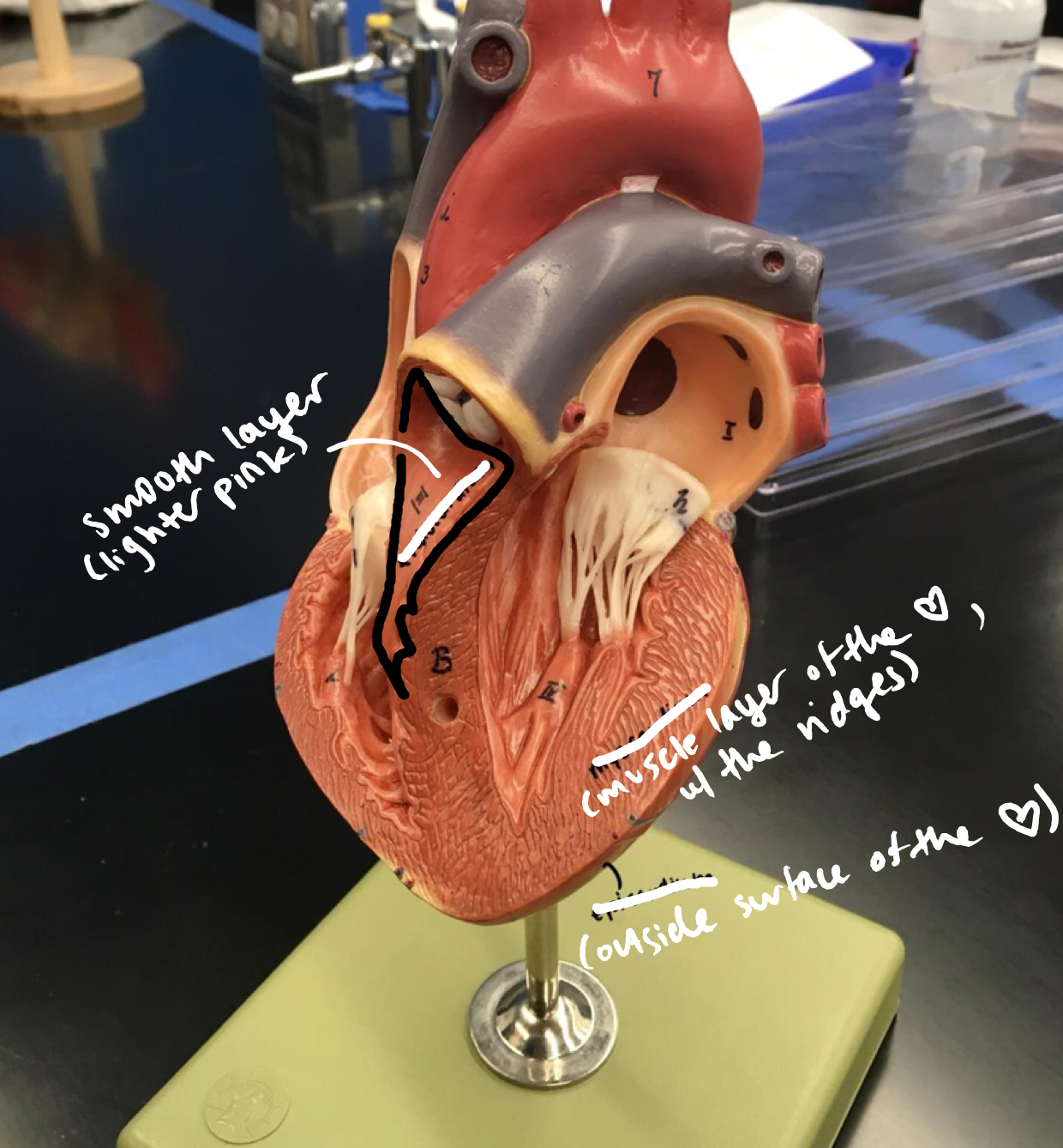
New cards
Layers of heart walls
- Epicardium (visceral pericardium)
- Epicardium (visceral pericardium)
Outside surface of the heart
19
New cards
Layers of heart walls
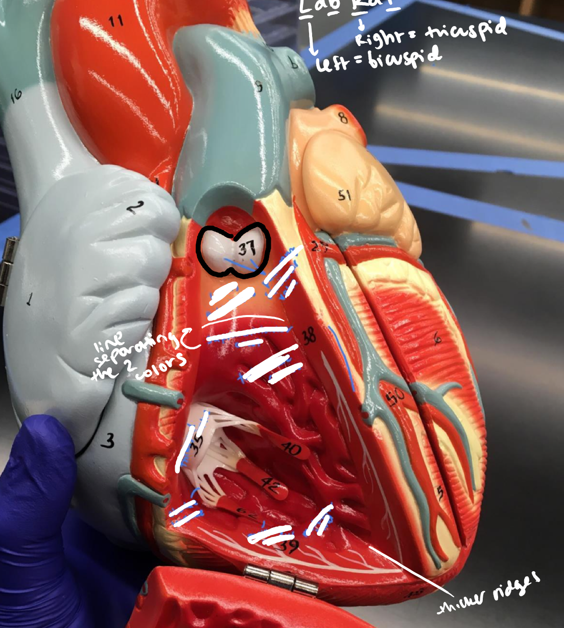
- Myocardium
- Myocardium
Muscle layer of the heart, with the ridges
20
New cards
Layers of heart walls
- Endocardium
- Endocardium
Smooth lighter pink layer of the heart
21
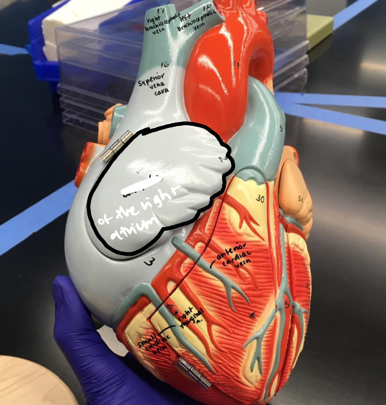
New cards
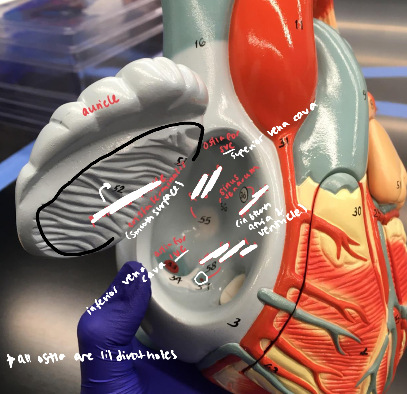
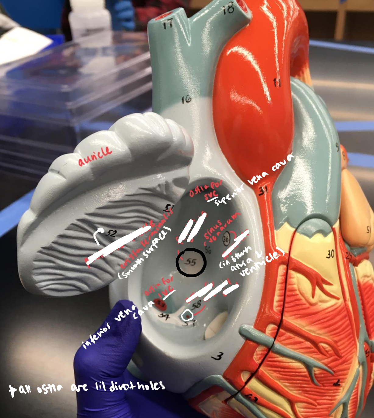
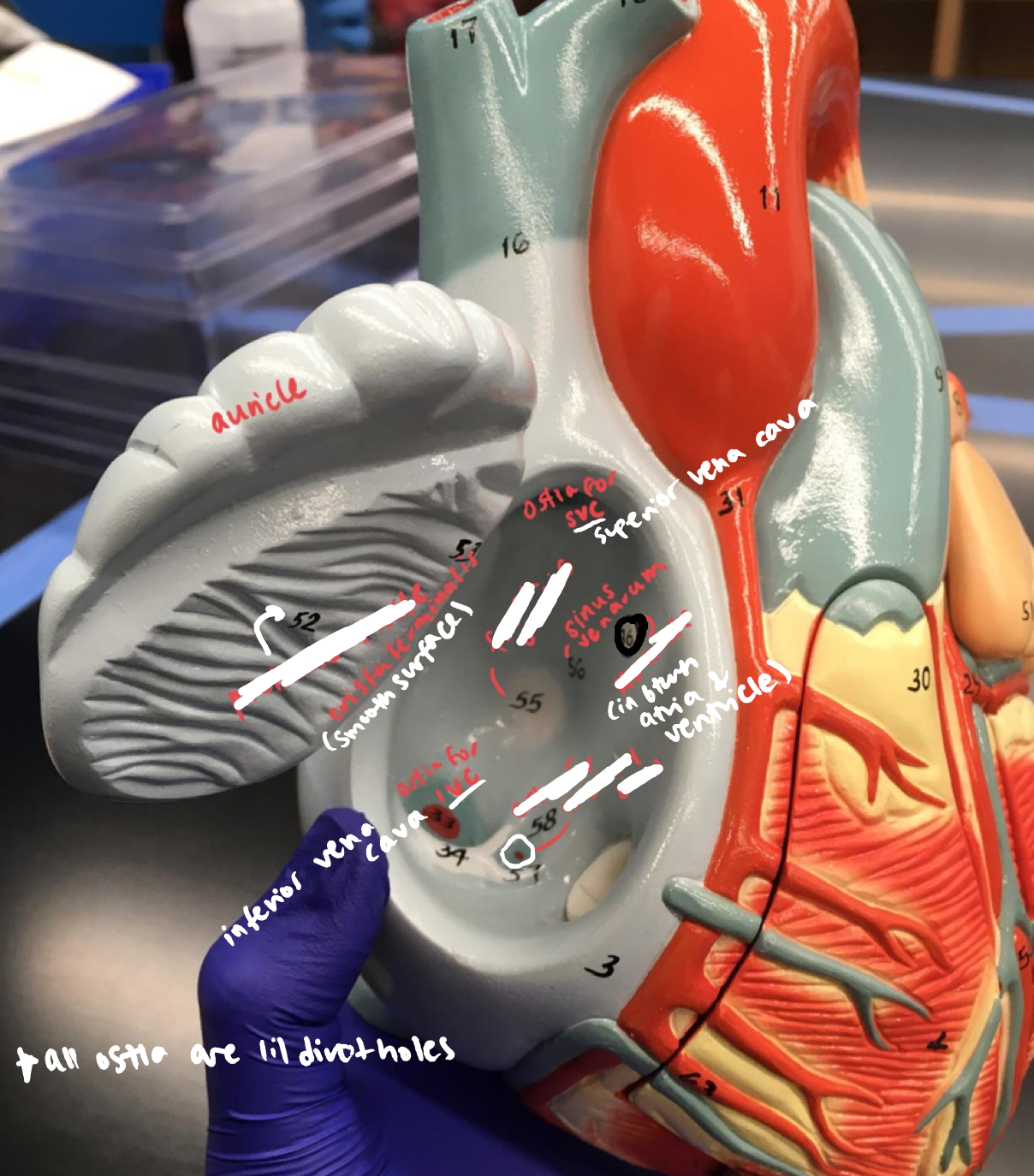
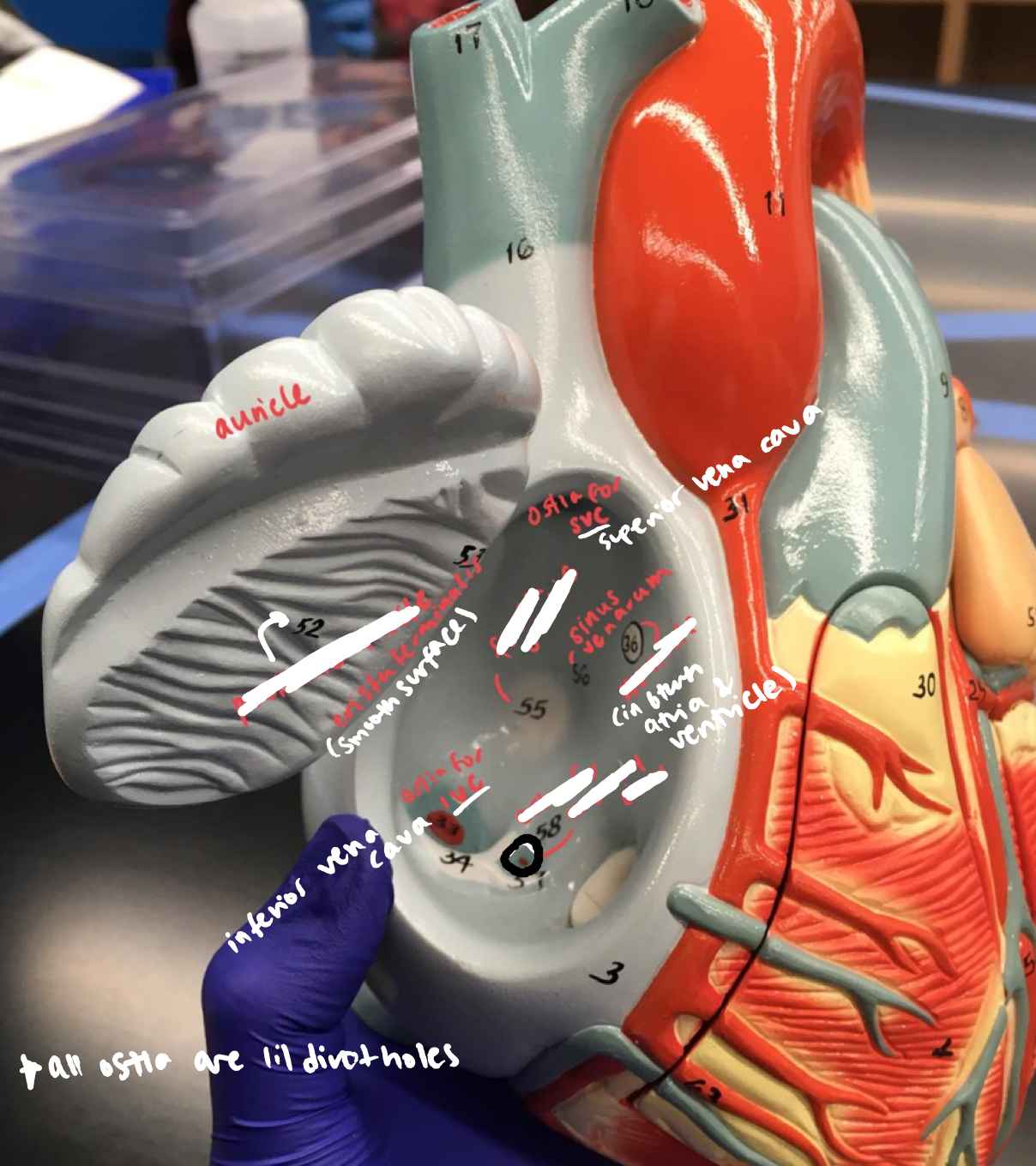
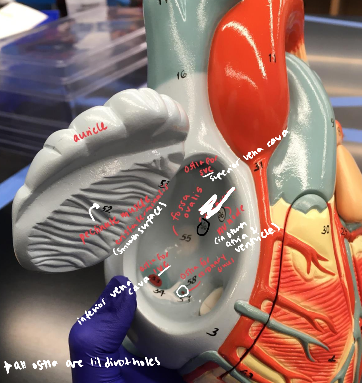
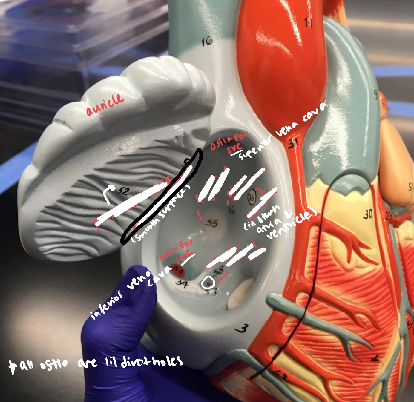
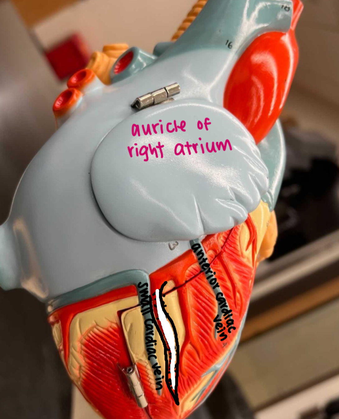
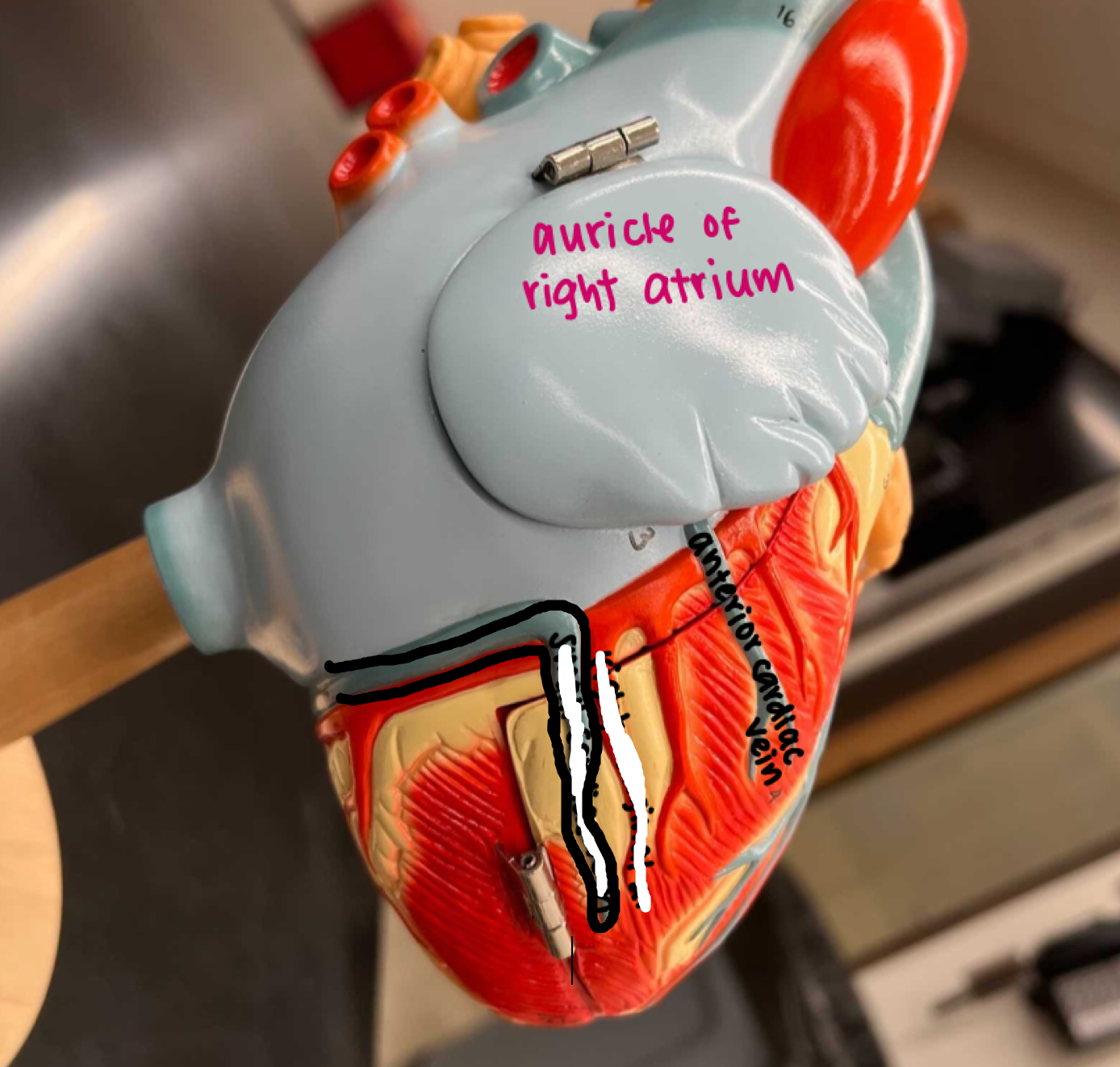
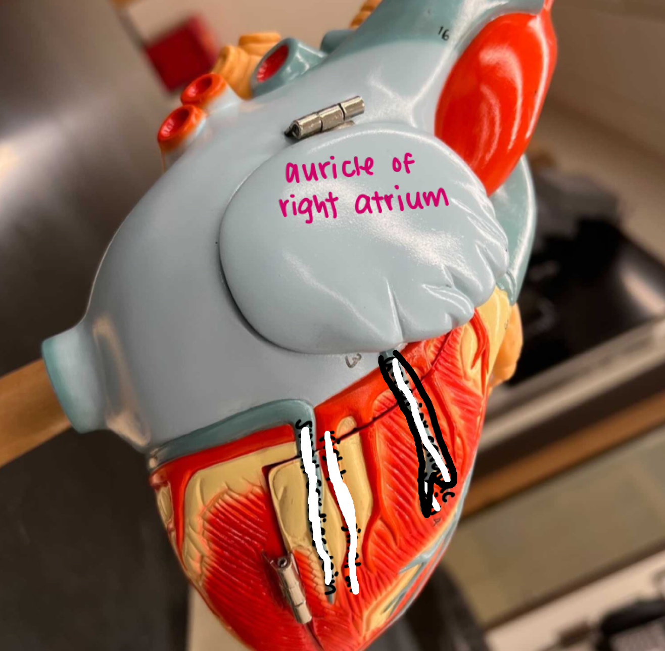
Chambers of Right Atrium
- Auricle
- Auricle
Blue wing
22
New cards
Chambers of Right Atrium
- Pectinate muscle
- Pectinate muscle
Inner ridged section of the blue wing
23
New cards
Chambers of Right Atrium
- Fossa ovalis (fetal remnant)
- Fossa ovalis (fetal remnant)
Inner circular ditch of the blue wing
24
New cards
Chambers of Right Atrium
- SA node
- SA node
The circular node on the edge of the blue wing

25
New cards
Chambers of Right Atrium
- AV node
- AV node
The circular node in between the atria and ventricle of the blue wing
26
New cards
Chambers of Right Atrium
- Ostia for coronary sinus
- Ostia for coronary sinus
Lower smaller circular ditch of the blue wing
27
New cards
Chambers of Right Atrium
- Superior vena cava
- Superior vena cava
Top light blue section of the root coming off the blue wing
28
New cards
Chambers of Right Atrium
- Inferior vena cava
- Inferior vena cava
Bottom blue holed part on the back side of the heart
29
New cards
Chambers of Right Atrium
- Sinus venarum
- Sinus venarum
Inner space of the right atrium
30
New cards
Chambers of Right Atrium
- Crista terminalis
- Crista terminalis
Smooth inner edged surface of the blue wing
31
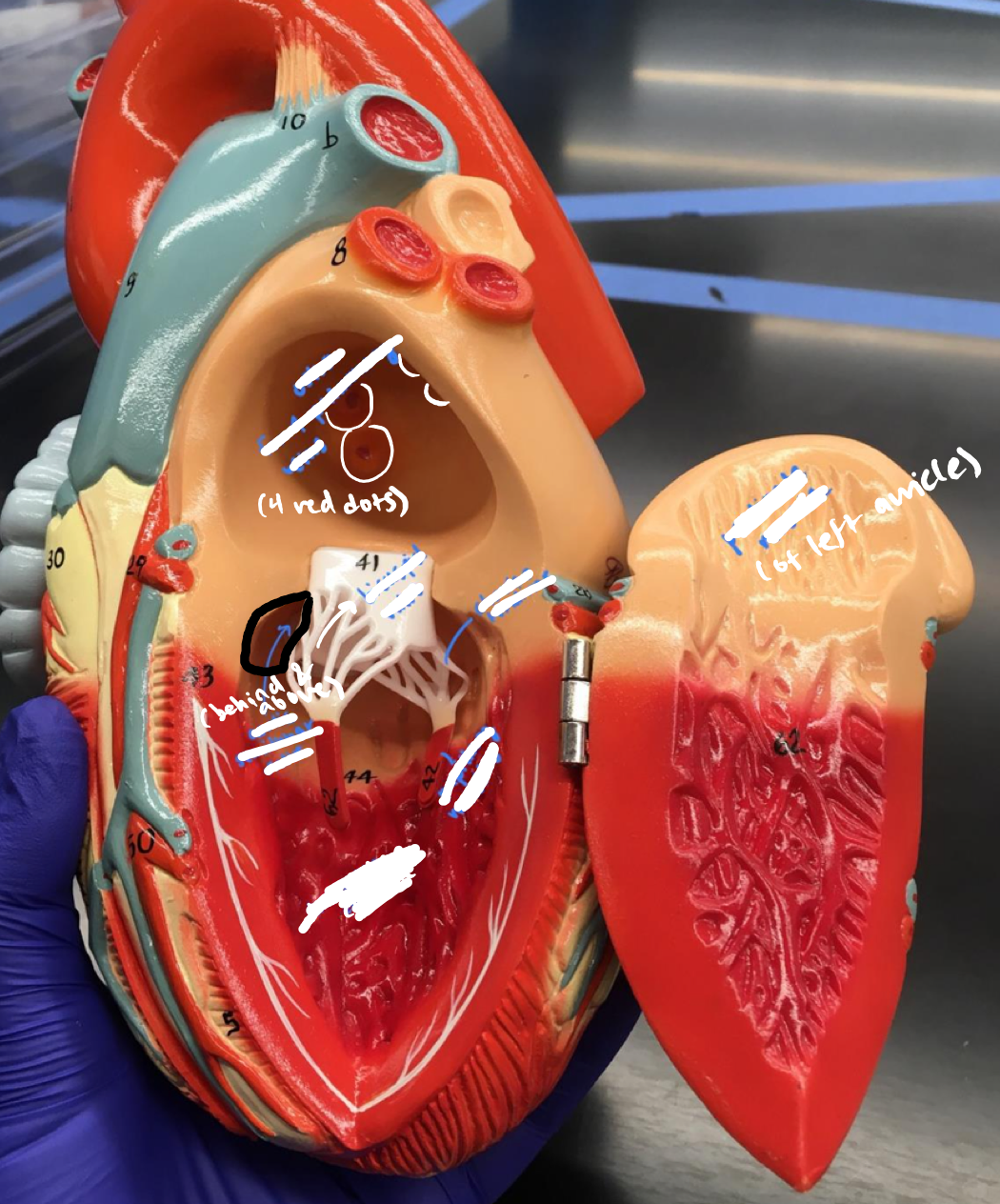
New cards
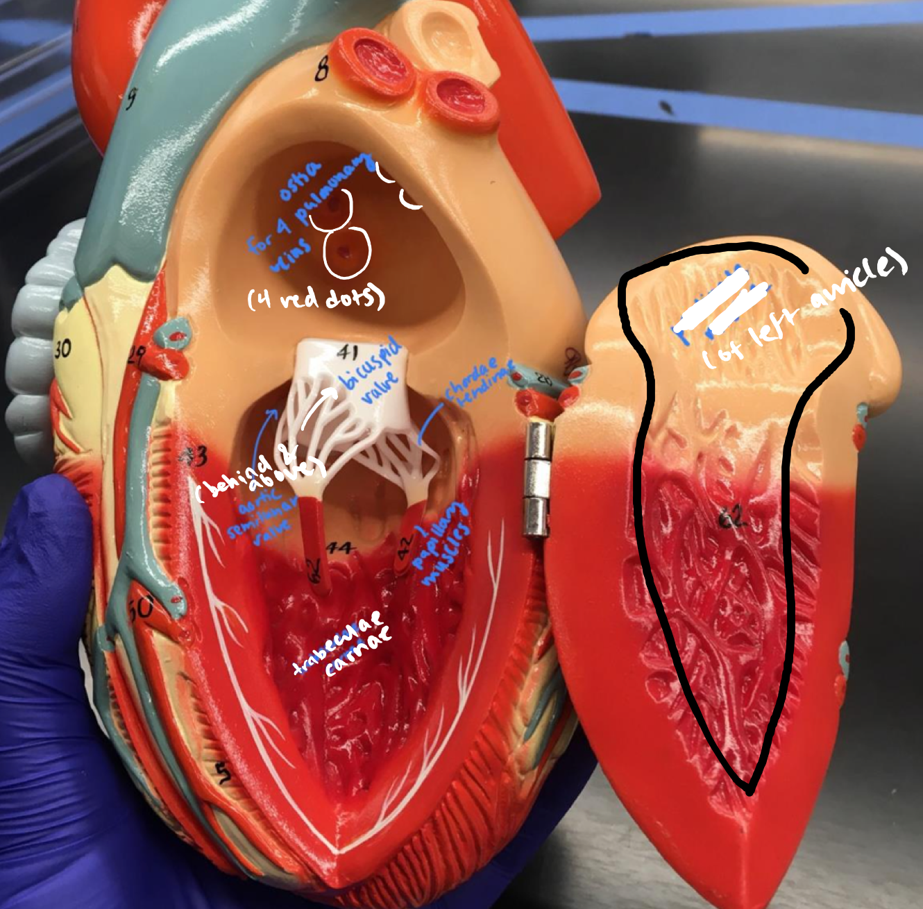
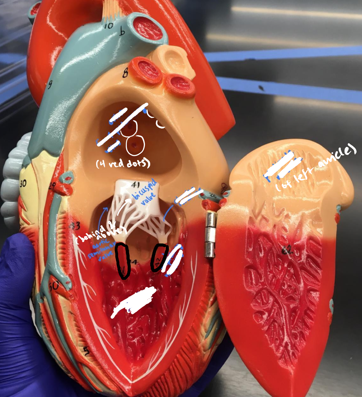
Chambers of Left Atrium
- Auricle
- Auricle
Beige wing
32
New cards
Chambers of Left Atrium
- Pectinate muscle
- Pectinate muscle
Inner veiny wall of the beige wing
33
New cards
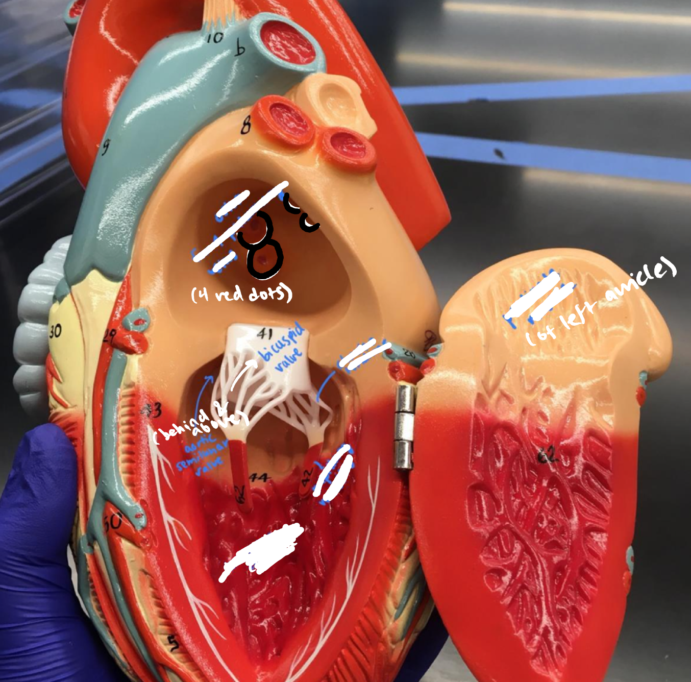
Chambers of Left Atrium
- Ostia for 4 pulmonary veins
- Ostia for 4 pulmonary veins
4 red dots in the upper deep triangular beige region
34
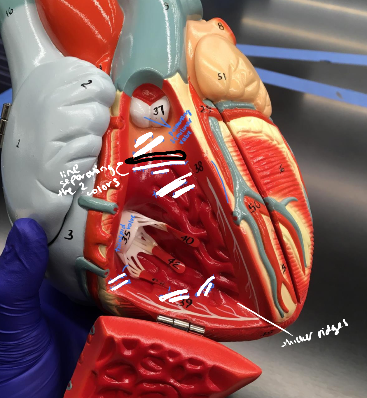
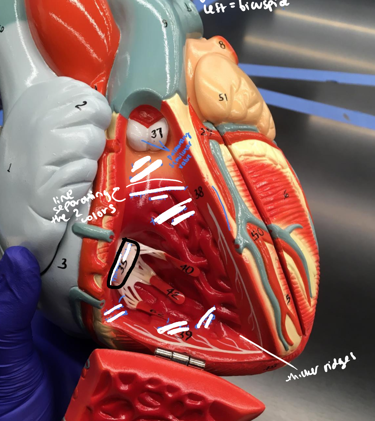
New cards
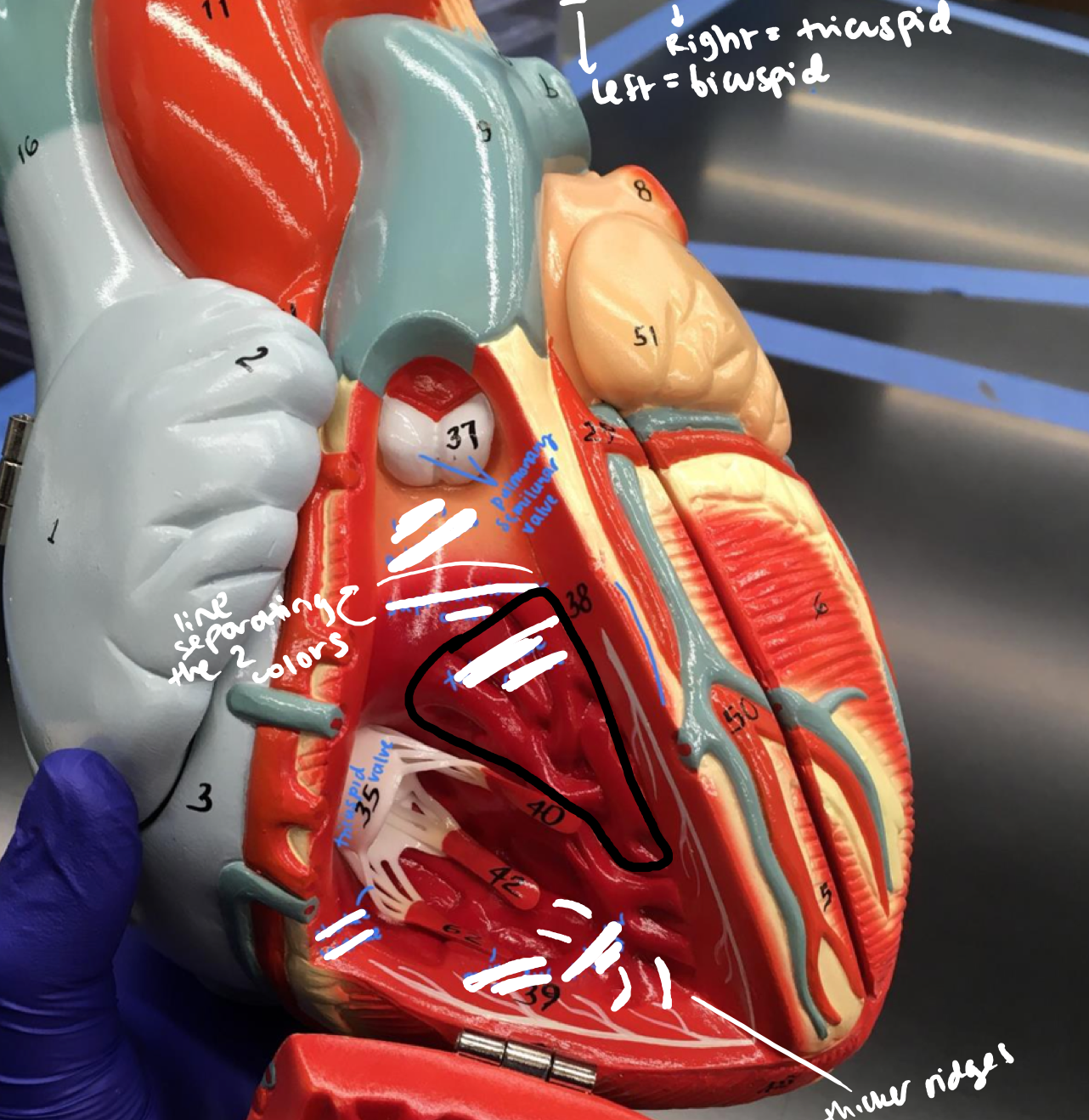
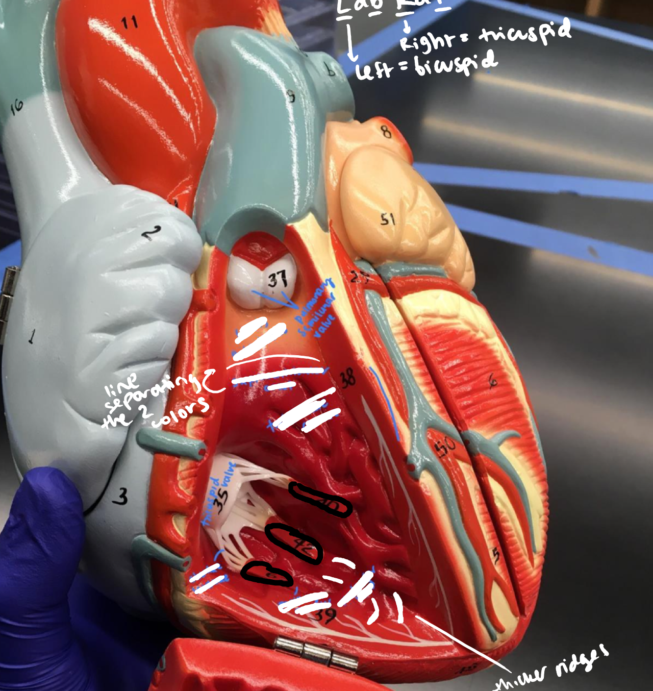
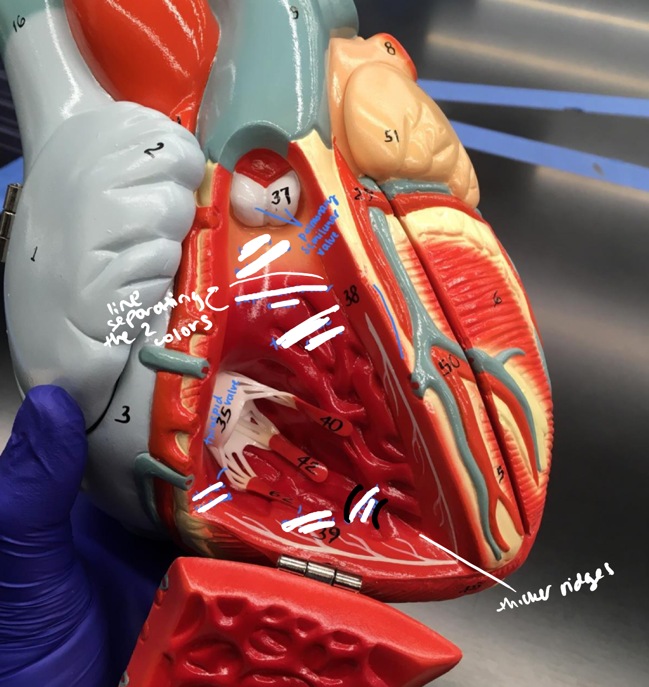
Right Ventricle
- Trabeculae carnae (myocardium)
- Trabeculae carnae (myocardium)
The veiny red meat under the pulmonary semilunar valve
35
New cards
Right Ventricle
- Chordae tendinae
- Chordae tendinae
The white connections from the papillary muscles to the valve under the pulmonary semilunar valve
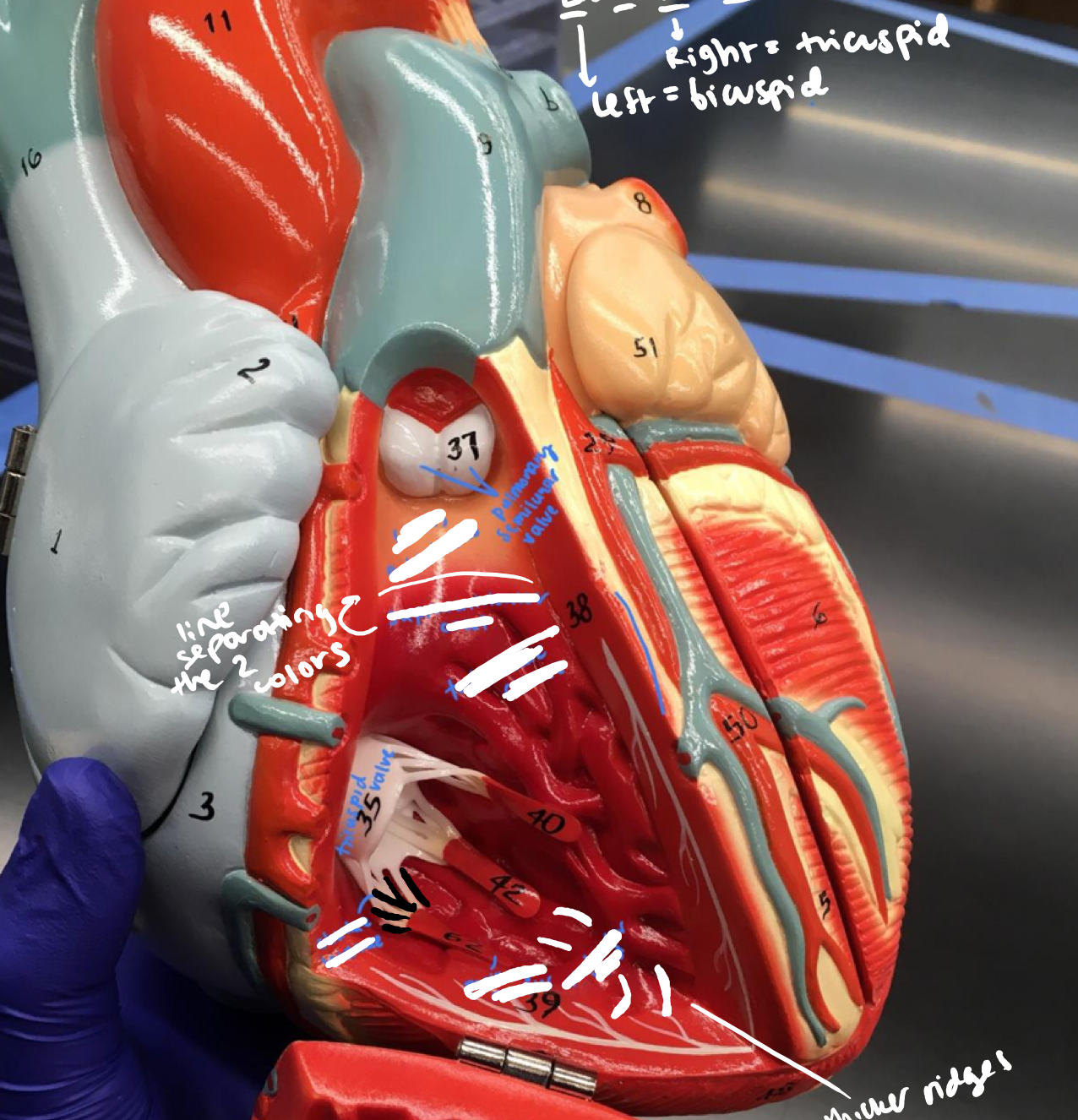
36
New cards
Right Ventricle
- Papillary muscle (myocardium)
- Papillary muscle (myocardium)
The red connections from the trabeculae carnae and the chordae tendinae under the pulmonary semilunar valve
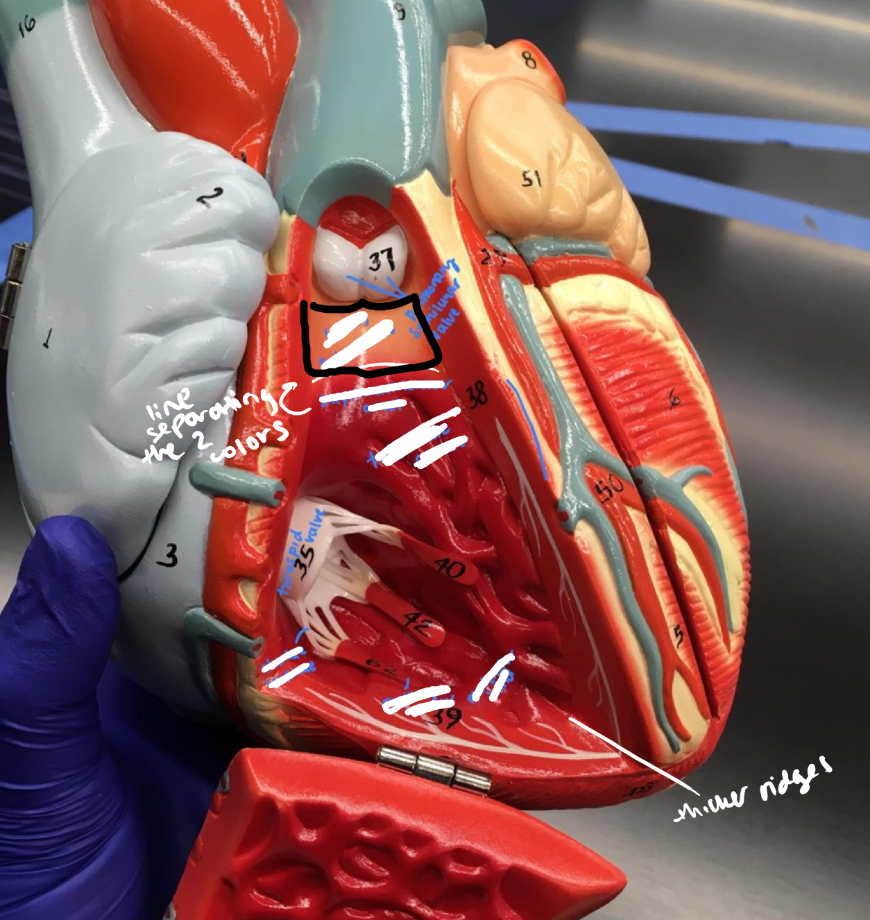
37
New cards
Right Ventricle
- Septomarginal trabeculae (moderator band)
- Septomarginal trabeculae (moderator band)
The band connecting the trabeculae carnae under the pulmonary semilunar valve
38
New cards
Right Ventricle
- Conus arteriosus
- Conus arteriosus
The lighter orange upper part of the right ventricle, just below the pulmonary semilunar valve
39
New cards
Right Ventricle
- Supraventricular crest
- Supraventricular crest
Crest in between the conus arteriosus and trabeculae carnae
40
New cards
Left Ventricle
- Trabeculae carnae (myocardium)
- Trabeculae carnae (myocardium)
The veiny red meat under the bicuspid valve
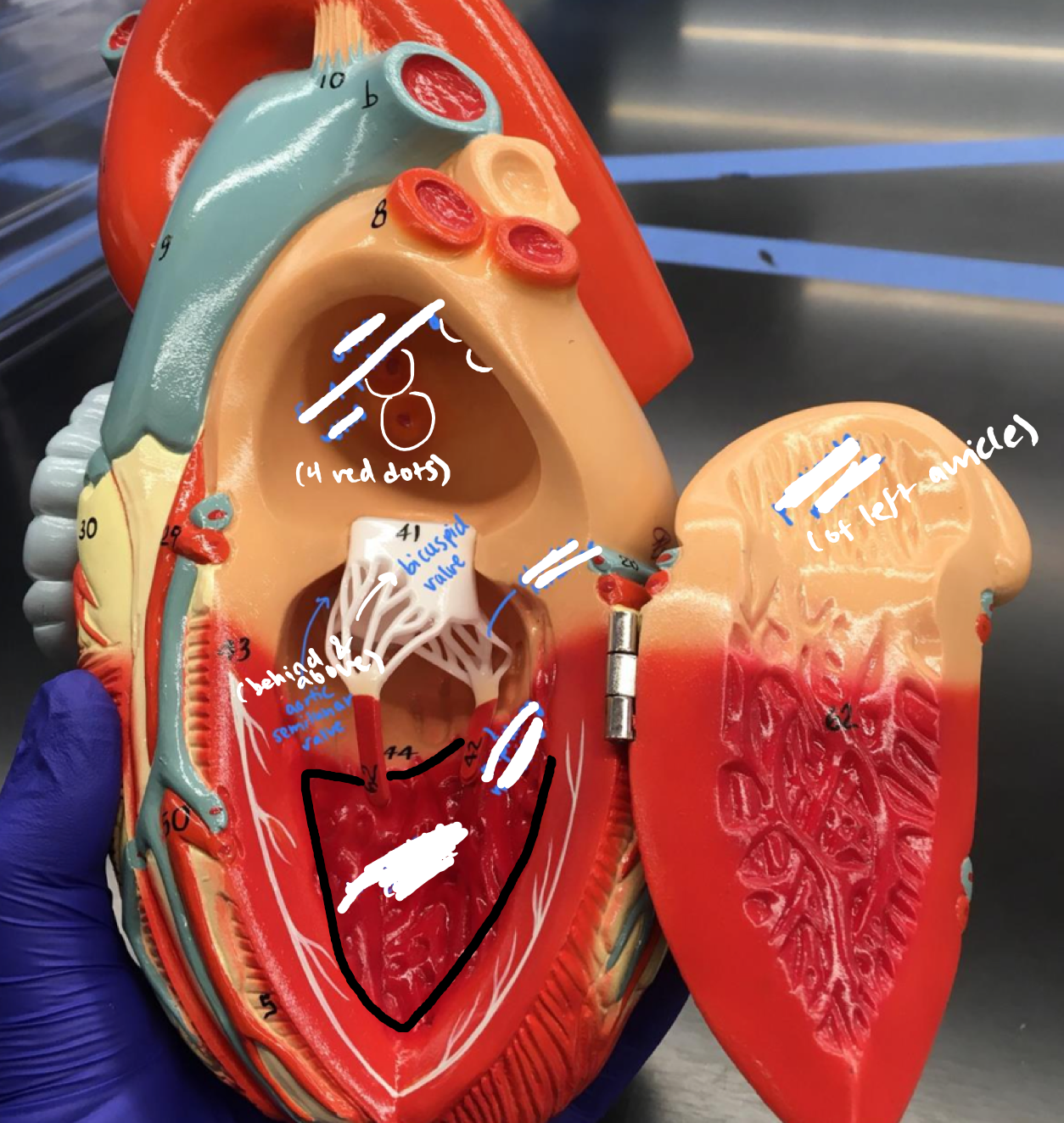
41
New cards
Left Ventricle
- Chordae tendinae
- Chordae tendinae
The white connections from the papillary muscles to the valve under the bicuspid valve
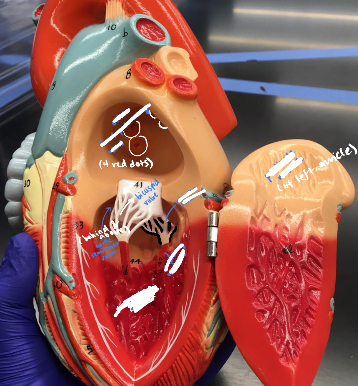
42
New cards
Left Ventricle
- Papillary muscle (myocardium)
- Papillary muscle (myocardium)
The red connections from the trabeculae carnae and the chordae tendinae under the bicuspid valve
43
New cards
D. Valves
- Tricuspid (right atrioventricular)
- Tricuspid (right atrioventricular)
The valve with 3 papillary muscle connections
44
New cards
D. Valves
- Bicuspid (left atrioventricular valve)
- Bicuspid (left atrioventricular valve)
The valve with 2 papillary muscle connections
45
New cards
D. Valves
- Pulmonary semilunar
- Pulmonary semilunar
The white butt in the right ventricle
46
New cards
D. Valves
- Aortic semilunar
- Aortic semilunar
The empty space behind and above the bicuspid valve
47
New cards
Great vessels
- Pulmonary trunk
- Pulmonary trunk
The big dark blue trunk
48
New cards
Great vessels
- Systemic aorta
- Systemic aorta
The big red overarching part
49
New cards
Great vessels
- Superior vena cava
- Superior vena cava
The half light blue half dark blue doubled part

50
New cards
Great vessels
- Inferior vena cava
- Inferior vena cava
The big blue holed part at the bottom
51
New cards
Coronary circulation: Left Coronary Artery
- Anterior interventricular artery
- Anterior interventricular artery
The middle red artery below the great cardiac vein leading to the apex
52
New cards
Coronary circulation: Left Coronary Artery
- Circumflex artery
- Circumflex artery
The red artery lining the front of the heart underneath the auricle of the left atrium
53
New cards
Coronary circulation: Left Coronary Artery
- Left (obtuse) marginal artery
- Left (obtuse) marginal artery
The artery extending below the circumflex artery to the lower right, right of the auricle of the left atrium
54
New cards
Coronary circulation: Right Coronary Artery
- Right (acute) marginal artery
- Right (acute) marginal artery
On the side of the auricle of the right atrium, the bottommost extension of the right artery, above the small cardiac vein
55
New cards
Coronary circulation: Right Coronary Artery
- Posterior interventricular artery
- Posterior interventricular artery
On the bottom of the heart, the spiky red extension coming off the circumflex artery and just below the coronary sinus
56
New cards
Venous circulation
- Coronary sinus
- Coronary sinus
The large thick blue on the bottom of the heart underneath the inferior vena cava
57
New cards
Venous circulation
- Great cardiac vein
- Great cardiac vein
The middle blue vein that splits into 2 on the front of the heart
58
New cards
Venous circulation
- Small cardiac vein
- Small cardiac vein
On the side with the auricle of the right atrium, the bottom-most blue vein, not touching the actual auricle
59
New cards
Venous circulation
- Middle cardiac vein
- Middle cardiac vein
On the bottom of the heart, the spiky blue extension coming off the coronary sinus
60
New cards
Venous circulation
- Anterior cardiac vein
- Anterior cardiac vein
On the side with the auricle of the right atrium, the top-most blue vein, touching the actual auricle
61
New cards
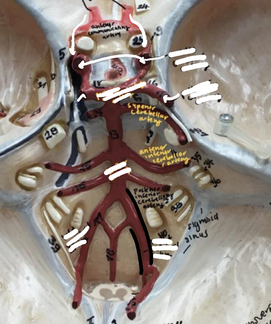
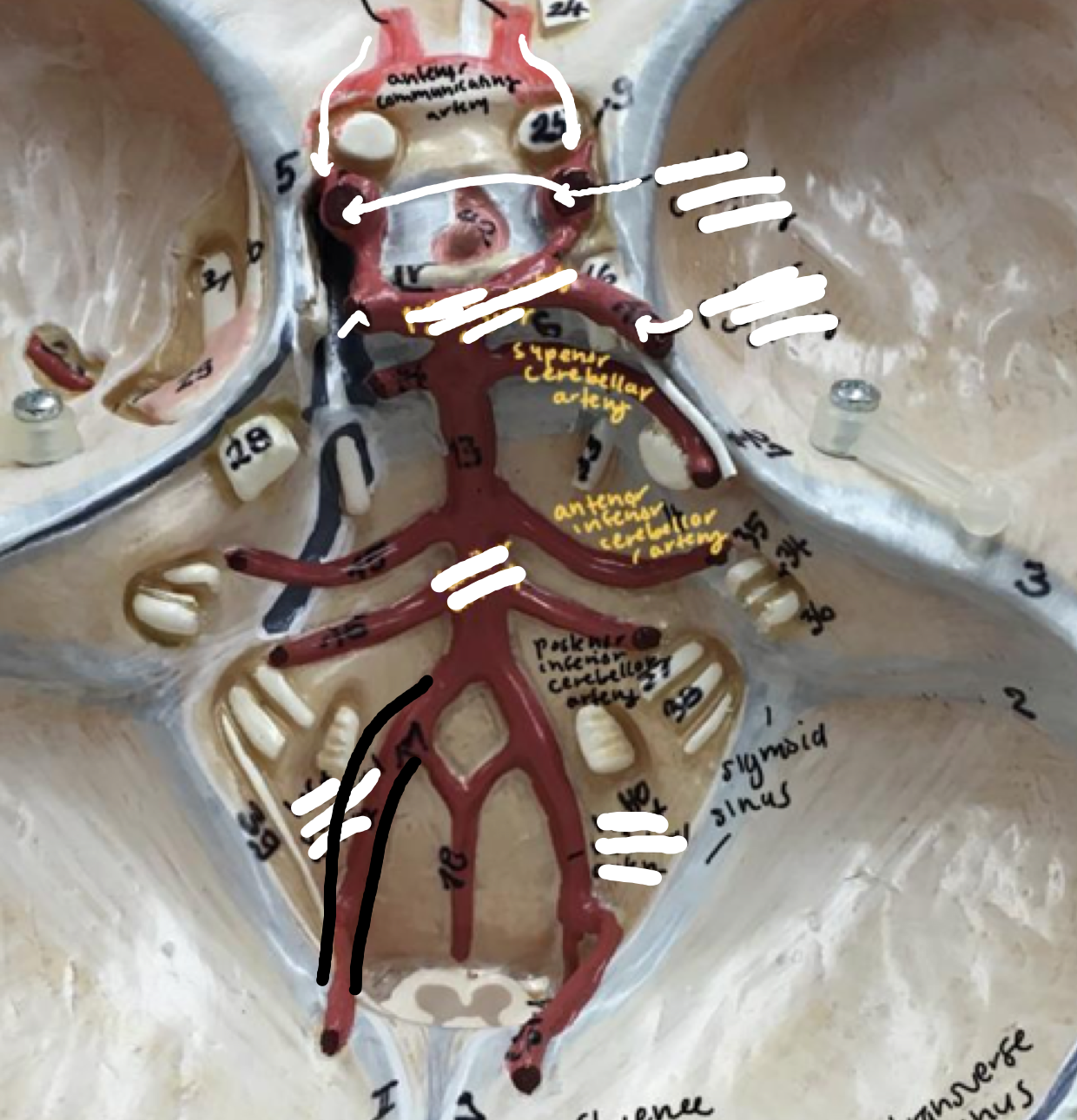
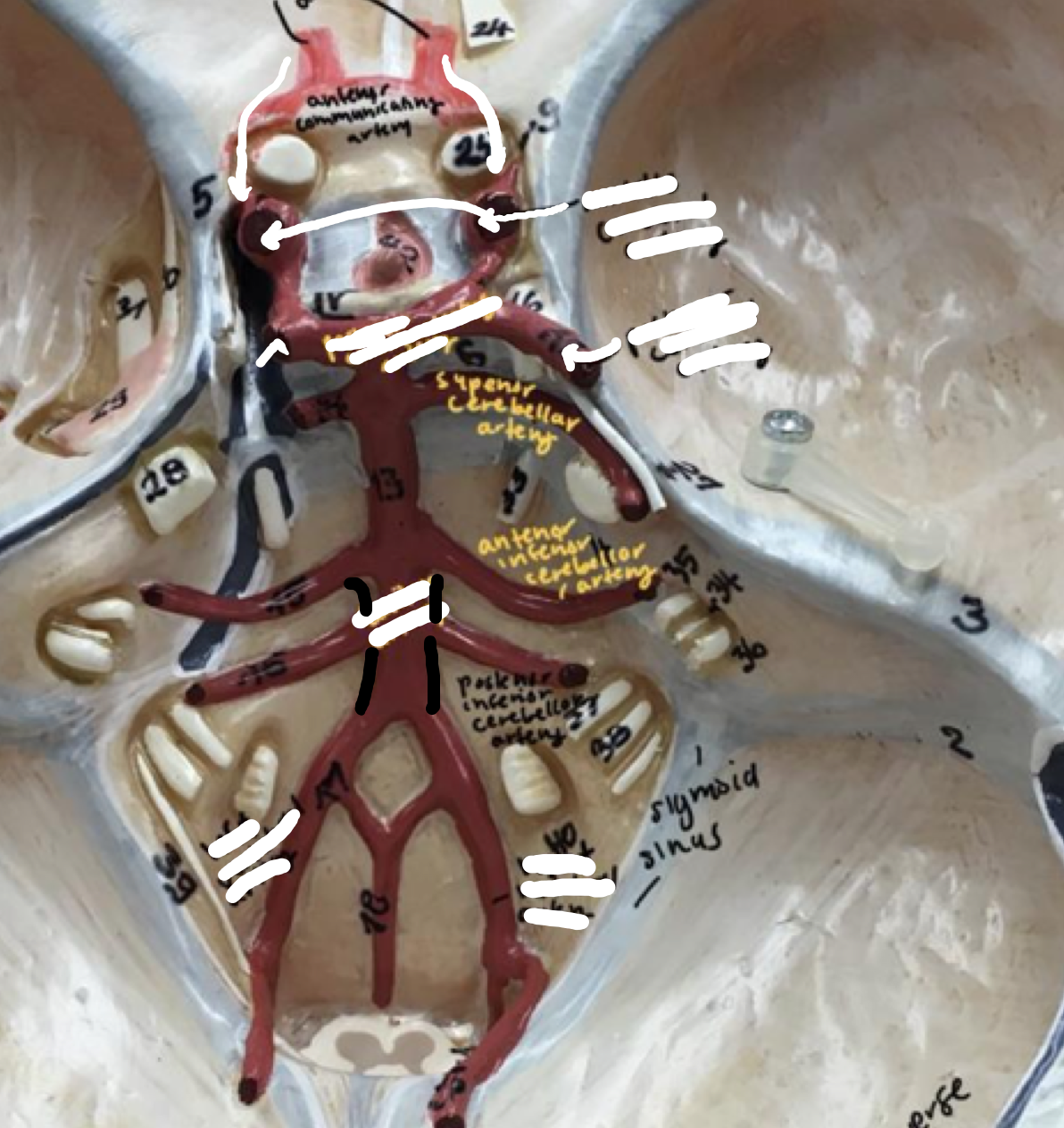
Circle of Willis
- Vertebral artery (right)
- Vertebral artery (right)
Right leg of stickman
62
New cards
Circle of Willis
- Vertebral artery (left)
- Vertebral artery (left)
Left leg of stickman
63
New cards
Circle of Willis
- Basilar artery
- Basilar artery
Body of the stick man
64
New cards
Circle of Willis
- Posterior cerebral artery
- Posterior cerebral artery
The arm like projections coming off the posterior communicating artery
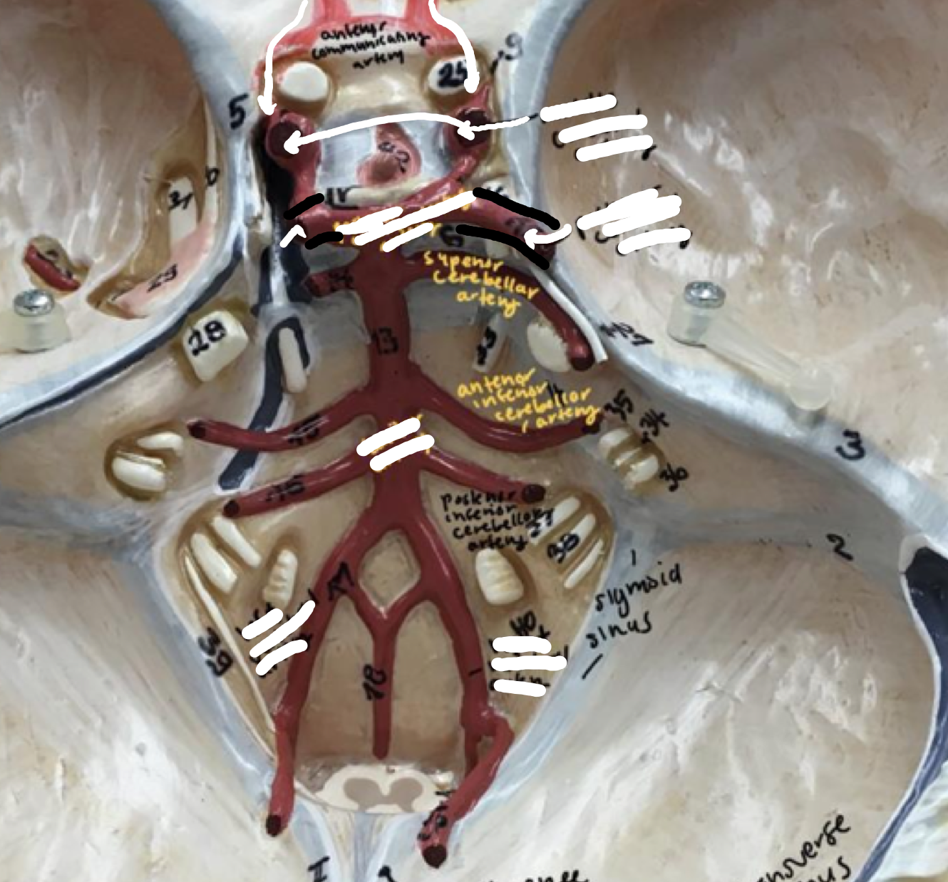
65
New cards
Circle of Willis
- Posterior communicating artery
- Posterior communicating artery
1st thick red artery around the head of the stick man
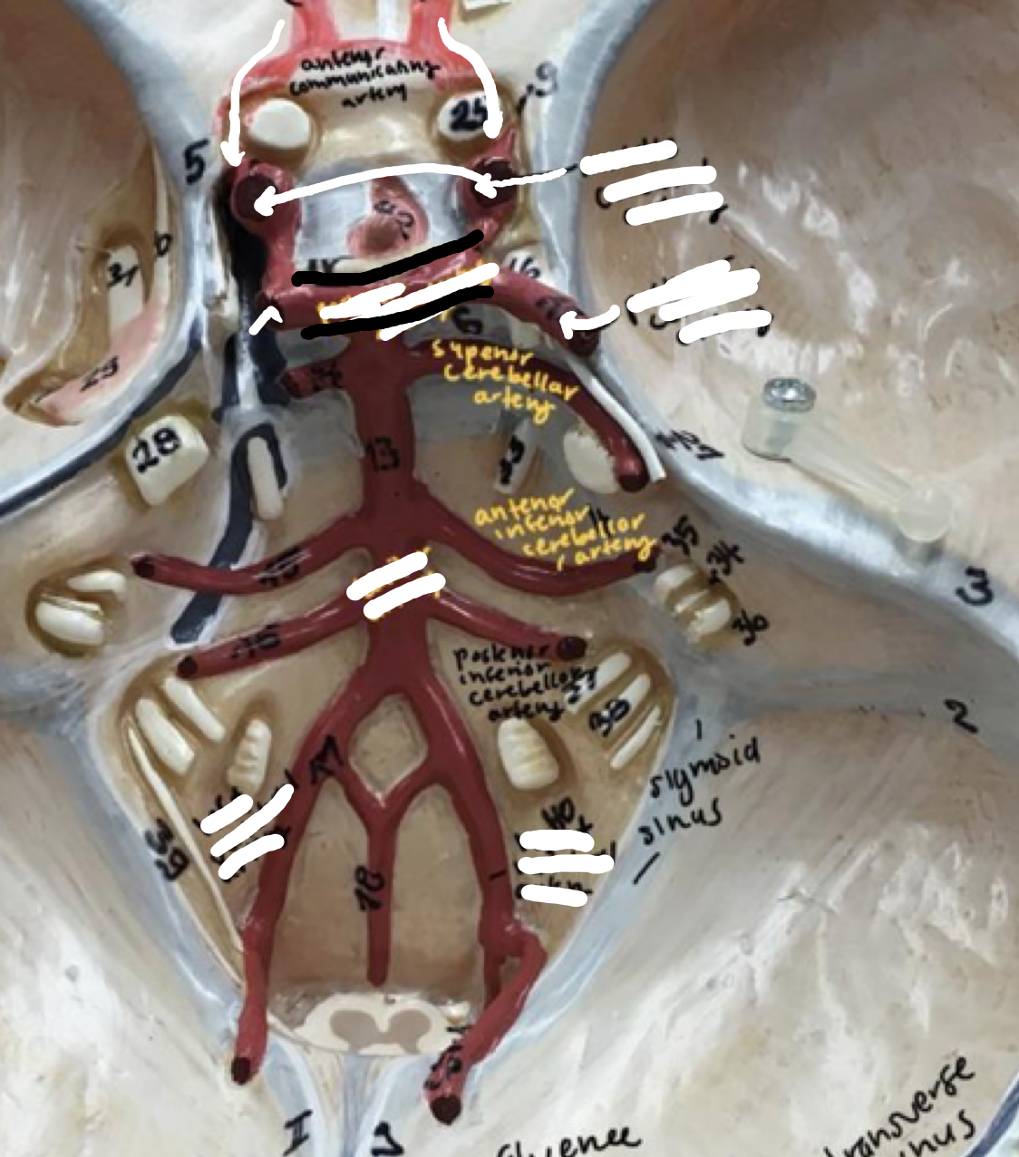
66
New cards
Circle of Willis
- Middle cerebral artery
- Middle cerebral artery
1st 2 hole like projections
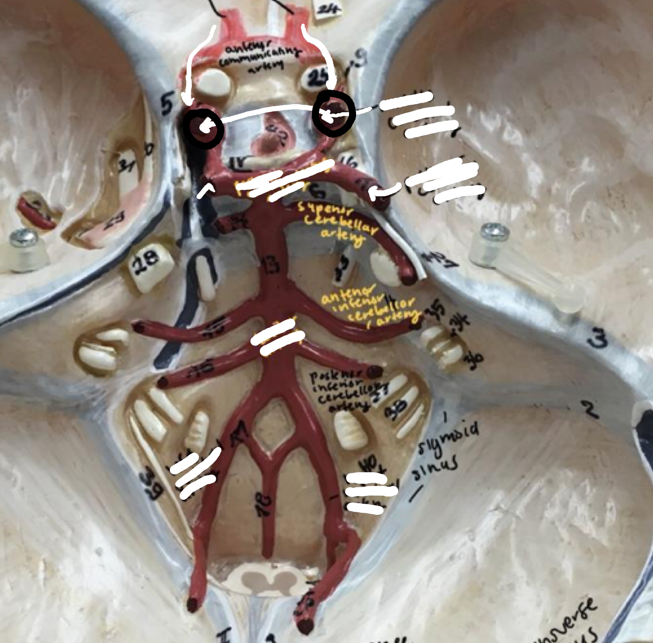
67
New cards
Circle of Willis
- Anterior cerebral artery
- Anterior cerebral artery
Top 2 red horns on the head of the stick man
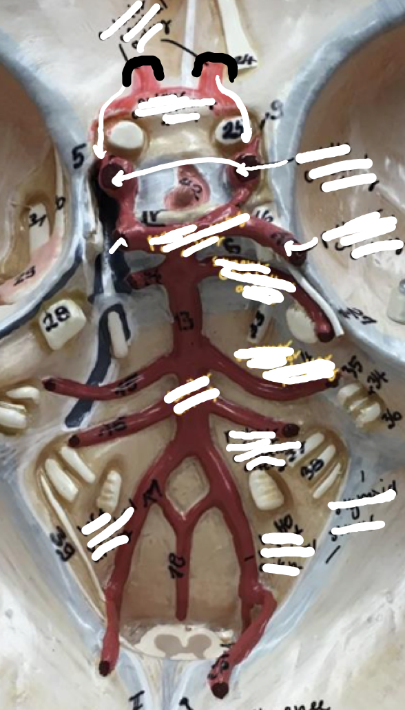
68
New cards
Circle of Willis
- Anterior communicating artery
- Anterior communicating artery
The red bridge in between the 2 horns of the anterior cerebral artery
69
New cards
Circle of Willis
- Posterior inferior cerebellar artery
- Posterior inferior cerebellar artery
3rd set of the stick man's arms
70
New cards
Circle of Willis
- Anterior inferior cerebellar artery
- Anterior inferior cerebellar artery
2nd set of the stick man's arms
71
New cards
Circle of Willis
- Superior cerebellar artery
- Superior cerebellar artery
- Artery just below the posterior communicating artery
- 1st set of the stick man's arms
- 1st set of the stick man's arms
72
New cards
Circle of Willis
- Superior sagittal sinus
- Superior sagittal sinus
Blue line near the inner top of the skull, the midline right underneath
73
New cards
Circle of Willis
- Transverse sinus
- Transverse sinus
Blue circular divot on the back of the skull
74
New cards
Circle of Willis
- Confluence of the sinuses
- Confluence of the sinuses
Raised meeting point of the sinuses
75
New cards
Circle of Willis
- Sigmoid sinus
- Sigmoid sinus
The inner light blue S-shaped curve just below the transverse sinus
76
New cards
Aorta
- Ascending aorta
- Ascending aorta
Thick front part coming off the heart
77
New cards
Aorta
- Aortic arch
- Aortic arch
Arch going up to the upper portions of the body
78
New cards
Aorta
- Descending thoracic aorta
- Descending thoracic aorta
Back bottom portion off the arch, where it starts to disappear
79
New cards
Aorta
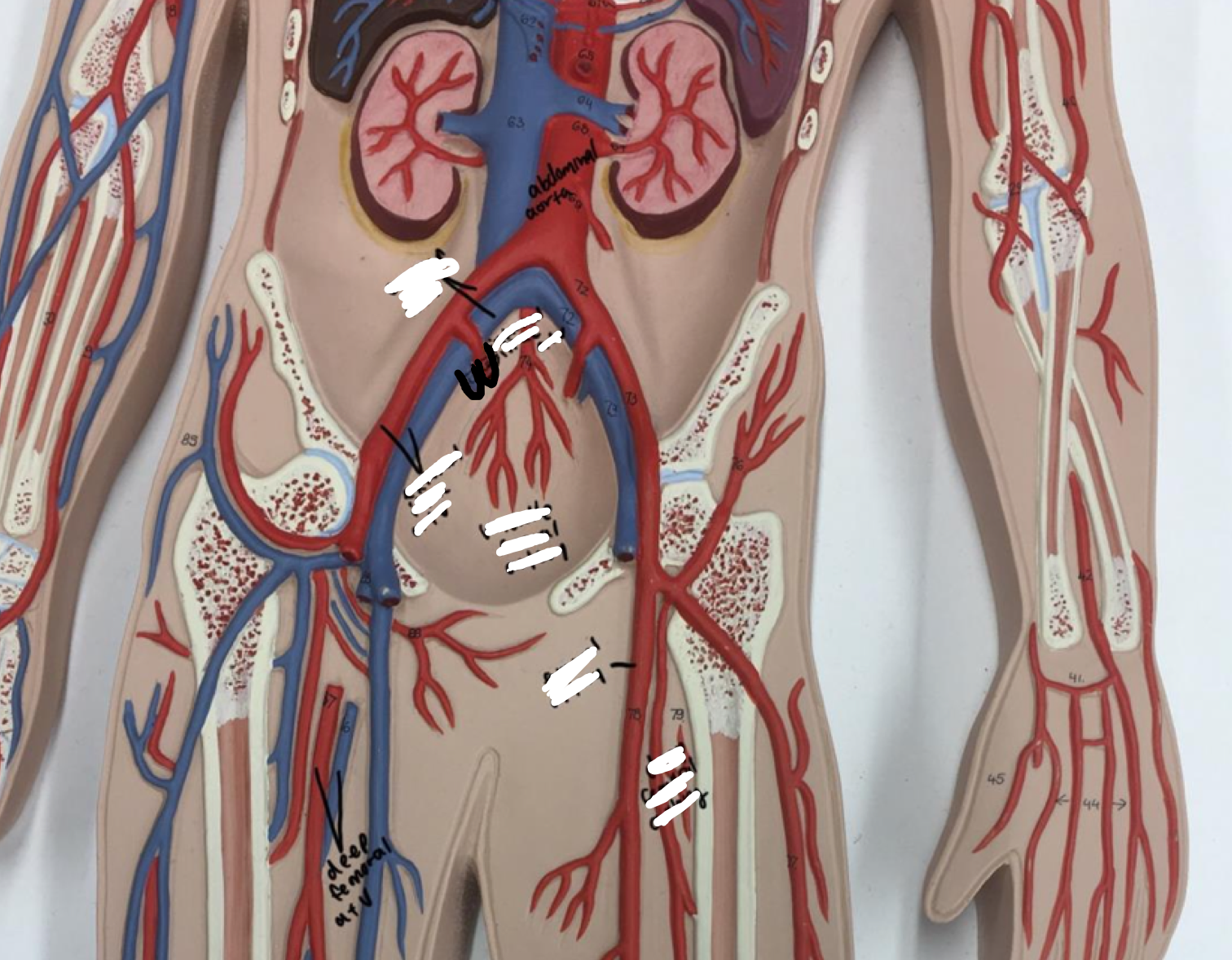
- Abdominal aorta
- Abdominal aorta
Thick long red arch coming down, splitting into 2
80
New cards
Head and neck
- Brachiocephalic trunk (artery)
- Brachiocephalic trunk (artery)
The red artery coming off the ascending arch
81
New cards
Head and neck
- Common carotid artery (right)
- Common carotid artery (right)
- Right (when looking through the body's POV)
- Coming off ascending aorta
- Coming off ascending aorta
82
New cards
Head and neck
- Common carotid artery (left)
- Common carotid artery (left)
- Left (when looking through the body's POV)
- Coming off aortic arch
- Coming off aortic arch
83
New cards
Head and neck
- Internal carotid artery
- Internal carotid artery
Upper part of the blue neck vein
84
New cards
Head and neck
- External carotid artery
- External carotid artery
Short upper part of the red neck artery
85
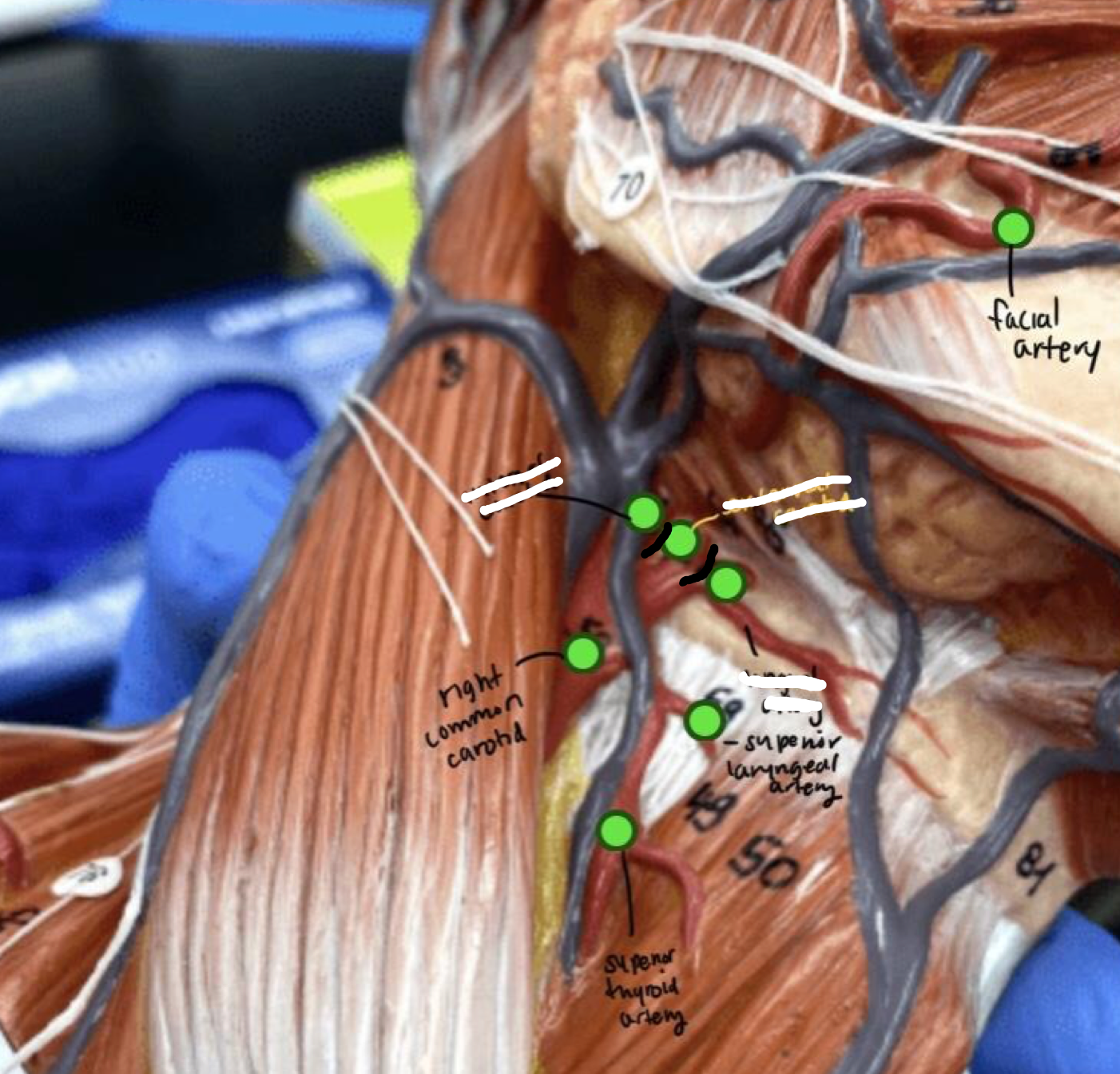
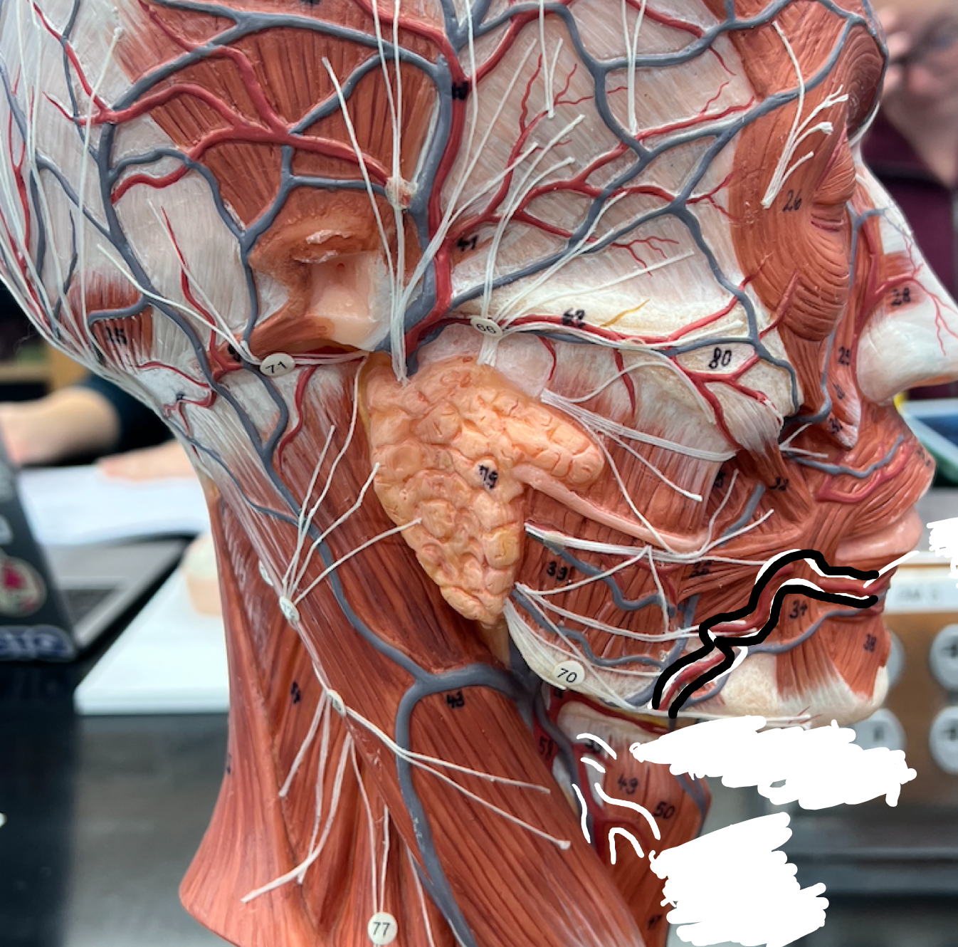
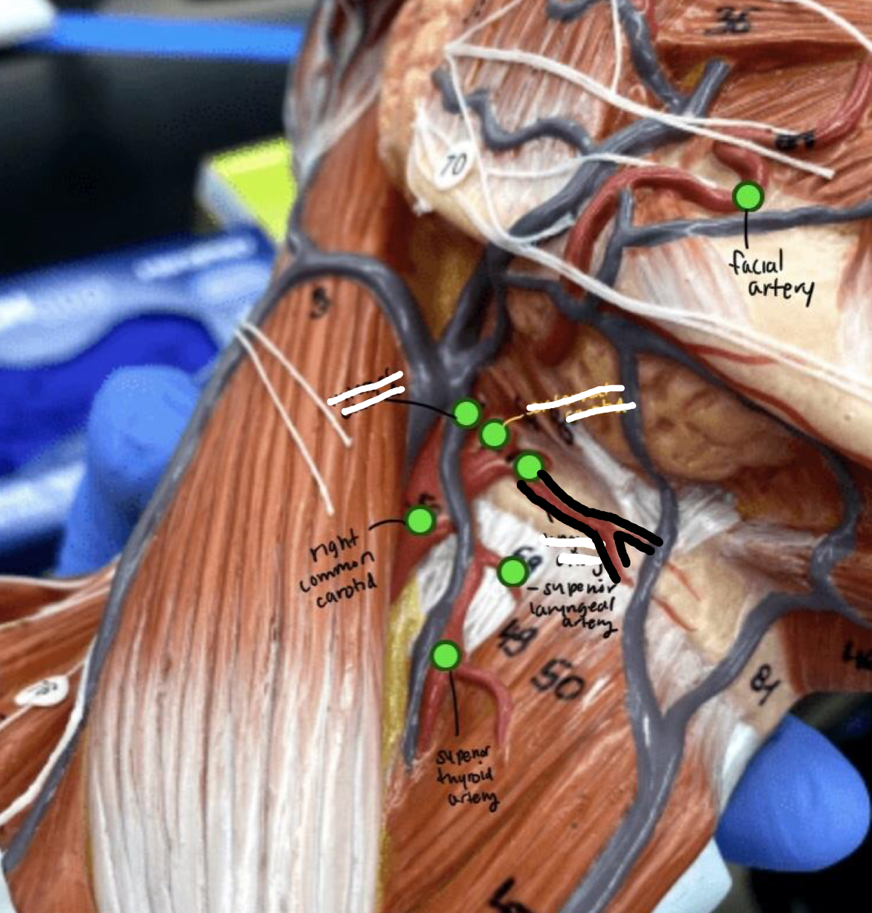
New cards
Head and neck
- Facial artery
- Facial artery
Nerve running along the cheek
86
New cards
Head and neck
- Lingual artery
- Lingual artery
The red artery forked off the external carotid artery
87
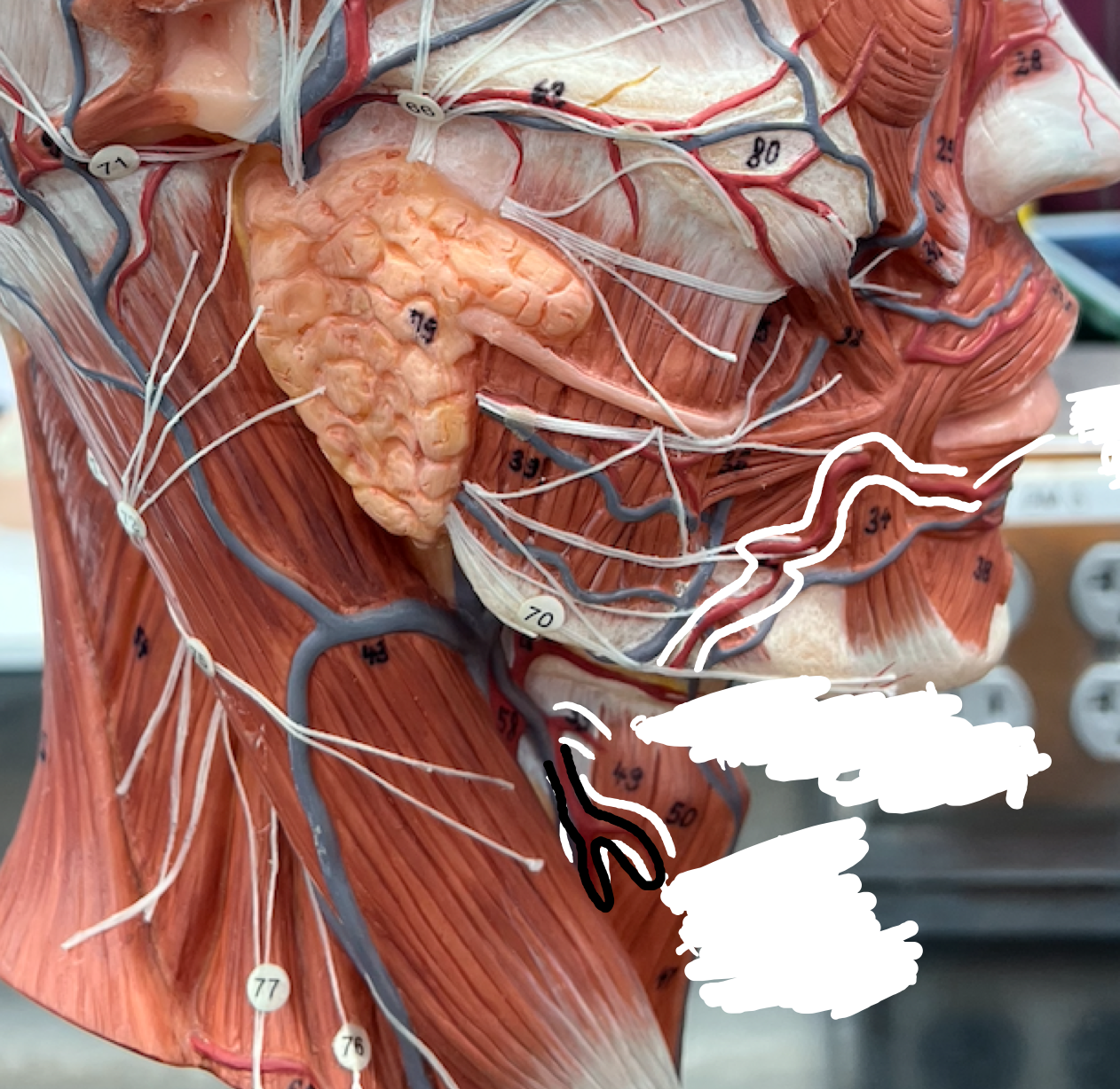
New cards
Head and neck
- Superior thyroid artery
- Superior thyroid artery
The downward forking red artery, coming down along the side neck muscle
88
New cards
Head and neck
- Superior laryngeal artery
- Superior laryngeal artery
The top forked part coming off the superior thyroid artery
89
New cards
Head and neck
- Subclavian arteries
- Subclavian arteries
The 2 red arteries starting near the clavicle and running down the shoulder area (left/right is always through the body's pov)
90
New cards
Head and neck
- Thyrocervical trunk: suprascapular artery
- Thyrocervical trunk: suprascapular artery
The one red artery on the body's right side, near the clavicle
91
New cards
Head and neck
- Brachiocephalic vein (right)
- Brachiocephalic vein (right)
- Upper thick part of the blue vein, left of the ascending aorta
- Superior of the heart
- Superior of the heart
92
New cards
Head and neck
- Brachiocephalic vein (left)
- Brachiocephalic vein (left)
A blue vein coming off of the superior vena cava
93
New cards
Thorax
- Internal thoracic (mammary) artery
- Internal thoracic (mammary) artery
Red lines above the diaphragms
94
New cards
Thorax
- Musculophrenic artery
- Musculophrenic artery
Arches across the top of the diaphragm
95
New cards
Thorax
- Superior epigastric artery
- Superior epigastric artery
The vertical red lines beneath the diaphragm and above the rectus sheath
96
New cards
Thorax
- Posterior intercostal artery
- Posterior intercostal artery
Red lines in between the intercostals
97
New cards
Thorax
- Azygous vein
- Azygous vein
Vein connected to the liver
98
New cards
Pelvic
- Common iliac artery and vein
- Common iliac artery and vein
The artery and vein closest to the arch of the pelvis
99
New cards
Pelvic
- External iliac artery and vein
- External iliac artery and vein
The downward sloping legs (artery + vein) of the pelvic arch
100
New cards
Pelvic
- Internal iliac artery and vein
- Internal iliac artery and vein
The little extensions coming off the artery/vein wishbone arch