Anatomy Exam 1
1/236
There's no tags or description
Looks like no tags are added yet.
Name | Mastery | Learn | Test | Matching | Spaced | Call with Kai |
|---|
No analytics yet
Send a link to your students to track their progress
237 Terms
neural tube, gut tube, urogenital system, body wall, cardiovascular loop
What are the 5 principle elements of chordates?
neural arch, vertebral body, costal process vertebral foramen
what are the four basic parts of all vertebrae?
7 cervical, 12 thoracic, 5 lumbar, 5 sacral, 3-5 coccyx
what are the 32 vertebrae?
the vertebral body gets bigger going down the column, until you reach the sacrum where it changes from bigger to smaller
what is the general trend for the vertebral body?
neural arches change orientation due to the regionalized movement requirements in each segment of the column
what is the general trend for the neural arch?
the costal processes are different at each level (ex. at thoracic its actually the rib)
what is the general trend for costal processes?
primary, convex out/hunchback, thoracic and sacral
what is kyphosis and where is it in the vertebral column?
seconday, concave in/swayback, cervical and lumbar
what is lordosis and where is it in the vertebral column?
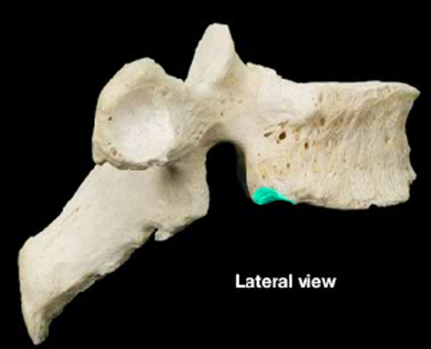
vertebral foramen
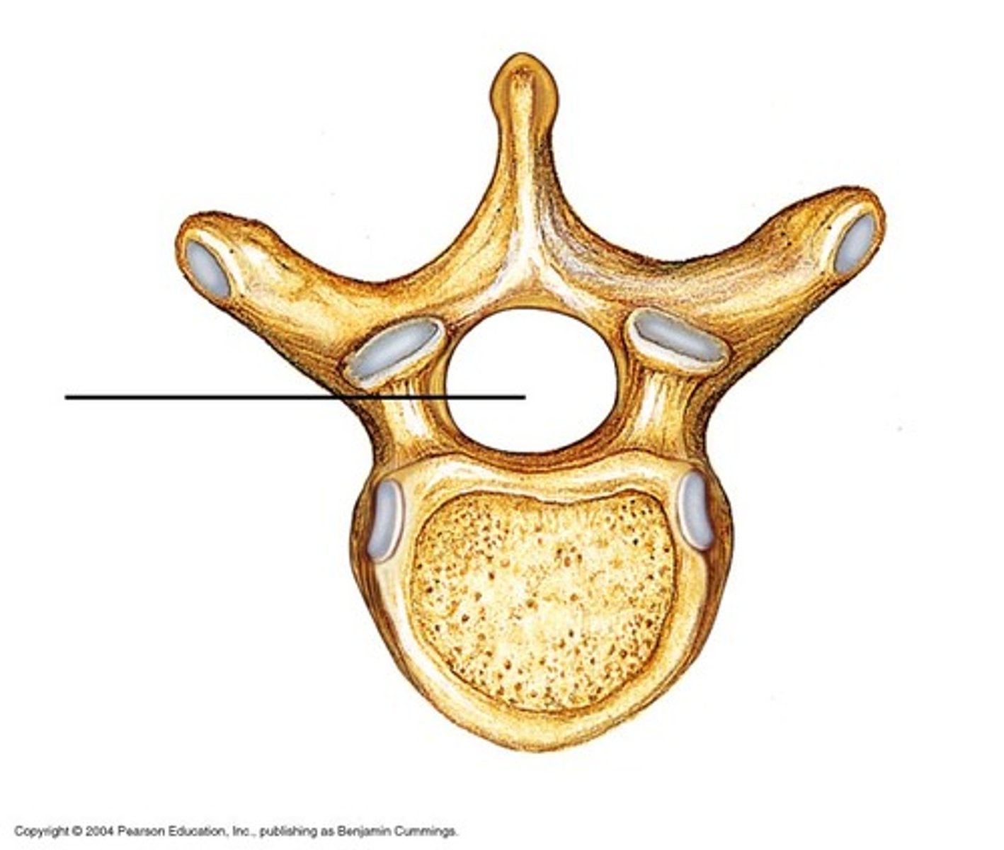
transverse process
lamina
pedicle
inferior articular facet
superior articular facet
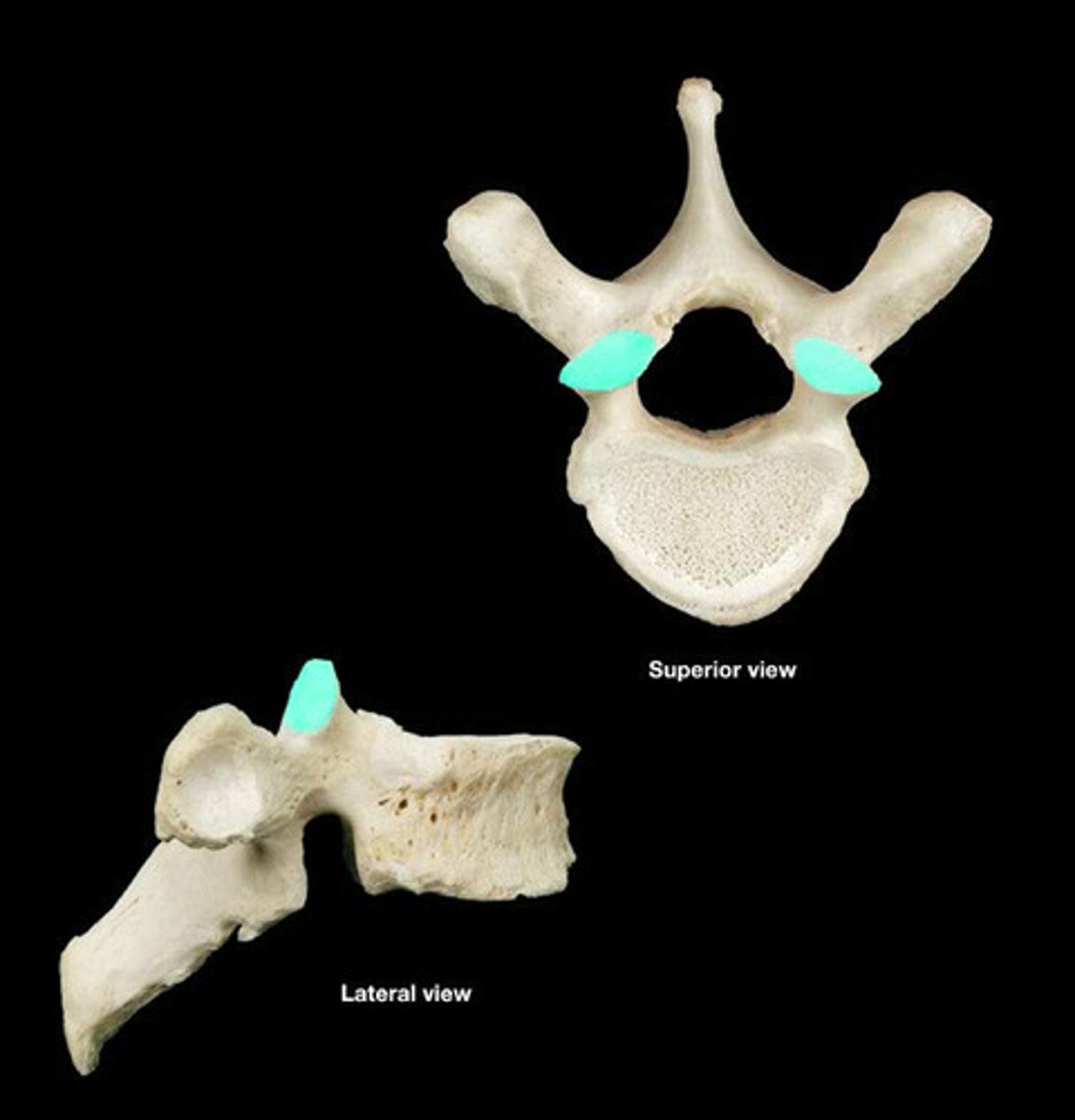
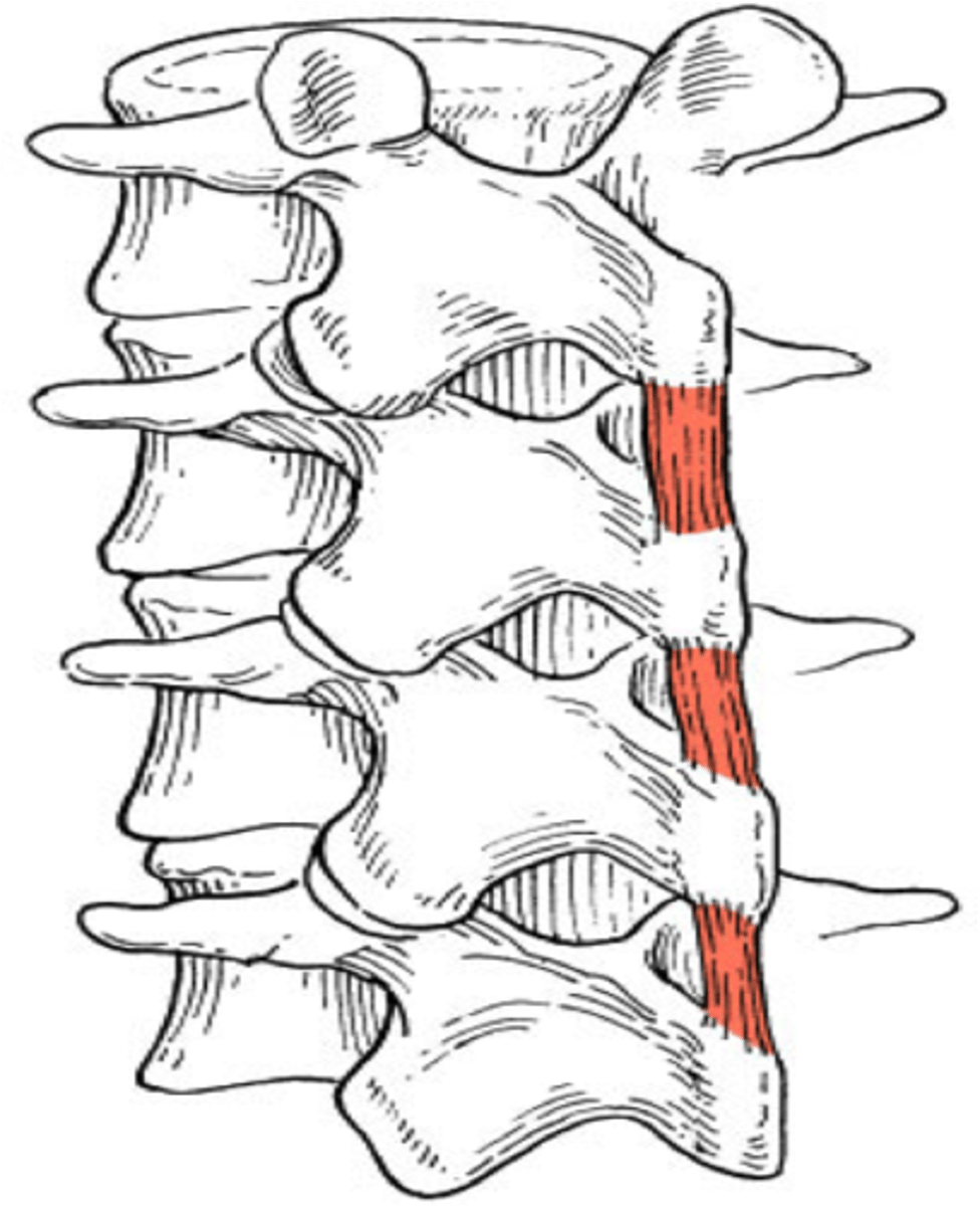
inferior vertebral notch
between vertebrae this forms the intervertebral foramen
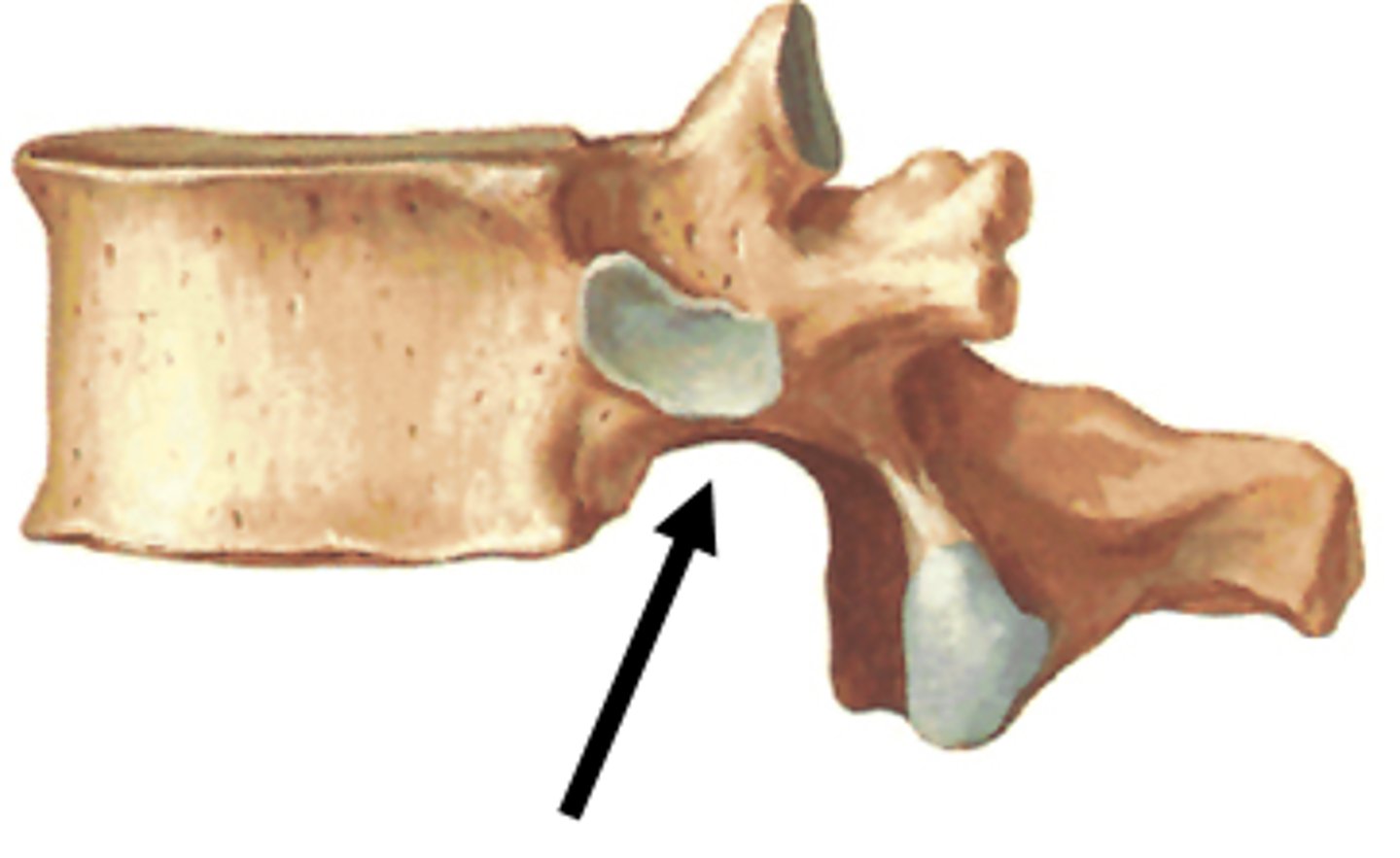
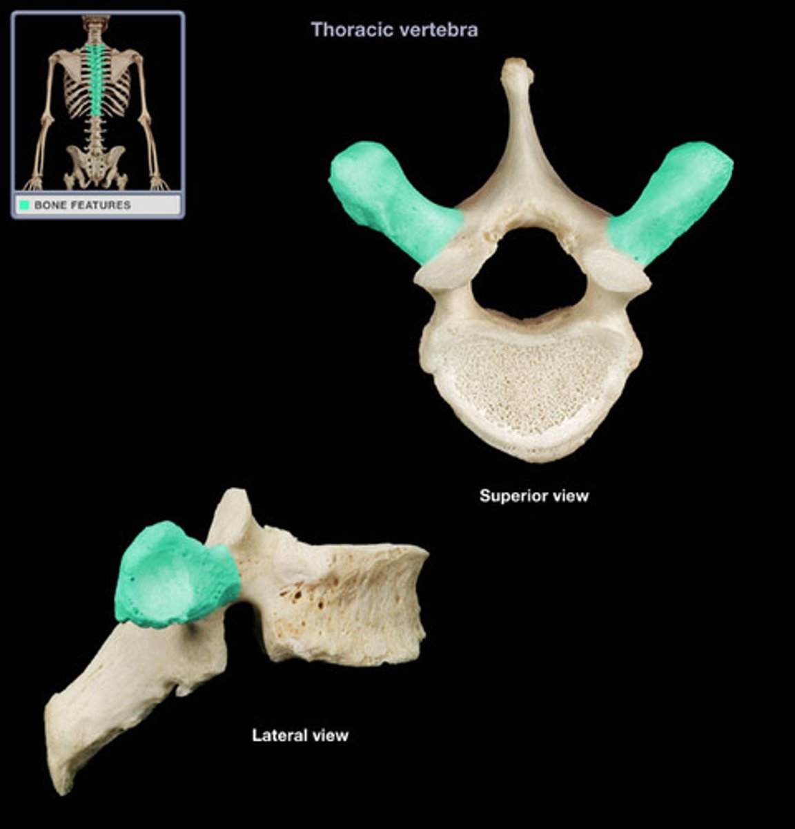
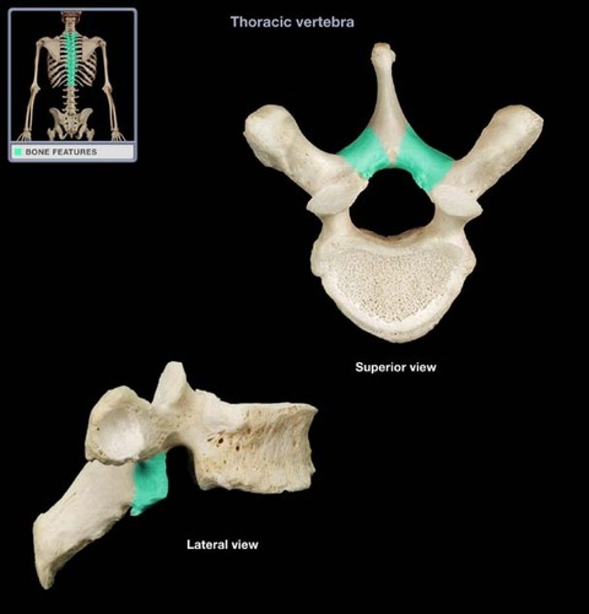
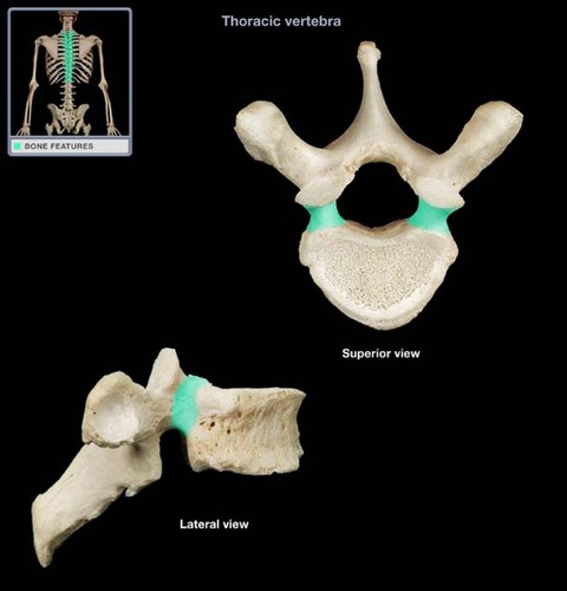
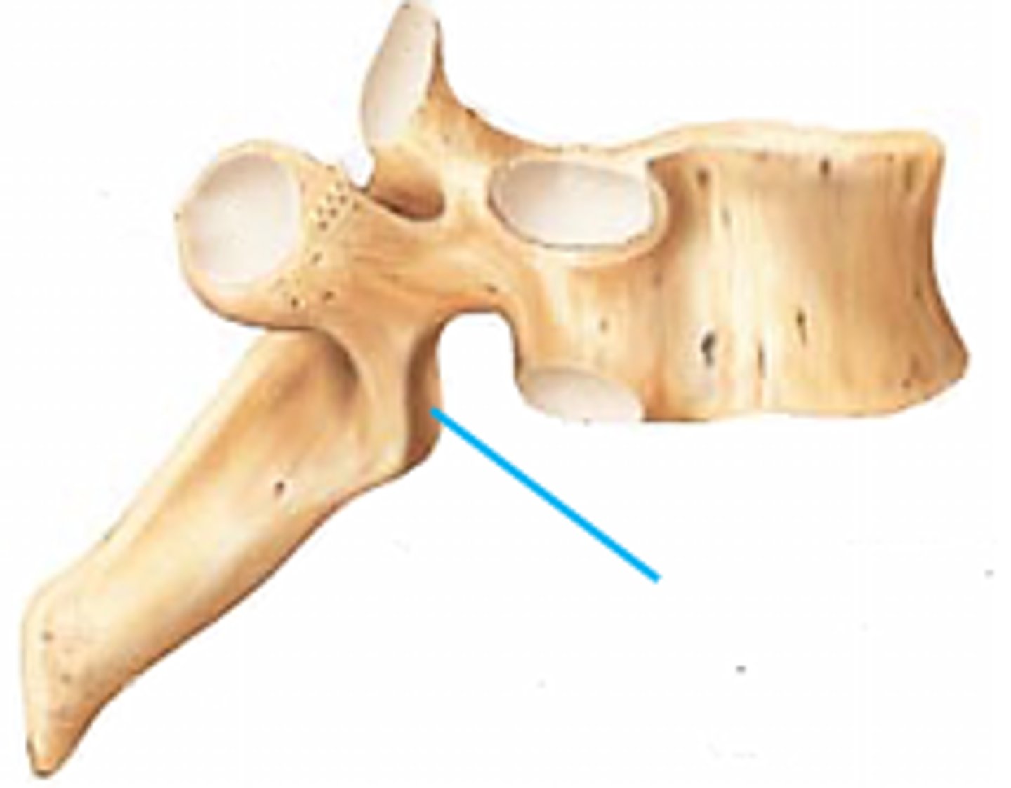
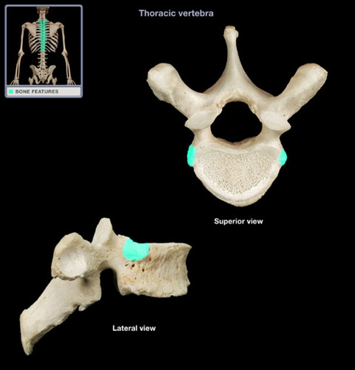
inferior costal facet of thoracic vertebrae
transverse costal facet of thoracic vertebrae
superior costal facet
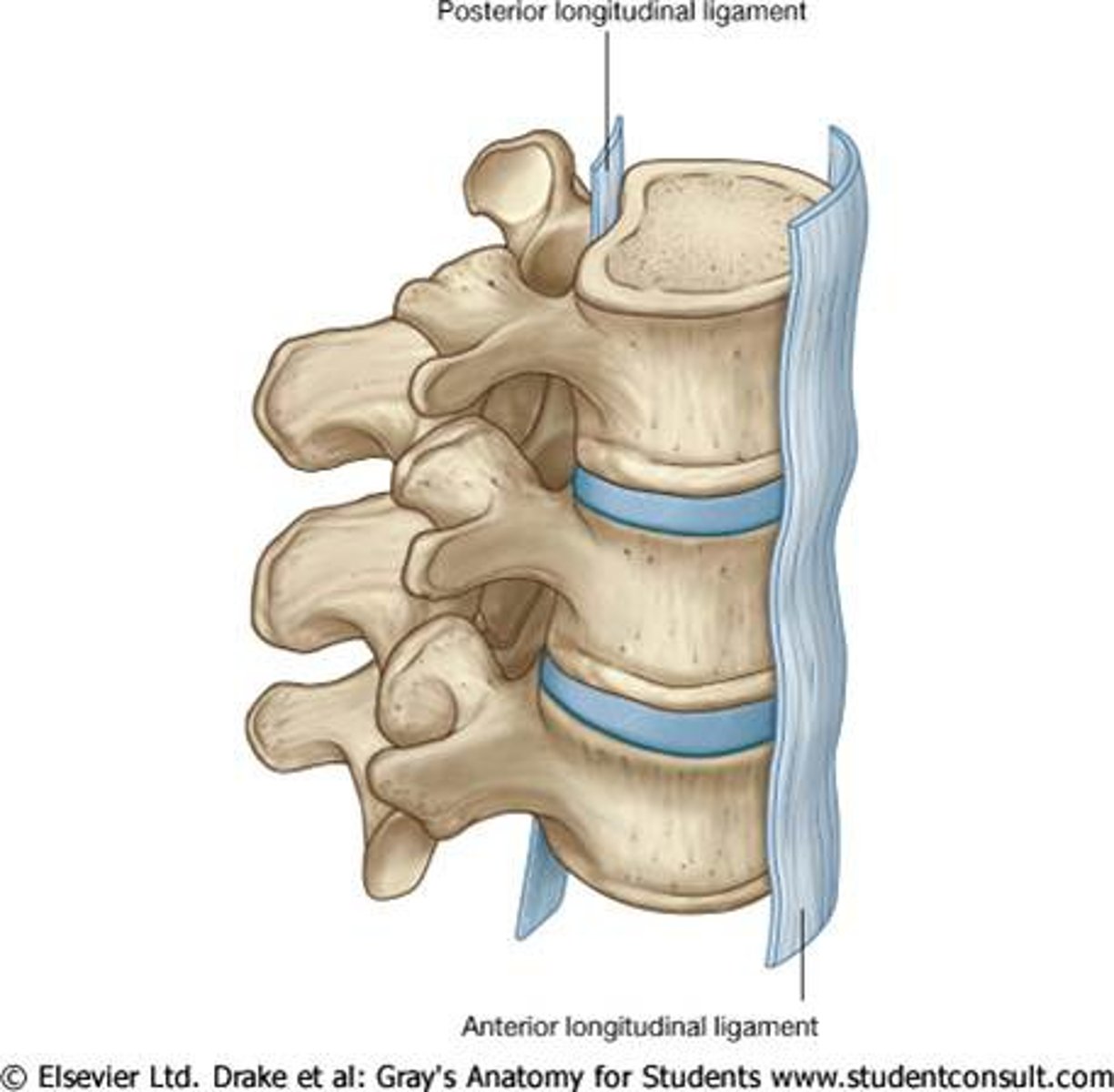
anterior/posterior longitudinal ligament
line the front/back of vertebral body
interspinous/spinous ligament
between the spinous processes
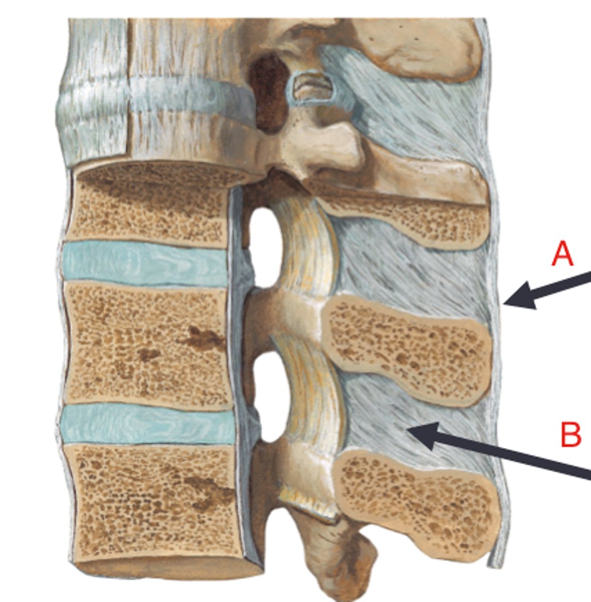
ligamentum flavum
connects the laminae of adjacent vertebrae, coated in fat
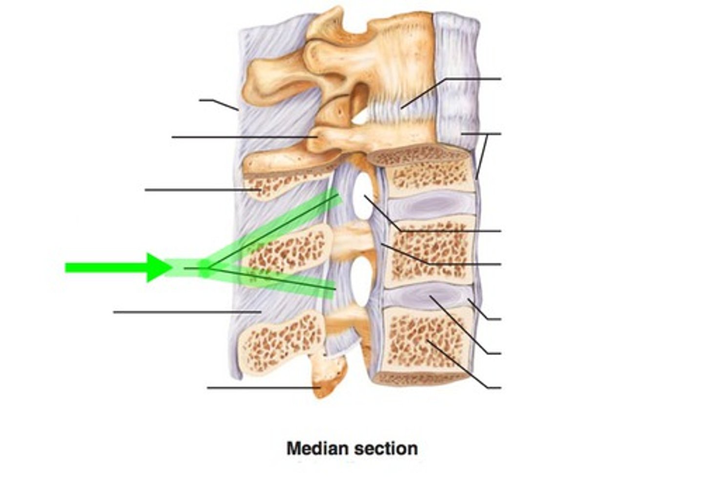
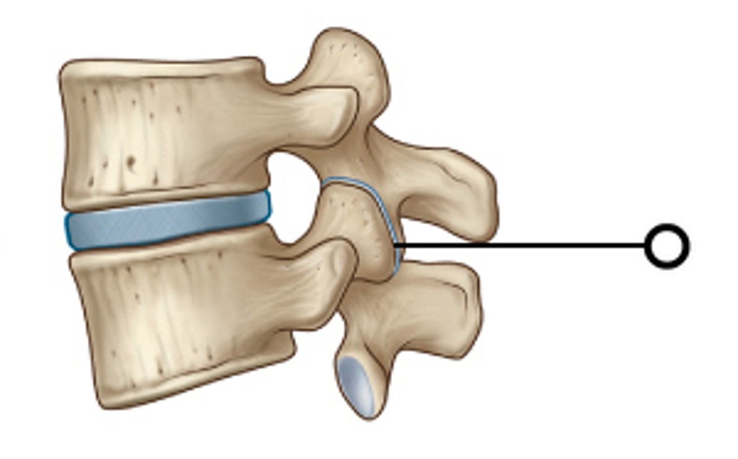
intervertebral facet joints
what are these synovial joints?
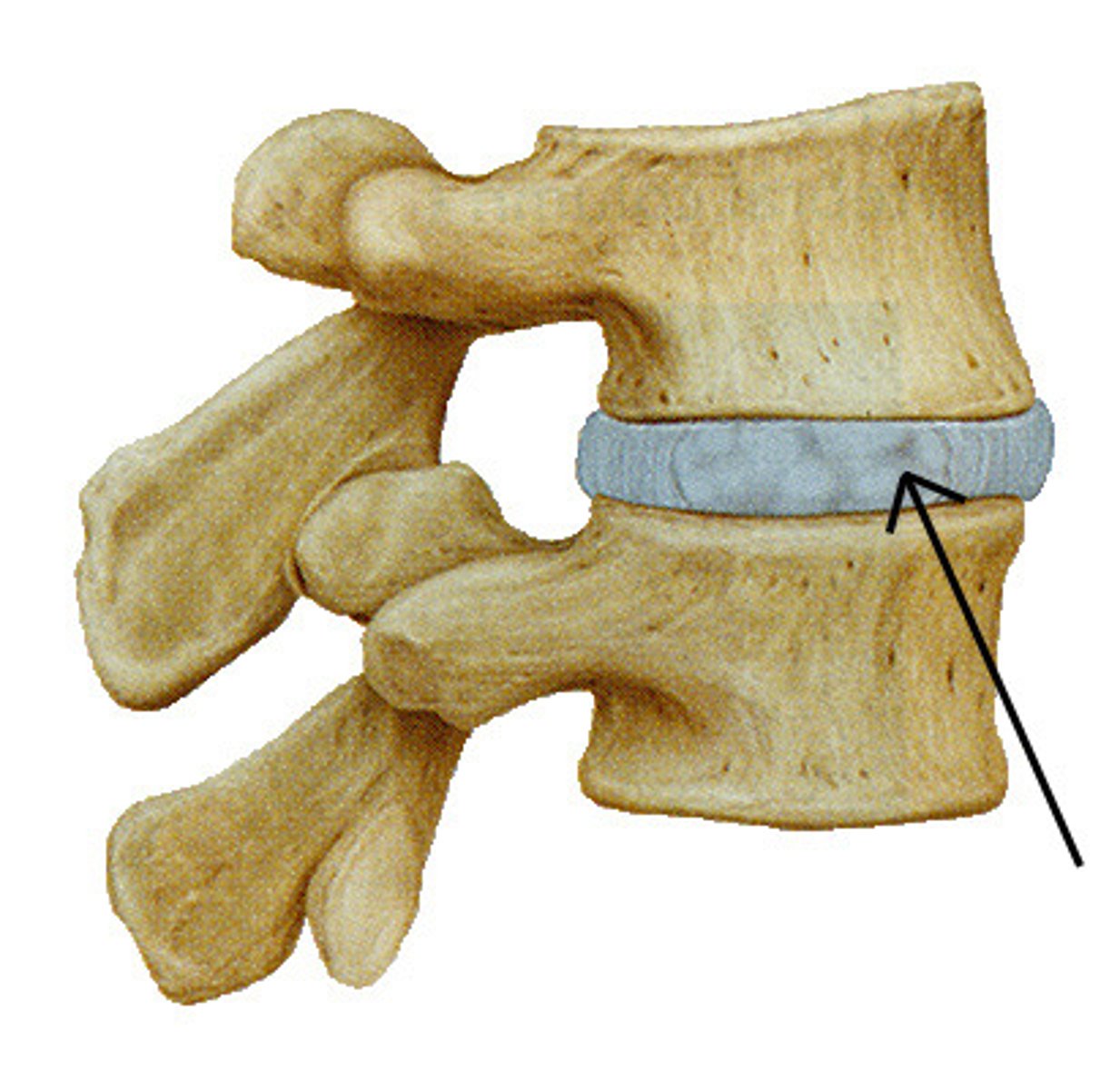
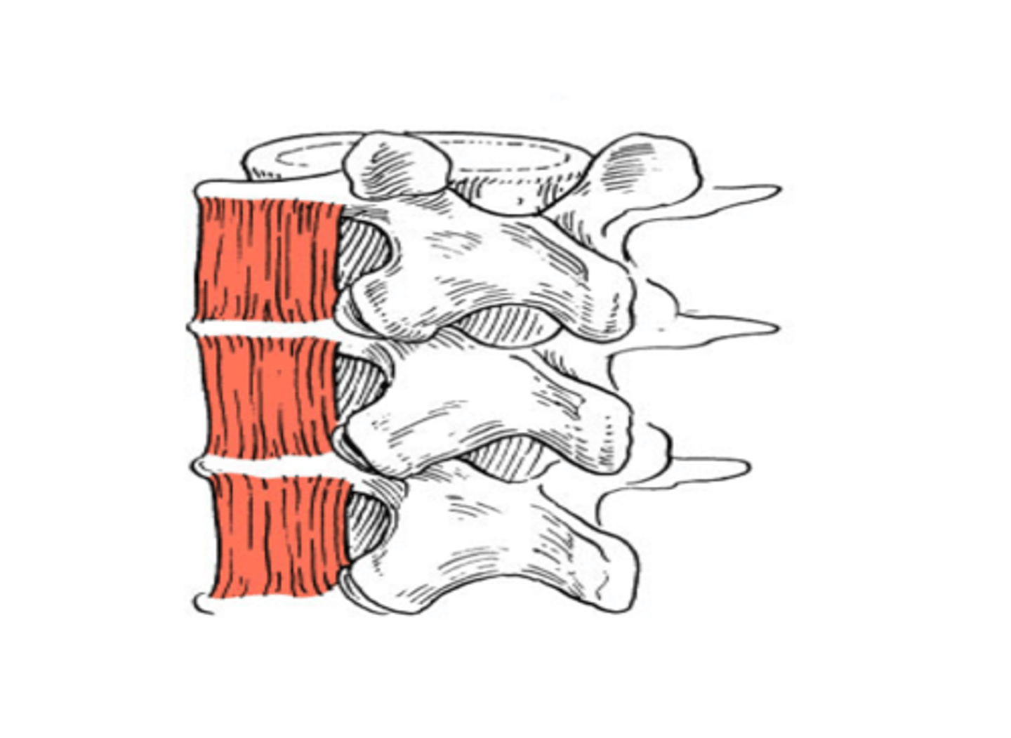
intervertebral discs (anulus fibrosus, nucleus polposus)
what are the fibrous "joints" between vertebral bodies
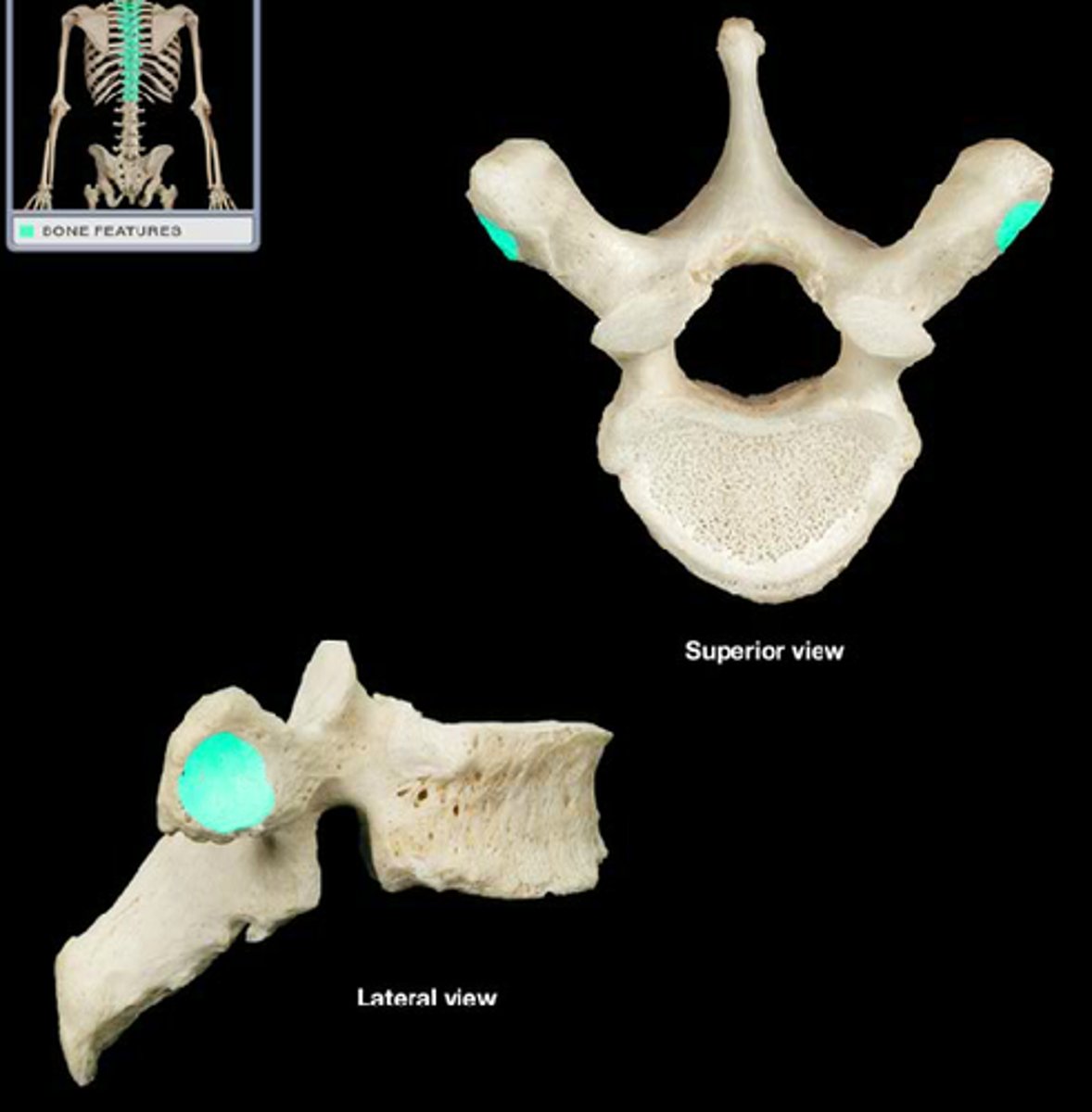
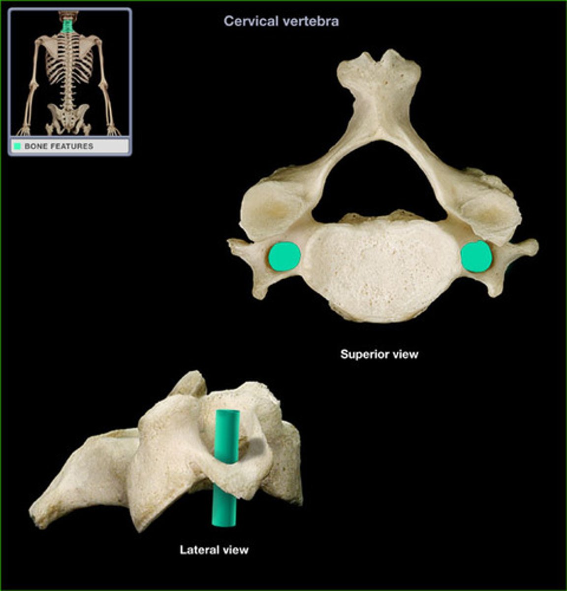
most mobile, least weight bearing, oblique articular facets, thick IV disc, forked spinous process, *transverse foramen*
what are the characteristics of cervical vertebrae?
transverse foramen (artery loops through except C7)
what foramen is unique to cervical vertebrae and what is its purpose?
least mobile (of 3), thin IV disc, AF nearly vertical
what are characteristics of the thoracic vertebrae?
permits rotation, limits flexion/extension (volume sensitive)
what are the movement qualities of thoracic vertebrae?
rib
what is the costal process of a thoracic vertebrae?
most weightbearing, cupped AF, broad SP (muscle attachment)
what are the characteristics of a lumbar vertebrae?
limited rotation, permits flexion and extension
what are the movement qualities of a lumbar vertebrae?
mammillary body
"original" transverse process is called what on a lumbar?
vertebral bodies fused together, extra foramina (sacral), transverse processes fuse to form wings (ala/CP)
what are the characteristics of the sacrum/coccyx?
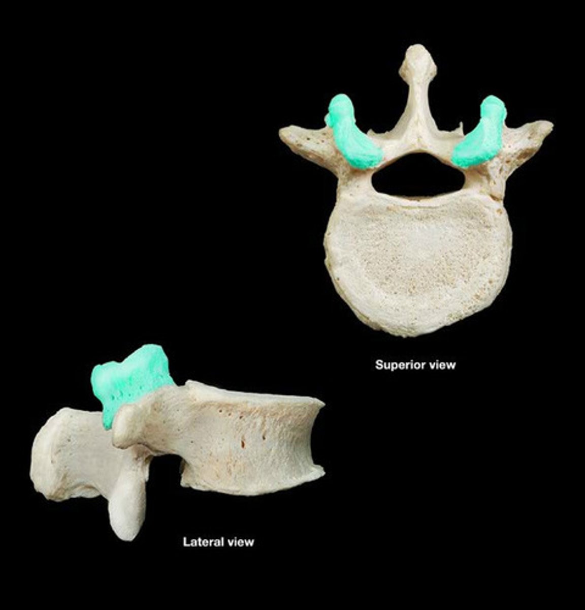
atlas
C1, no vertebral body
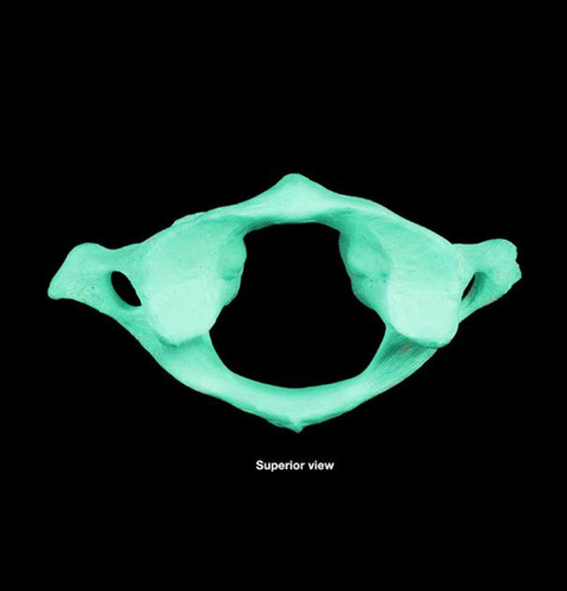
occipital bone, yes motion
what does atlas articulate with superiorly and what is its purpose?
dens
stolen verterbral body from atlas
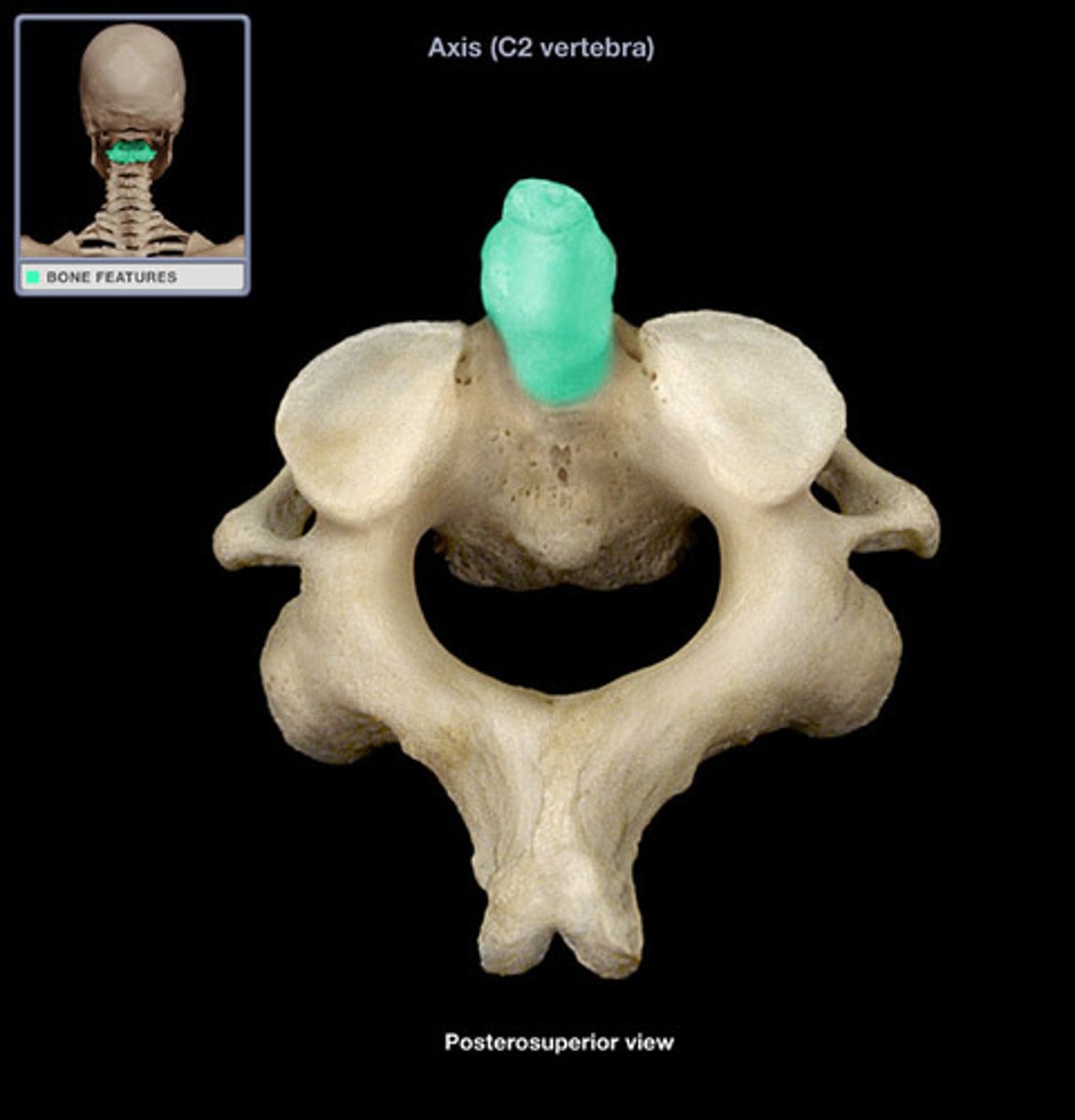
axis
C2
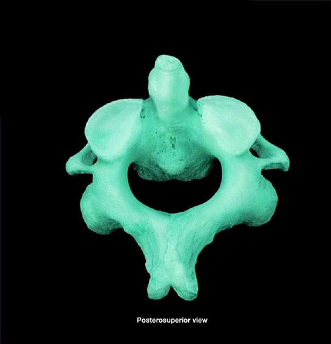
no motion
articulation of atlas and axis
alar ligament
modification of posterior longitudinal ligament for the dens of axis
epidermis/dermis
what are the two parts of the skin?
living layer, produces glands sends down to the dermis
what is the function of the epidermis?
connective tissue, holds glands and sensory neurons
what is the function of the dermis?
superficial fascia (hypodermis)
found between skin and muscles, contains adipose tissue, allows skin to glide
deep fascia
dense connective tissue on the surface of individual muscles, forms tendons
epaxial muscles
The muscles located posterior to the spinal cord, innervated by dorsal rami
hypaxial muscles
limb and superficial back muscles, invervated by ventral rami
bilateral and unilateral flexion
function of epaxial muscles of the back
dorsal rami
innervate epaxial muscles
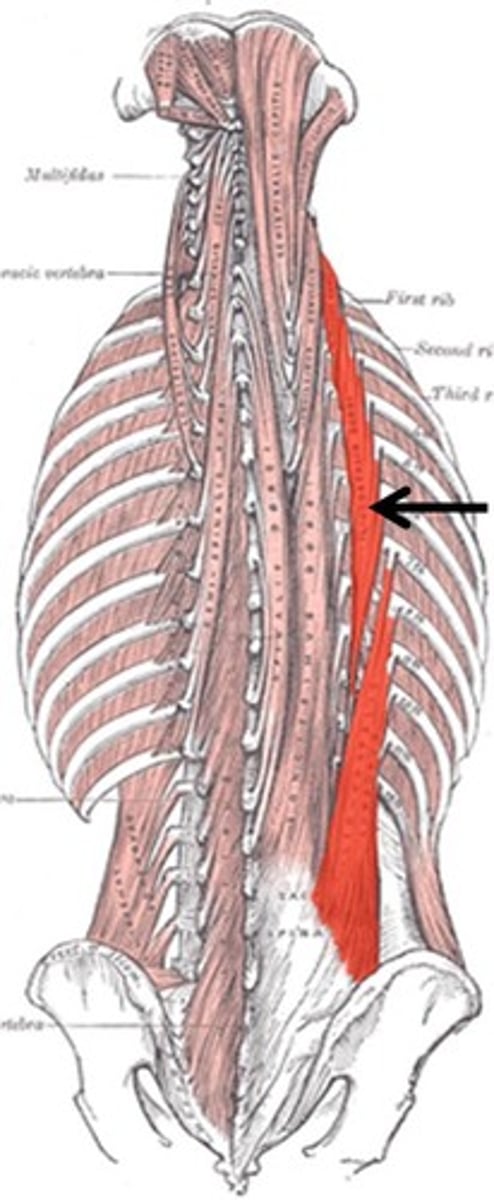
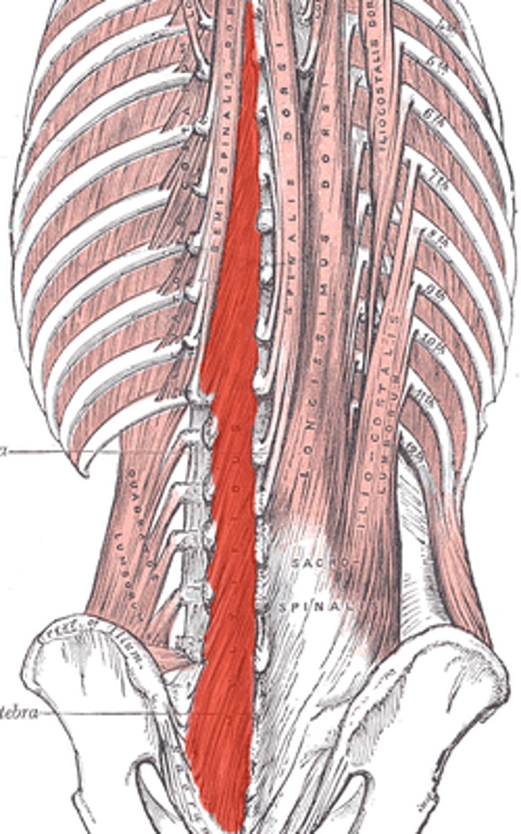
iliocostalis
O: sacrum, iliac crest, lumbar TP
I: angle of rib higher in the column
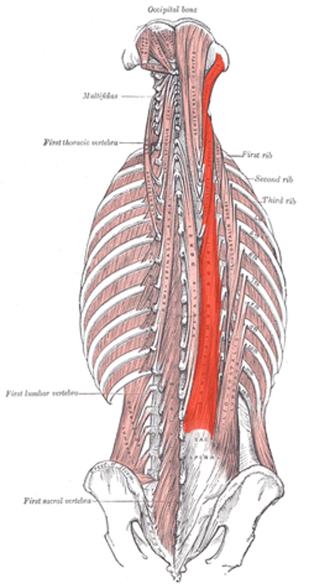
longissimus
O: sacrum, iliac crest, TP
I: TP up the column (till axis)
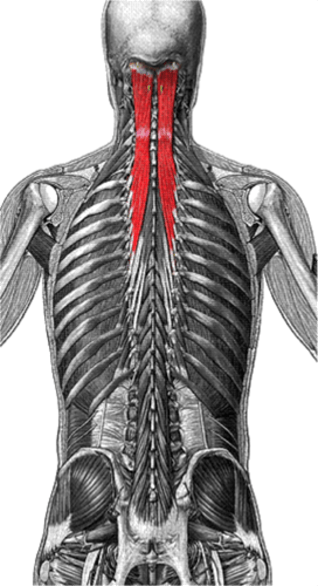
spinalis
O: lateral surface of SP
I: lateral surface of SP
iliocostalis, longissimus, spinalis
erector spinae group
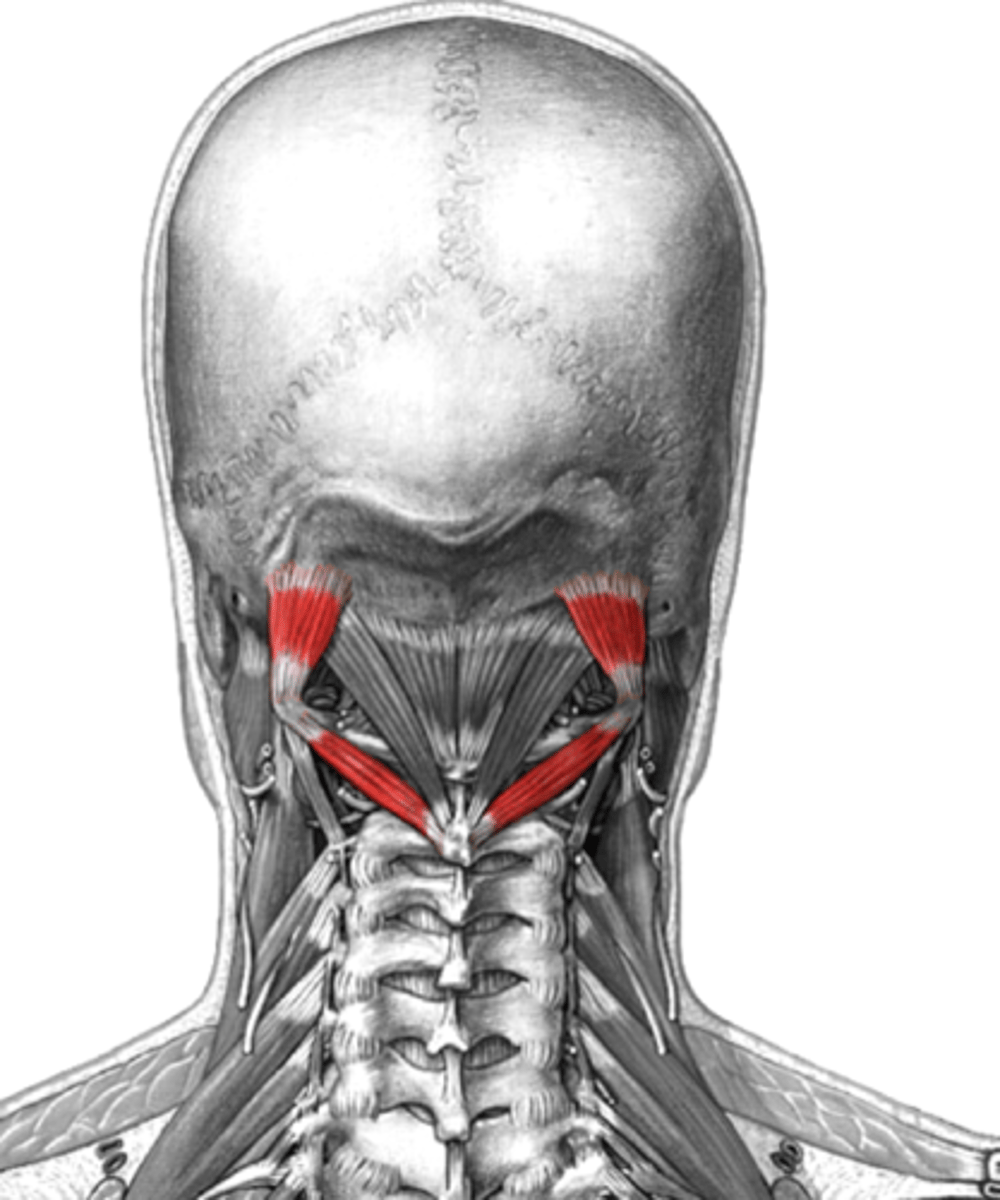
splenius
o: lateral surface of SP
i: TP, nuchal line, mastoid
downward chevron
semispinalis
o: TP
i: SP several vertebrae up the column
upward chevron
multifundus
o: sacrum, ilium, lumbar TP
i: SP at least 3 vertebrae away
Intertransversarii
o: lumbar TP
i: lumbar TP directly above
HYPAXIAL
interspinales
bridge between SPs of vertebrae
rotares
o: TP lower vertebrae
i: SP of 1 or 2 vertebrae above
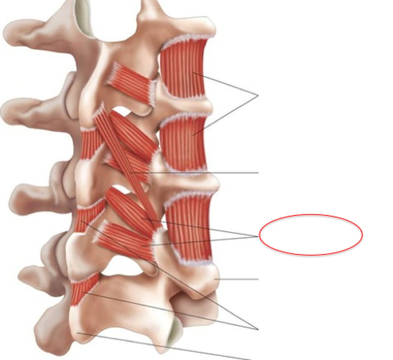
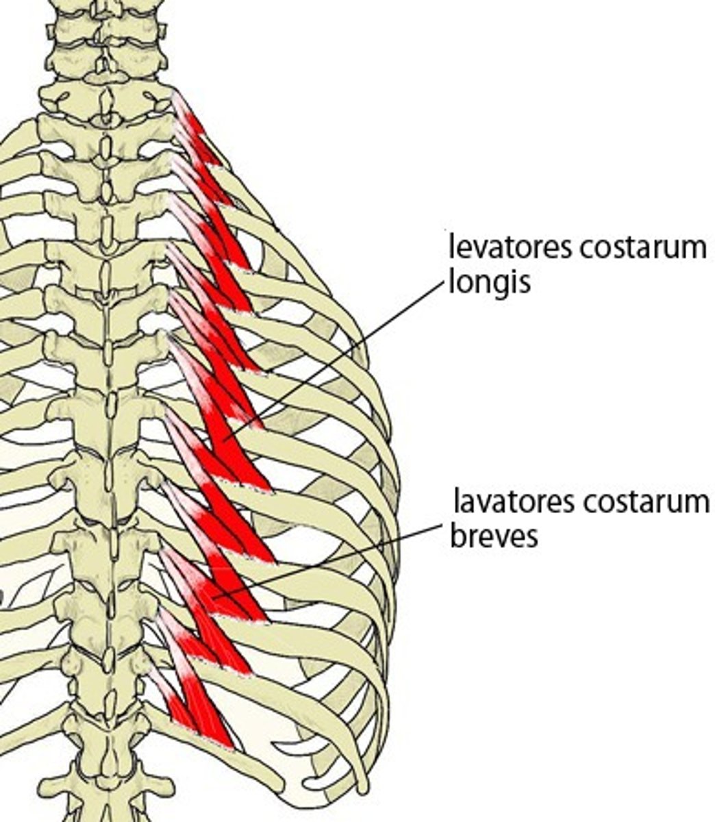
levatores costarum
o: thoracic TP
i: 1 or two ribs below
a: lift rib
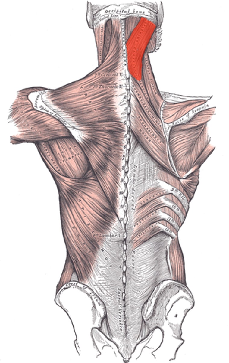
rectus capitis major
axis to skull, rocks head backwards
rectus capitis major and minor
rocks head backwards
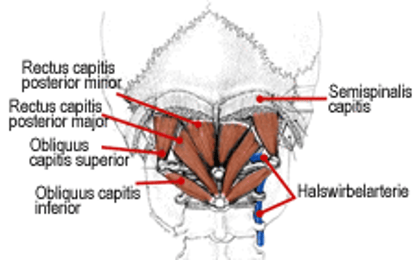
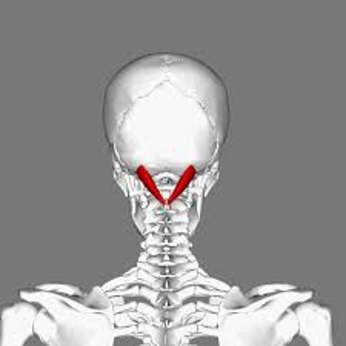
oliquus capitis superior and inferior
rotate head
the embryo
the junction between the endoderm and the ectoderm become?
somites, intermediate mesoderm, lateral plate mesoderm
three types of mesoderm
dermamyotome and sclerotome
somites differentiate into
dermatome (forming the dermis), myotome
dermamyotome differentiates into
epimere (epaxial) and hypomere (hypaxial)
what are the two forms of myotome
binds to notochord to make vertebrae
how do scleretomes develop further
partial superior sclerotome and partial inferior sclerotome fuse back together once developing neural tube punches through sclerotome and attach to myotome
why are there uneven number of spinal nerves to spinal vertebrae?
segmented spinal nerve
what is formed when developing neural tube punches through sclerotome?
dorsal ramus
what is the connection from neural tube to epimere called (innervating epaxial muscles)?
ventral ramus
what is the connection from neural tube to hypomere called (innervating hypaxial muscles)?
intermediate mesoderm
interacts with aorta to form 6 tubes that become kidneys, part of urinary system, and nongonads
somatic mesoderm, intraembryonic celom, and splanchnic mesoderm
what are the three aspects of lateral plate mesoderm
somatic mesoderm
pushed into body wall, becomes connective tissue (hypodermis), limb bones
intraembryonic celom
becomes the body cavity for internal organs, space between the somatic and splanchnic mesoderm
splanchnic mesoderm
becomes everything for organ other than inner lining (smooth muscle, mesentary, connective tissues, heart)
mesentery
double fold of the splanchnic mesoderm that encases gut tube and tethers it to the posterior body wall
somatic and visceral (autonomic)
subdivisions of the peripheral NS
somatic nervous system
conscious control of skeletal muscles, innervates body wall only, derived from SOMITES
somatic motor neurons
myelinated throughout, singular neuron from CNS to muscle, stimulates conscious motor function, travel down dorsal or ventral ramus
visceral nervous system
innervates unconscious part of body wall and internal organs
motor and sensory neurons
both visceral and somatic NS have
sympathetic and parasympathetic
subdivisions of the visceral nervous system
visceral motor nuerons
autonomic, always have two neurons involved, second neuron is unmyelinated
adrenal gland
what is the one exception to the two-neuron rule for visceral motor neurons
dorsal root ganglion
contains cell bodies of sensory neurons with fibers growing out
sensory neurons
to viscera: follow pathways laid out by sympathetic flow
to body walls: follow pathways of segmented spinal nerve to dermatome of epaxial and hypaxial
sympathetic chain ganglia, subdiaphragmatic/preaortic, and enteric
three types of visceral motor neurons
sympathetic, turn on cardiovascular loop and body wall, right next to vertebrae
sympathetic chain ganglion
lateral horn
what triggers the sympathetic chain ganglia
myelinated and preganglionic along segmental spinal nerve
connections from lateral horn to ganglia targets are
unmyelinated along segmental spinal nerve
chain ganglia grow connections to target that are
sympathetic, turn off gut tube and urogenital system, close to aorta
subdiaphragmatic/preaortic ganglion
lateral horn
what triggers subdiaphragmatic/preaortic ganglion
unmyelinated along blood vessels to these organs
subdiaphragmatic/preaortic ganglion grow connections to target organs that are
parasympathetic, turn on urine/digestion and turns off CV loop, in organ walls
enteric ganglion
vagus nerve
enteric ganglion are triggered by
myelinated and follow esophagus down to ganglion
connections from vagus nerve to enteric ganglion are
unmyelinated and very short (since already in organ walls)
enteric ganglion grow connections to target organs that are
releasing the egg, cleavage, blastocyst, gastrula, trilaminar, folding, amnion
what are the "stages" of embryogenesis