Nucleotides and nucleic acids
1/57
There's no tags or description
Looks like no tags are added yet.
Name | Mastery | Learn | Test | Matching | Spaced | Call with Kai |
|---|
No analytics yet
Send a link to your students to track their progress
58 Terms
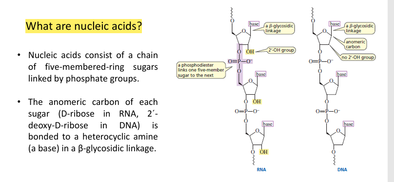
What are nucleic acids?
Nucleic acids are essential biomolecules present in all living organisms.
The term includes both DNA and RNA.
They store and transmit genetic information.
They control the synthesis of proteins and determine biological characteristics.
They are organic polymers made of nucleotides.
What is the basic structure of nucleic acids?
Nucleic acids are made of chains of nucleotides.
Each nucleotide contains a five-membered sugar linked by phosphate groups (phosphodiester bonds).
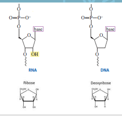
The sugars are:
D-ribose in RNA
2′-deoxy-D-ribose in DNA
The nitrogenous base is attached to the sugar at the anomeric carbon via a β-glycosidic bond.
What are the types of nucleic acids?
RNA (ribonucleic acid):
Contains ribose sugar
DNA (deoxyribonucleic acid):
Contains deoxyribose sugar
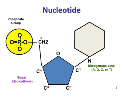
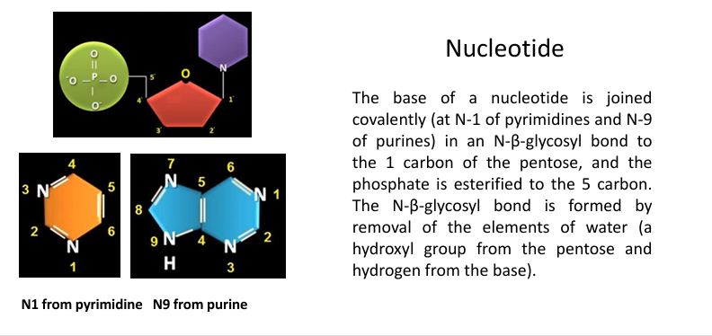
What is a nucleotide?
A nucleotide is an organic molecule composed of:
A nitrogenous base
A pentose sugar (ribose or deoxyribose)
A phosphate group
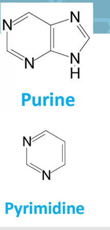
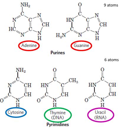
What are the nitrogenous bases of nucleotides?
Nitrogenous bases are derived from two groups:
Purines → Adenine (A), Guanine (G)
Pyrimidines → Cytosine (C), Thymine (T)
They are planar, aromatic, and heterocyclic molecules.
They have basic character due to nitrogen atoms with lone pairs.
The sequence of these bases determines genetic information.
What nitrogenous bases are found in DNA and RNA?
Both DNA and RNA contain the purines:
Adenine (A)
Guanine (G)
Both also contain the pyrimidine:
Cytosine (C)
The difference is:
DNA → Thymine (T)
RNA → Uracil (U)
What are the nitrogenous bases present in RNA and DNA?
RNA:
Adenine (A)
Guanine (G)
Cytosine (C)
Uracil (U)
DNA:
Adenine (A)
Guanine (G)
Cytosine (C)
Thymine (T)
👉 The key difference:
RNA uses uracil (U)
DNA uses thymine (T)
Which nucleotide bases are pyrimidines and which are purines?
Pyrimidines (single ring):
Cytosine (C)
Uracil (U)
Thymine (T)
Purines (double ring):
Adenine (A)
Guanine (G)
💡 Memory tip:
👉 “CUT = Pyrimidines | AG = Purines”
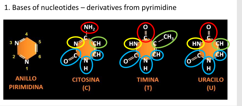
1. Bases of nucleotides – derivatives from pyrimidine
imagen
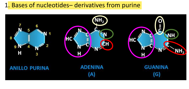
1. Bases of nucleotides– derivatives from purine
imagen
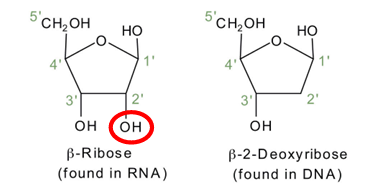
What sugars are found in nucleotides?
Nucleotides contain pentose sugars in a β-furanose (5-membered ring) form.
There are two types:
RNA → contains D-ribose
DNA → contains 2′-deoxy-D-ribose
👉 The key difference is that deoxyribose lacks an oxygen at the 2′ carbon.
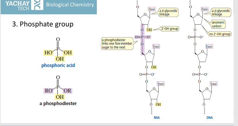
3. Phosphate group
imagen
How are the components of a nucleotide connected?
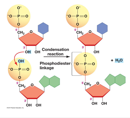
The nitrogenous base is attached to the sugar by an N-β-glycosidic bond:
At N-1 in pyrimidines
At N-9 in purines
This bond connects the base to the C-1′ of the pentose sugar.
The phosphate group is attached to the C-5′ of the sugar by an ester bond.
The glycosidic bond forms through a condensation reaction (loss of water).
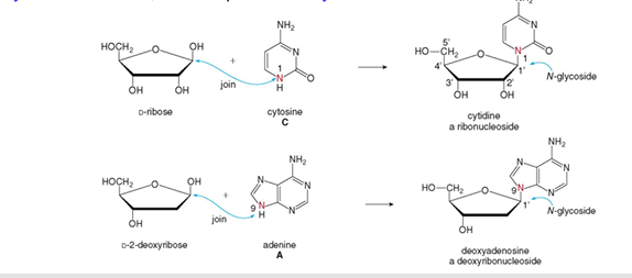
What is a nucleoside?
A nucleoside is composed of:
A nitrogenous base
A pentose sugar (ribose or deoxyribose)
It does NOT contain a phosphate group.
Types:
Ribonucleoside → contains ribose (RNA)
Deoxyribonucleoside → contains deoxyribose (DNA)
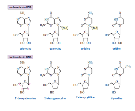
How are nucleosides named?
Nucleosides derived from pyrimidine bases use the suffix:
👉 “-idine” (e.g., cytidine, uridine)Nucleosides derived from purine bases use the suffix:
👉 “-osine” (e.g., adenosine, guanosine)For DNA forms, add the prefix:
👉 “deoxy-” (e.g., deoxyadenosine)
How are nucleotides formed from nucleosides?
A nucleoside (base + sugar) becomes a nucleotide when phosphate groups are added.
This process is called phosphorylation and is catalyzed by kinases.
Phosphate groups are attached to the –OH group of the sugar (usually at the 5′ carbon).
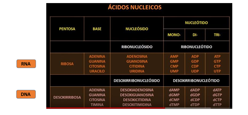
Depending on the number of phosphates:
Monophosphate (1) → e.g., AMP
Diphosphate (2) → e.g., ADP
Triphosphate (3) → e.g., ATP
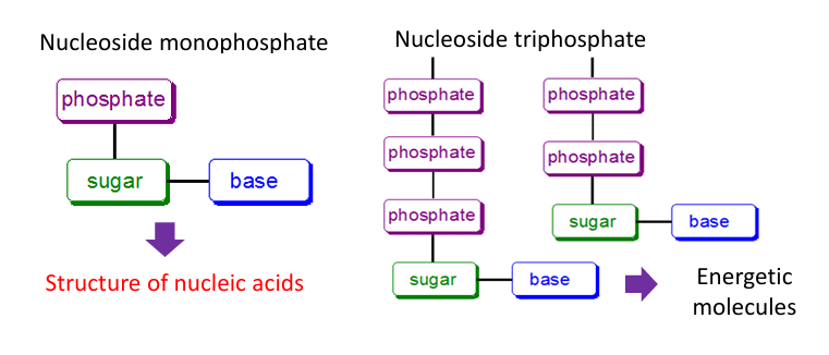
What are the different forms of nucleotides and their functions?
Nucleoside monophosphate (NMP):
Contains 1 phosphate group
Main role → structure of nucleic acids (DNA & RNA)
Nucleoside triphosphate (NTP):
Contains 3 phosphate groups
Main role → energy molecules (e.g., ATP)
👉 The extra phosphate bonds store high energy used in cellular processes.
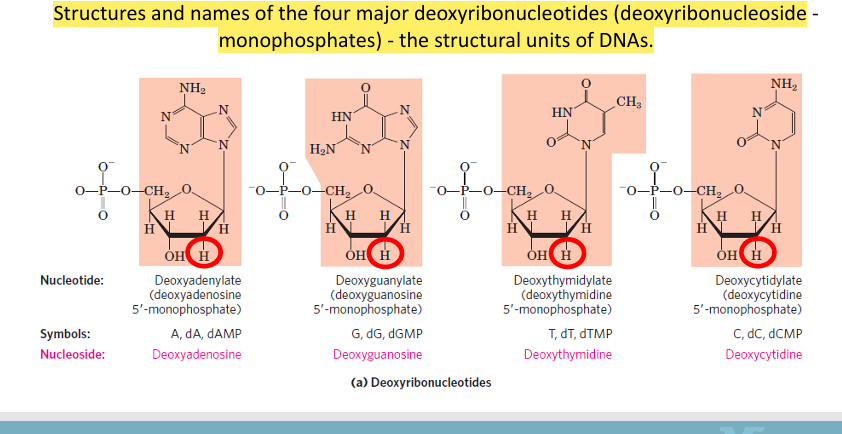
What are the four major deoxyribonucleotides in DNA?
The four structural units of DNA are:
dAMP (Deoxyadenylate) → Adenine (A)
dGMP (Deoxyguanylate) → Guanine (G)
dTMP (Deoxythymidylate) → Thymine (T)
dCMP (Deoxycytidylate) → Cytosine (C)
👉 All contain:
Deoxyribose sugar (with H at 2′ instead of OH)
A phosphate group
A nitrogenous base
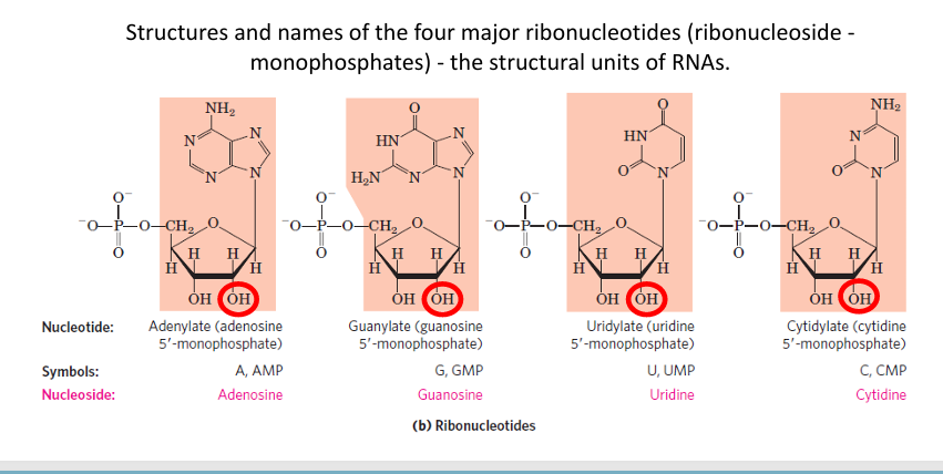
What are the four major ribonucleotides in RNA?
The four structural units of RNA are:
AMP (Adenylate) → Adenine (A)
GMP (Guanylate) → Guanine (G)
UMP (Uridylate) → Uracil (U)
CMP (Cytidylate) → Cytosine (C)
👉 All contain:
Ribose sugar (with OH at 2′ carbon)
A phosphate group
A nitrogenous base
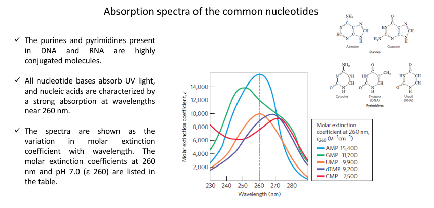
Why do nucleotides absorb UV light and at what wavelength?
The nitrogenous bases (purines and pyrimidines) are highly conjugated molecules.
Because of this conjugation, they absorb UV light strongly.
Nucleic acids show a characteristic absorption at:
👉 ~260 nmThis property is used to:
Quantify DNA and RNA
Study their purity and concentration
What are the biological functions of nucleotides?
Structural role → building blocks of DNA and RNA
Energy transfer → e.g., ATP (energy coupling in metabolism)
Cell signaling → e.g., cAMP (second messenger)
Coenzymes components → e.g., NAD⁺, FAD
How are bases, nucleosides, and nucleotides named in DNA and RNA?
🔹 Purines:
Adenine (A)
Nucleoside: Adenosine (RNA) / Deoxyadenosine (DNA)
Nucleotide: Adenylate (AMP) / Deoxyadenylate (dAMP)
Guanine (G)
Nucleoside: Guanosine / Deoxyguanosine
Nucleotide: Guanylate (GMP) / Deoxyguanylate (dGMP)
🔹 Pyrimidines:
Cytosine (C)
Nucleoside: Cytidine / Deoxycytidine
Nucleotide: Cytidylate (CMP) / Deoxycytidylate (dCMP)
Thymine (T) (DNA only)
Nucleoside: Thymidine (deoxythymidine)
Nucleotide: Thymidylate (dTMP)
Uracil (U) (RNA only)
Nucleoside: Uridine
Nucleotide: Uridylate (UMP)
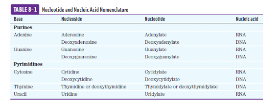
Nomenclature of nucleotides and nucleosides
imagen
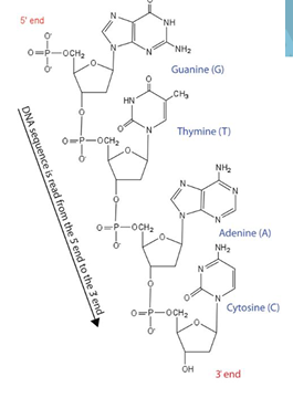
What are phosphodiester bonds in nucleic acids?
Phosphodiester bonds are covalent bonds that link nucleotides together in DNA and RNA.
They form when the 5′ phosphate group of one nucleotide connects to the 3′ hydroxyl group of the next nucleotide.
This creates the sugar-phosphate backbone of nucleic acids.
How are nucleic acids classified according to the number of nucleotide subunits?
Nucleic acids are long strands made of nucleotide subunits.
Dinucleotide → 2 nucleotides
Oligonucleotide → 3 to 10 nucleotides
Polynucleotide → many nucleotides
DNA and RNA are polynucleotides.
What are the four levels of nucleic acid structure?
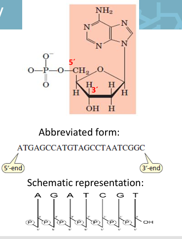
Primary structure: linear sequence of nucleotides linked by 3′–5′ phosphodiester bonds. It carries genetic information.
Secondary structure: regular folding or helix formed mainly by hydrogen bonding between bases.
DNA → usually double helix
RNA → hairpin loops, stems, and bulges
Tertiary structure: full 3D folding of the molecule, such as the L-shape of tRNA.
Quaternary structure: interactions with other molecules, such as DNA + histones = chromatin.
What is the primary structure of a nucleic acid?
The primary structure is the linear sequence of nucleotides.
Nucleotides are linked by phosphodiester bonds forming a polymer.
The order of bases determines the genetic information.
The sequence is always written in the 5′ → 3′ direction.
What defines the primary structure of a nucleic acid and why is it important?
The primary structure is the linear sequence of nucleotides (A, G, C, T/U).
Nucleotides are linked by 3′–5′ phosphodiester bonds.
This sequence:
Stores genetic information
Determines all higher levels of structure (secondary, tertiary, quaternary)
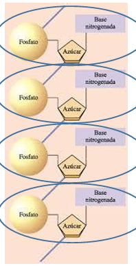
What is the backbone of nucleic acids like?
Nucleotides are linked by phosphodiester bonds.
Each phosphodiester bond connects the 5′ carbon of one sugar to the 3′ carbon of the next sugar.
This forms an asymmetric sugar-phosphate backbone.
The nitrogenous bases project outward as side groups and define the sequence.
DNA and RNA backbones are hydrophilic.
At physiological pH, phosphate groups are negatively charged, so nucleic acids are polyanions.
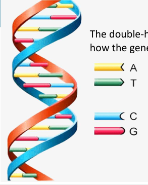
What is the secondary structure of DNA?
DNA forms a right-handed double helix.
It has two DNA strands wound around the same axis.
The sugar-phosphate backbone is on the outside and is hydrophilic.
The nitrogenous bases are stacked inside the helix and are mostly hydrophobic.
Bases from opposite strands pair with each other.
The two strands are antiparallel and complementary, not identical.

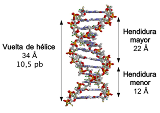
What are the key structural features of the DNA double helix?
DNA strands form a right-handed double helix.
The helix has a diameter of about 20 Å.
The bases are almost perpendicular to the helix axis.
There are about 10.5 base pairs per turn.
The distance between stacked bases is 3.4 Å.
One full turn has a pitch of about 34 Å.
DNA has a major groove where backbones are far apart and a minor groove where they are closer.
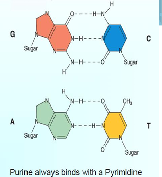
How do bases pair in the secondary structure of DNA?
DNA strands are complementary.
Adenine (A) pairs with Thymine (T).
Guanine (G) pairs with Cytosine (C).
A–T forms 2 hydrogen bonds.
G–C forms 3 hydrogen bonds.
A higher G:C ratio makes DNA harder to separate because G–C pairs are more stable.
What are Chargaff’s rules and why are they important for DNA structure?
Chargaff’s rules describe important patterns in DNA base composition:
DNA base composition varies between species.
DNA from different tissues of the same species has the same base composition.
Base composition does not change with age, nutrition, or environment.
In cellular DNA:
A = T
G = C
The DNA sequence varies among species and individuals.
These rules helped explain the complementary base pairing in DNA.
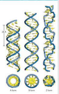
What are the different forms of DNA?
DNA can occur in different structural forms:
B-DNA:
Most common and stable form under physiological conditions
Right-handed double helix
About 10.5 base pairs per turn
A-DNA:
Favored in low-water conditions
Right-handed helix
Wider than B-DNA
About 11 base pairs per turn
Bases are tilted about 20°
Z-DNA:
Left-handed helix
More slender and elongated
About 12 base pairs per turn
Backbone has a zigzag shape
May be involved in gene regulation or recombination
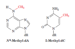
How can nucleic acid bases be modified?
Some DNA bases can be chemically modified by methylation.
Examples:
Adenine (dA) → N6-methyl-dA
Cytosine (dC) → 5-methyl-dC
DNA methylation can affect gene function, especially when it occurs in promoter regions.
Usually, methylation in promoters represses gene transcription.
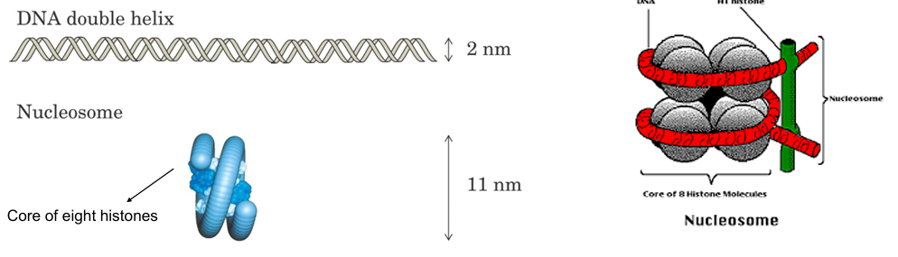
Where are DNA and RNA located in the cell?
DNA and RNA are polymers found mainly in the nucleus of cells.
In human cells, about 2 meters of DNA are packed inside the nucleus.
DNA is packaged into nucleosomes.
Nucleosomes are further compacted with histone proteins into larger 30 nm fibers.
This packaging helps organize DNA while allowing replication and transcription.
What is the higher structure of DNA?
DNA coils around proteins called histones.
Histones are rich in basic amino acids.
DNA is acidic/negatively charged because of its phosphate groups.
Basic histones and acidic DNA attract each other.
This interaction forms a chain of nucleosomes, helping compact DNA inside the nucleus.
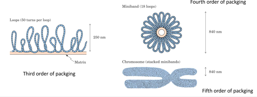
Higher structure of DNA
Chromatin: condensed nucleosomes
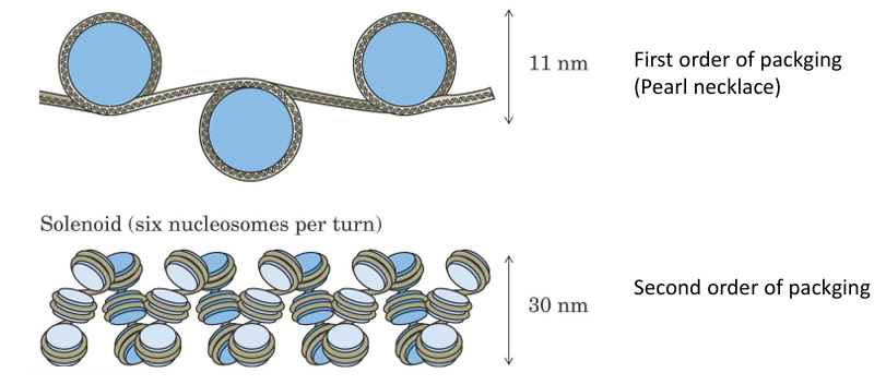
Higher structure of DNA
Chromatin fibers are organized into loops, and the loops into the bands that provide the superstructure of chromosomes.
What is the difference between a chromosome and a gene?
Humans have 46 chromosomes, organized into 23 pairs.
Each chromosome is made of DNA and contains many genes.
A gene is a specific segment of DNA that carries the information needed to synthesize one protein.
What are the main biological functions of DNA?
DNA has four main biological functions:
Replication: DNA copies itself before cell division.
Coding: DNA contains the instructions to make proteins.
Cellular differentiation: DNA helps cells specialize into different types.
Evolution and adaptation: DNA changes through mutations, allowing variation and adaptation over time.
What are the main biological functions of DNA?
Replication: DNA makes nearly error-free copies of itself before cells divide.
Coding: DNA contains codons, groups of three bases, that direct protein synthesis by coding for amino acids.
Cellular differentiation: DNA regulates which proteins are produced, allowing cells with the same genetic material to become different cell types.
Evolution and adaptation: DNA can mutate, and some mutations may be inherited, producing genetic variation over time.
What is genetic inheritance?
Genetic inheritance is the transmission of hereditary information from parents to offspring through the genetic material found in the cell nucleus.
This information determines anatomical, physiological, and other characteristics.
Genotype: the complete set of transmissible genetic traits fixed in the genes.
Phenotype: the visible or external expression of those traits in the individual.
How does the double-helical structure of DNA explain replication?
The DNA double helix explains replication because each strand acts as a template for a new complementary strand.
The base sequence of one strand determines the sequence of the other strand.
When the two strands separate, each original strand guides the formation of a new strand.
Each daughter DNA molecule contains:
One original parental strand
One newly synthesized strand
This is called semiconservative replication.
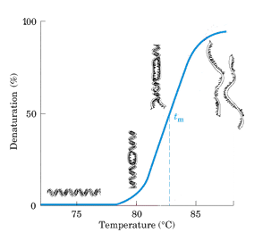
What is DNA melting?
DNA melting is the separation of the two DNA strands when hydrogen bonds between base pairs are disrupted.
It can occur by:
Heating
Adding acid or alkali
Action of helicase enzymes inside cells
Key terms:
Tm: temperature at which half of the DNA double helix is separated.
Annealing: renaturation process where complementary strands rejoin when temperature decreases.
Helicases: proteins that use ATP to separate DNA strands in cells.
What happens during DNA replication by polymerases?
During DNA replication, the two strands of the DNA double helix must be separated locally so each strand can act as a template.
DNA polymerases synthesize the new complementary DNA strands.
The process occurs in a region called the replication fork.
The replisome is the molecular machine that carries out DNA replication.
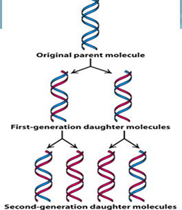
What are the main characteristics of DNA replication?
DNA replication is:
Semiconservative: each daughter DNA has one old strand and one newly synthesized strand.
Bidirectional: replication starts at the origin of replication (ORI) and proceeds in two opposite directions, forming a replication bubble and forks.
Semidiscontinuous: DNA polymerase only synthesizes in the 5′ → 3′ direction.
This produces:
Leading strand: synthesized continuously toward the replication fork.
Lagging strand: synthesized discontinuously away from the fork in Okazaki fragments, each starting with an RNA primer.
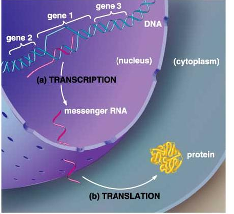
What is RNA in gene expression?
RNA is a polymer made of four ribonucleotides:
Adenine (A)
Cytosine (C)
Guanine (G)
Uracil (U)
RNA participates in gene expression, helping transfer genetic information from DNA to protein synthesis.
What are the main types of RNA and their functions?
There are three main types of RNA:
Messenger RNA (mRNA): carries the instructions from DNA to synthesize a protein.
Transfer RNA (tRNA): brings amino acids to the ribosome during protein synthesis.
Ribosomal RNA (rRNA): forms part of the ribosome and helps in protein synthesis.
What is mRNA and what is its function?
Messenger RNA (mRNA) is produced by transcription of a gene and carries the information needed to synthesize a specific polypeptide chain.
Each gene can produce a specific mRNA.
mRNA contains a sequence of codons.
Codons are read during translation to join amino acids in the correct order.
mRNA is metabolically unstable, so it has a short life inside the cell.
Continuous protein synthesis requires continuous mRNA production by transcription
How does transcription produce mRNA from DNA?
RNA polymerase binds to the promoter at the beginning of a gene.
DNA unwinds locally, forming two single strands.
The template strand (antisense) is read in the 3′ → 5′ direction.
RNA is synthesized in the 5′ → 3′ direction.
The mRNA sequence is complementary to the template strand and similar to the coding strand, but RNA uses U instead of T.
After mRNA is released, the DNA double helix reforms.
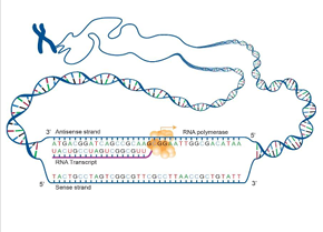
How is mRNA complementary to DNA during transcription?
During transcription, mRNA is complementary to the template strand of DNA.
Because the coding strand and the mRNA are both complementary to the template strand, they have almost the same sequence.
The only difference is:
DNA coding strand uses T (thymine)
mRNA uses U (uracil) instead of T
What do you need to understand translation?
To understand translation from mRNA to protein, you must know:
Genetic code: the set of rules that explains how mRNA codons correspond to specific amino acids.
tRNA: transfer RNA that carries specific amino acids to the ribosome and matches them with the correct mRNA codons.
During translation, the ribosome reads the mRNA codons and tRNA brings the correct amino acids to build a protein.
What is the genetic code?
The genetic code is the set of rules that cells use to translate mRNA sequences into proteins.
Every 3 bases in mRNA form a codon.
Each codon specifies one amino acid.
Codons are read in the 5′ → 3′ direction.
The code has 64 codons.
AUG is the start codon and codes for methionine (Met).
UAA, UAG, and UGA are stop codons and signal the end of protein synthesis.
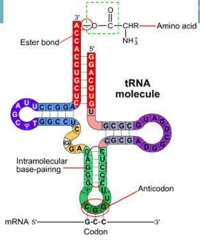
What is tRNA and what is its function?
tRNA (transfer RNA) is a small RNA molecule that carries specific amino acids to the ribosome during protein synthesis.
Key points:
It has about 70–90 nucleotides.
It folds into a structure with three loops and paired regions.
The 3′ end has ACC and a free –OH group, where the amino acid attaches.
The anticodon is a sequence of three nucleotides that pairs with the complementary mRNA codon.
This ensures the correct amino acid is added to the growing protein.
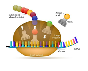
What is translation and how does it produce proteins?
Translation is the process by which mRNA is decoded into a protein at the ribosome (in the cytoplasm).
The ribosome reads mRNA codons (3 bases at a time).
tRNA brings the correct amino acids based on codon–anticodon pairing.
The ribosome links amino acids together forming a polypeptide chain.
This results in the synthesis of a specific protein.
What are ribosomes and what is their function?
Ribosomes are macromolecular complexes made of proteins and ribosomal RNA (rRNA).
They are the translation centers of the cell because they synthesize proteins using the information carried by mRNA.
DNA information is transcribed into mRNA.
mRNA goes to the ribosome.
The ribosome reads the mRNA and builds a protein.
What are the three phases of translation?
Initiation: the ribosome assembles around the mRNA, and the first tRNA binds to the start codon (AUG).
Elongation: new tRNAs bring amino acids, and peptide bonds form between them.
Termination: translation stops when a stop codon is reached: UAA, UAG, or UGA, and the completed protein is released.