Anesthesia and Pain Control 1: Anatomical Considerations for Local Anesthesia- Dr. Synan
1/72
There's no tags or description
Looks like no tags are added yet.
Name | Mastery | Learn | Test | Matching | Spaced | Call with Kai |
|---|
No analytics yet
Send a link to your students to track their progress
73 Terms
Olfactory
Sensory
CN I
Optic
Sensory
CN II
Oculomotor
Motor
CN III
Trochlear
Motor
CN IV
Trigeminal
Mixed:
- V1: Sensory
- V2: Sensory
- V3: Sensory, Motor
CN V
Abducens
Motor
CN VI
Facial
Motor
CN VII
Auditory
Sensory
CN VIII
Glossopharyngeal
Mixed
CN IX
Vagus
Mixed
CN X
Accessory
Motor
CN XI
Hypoglossal
Motor
CN XII
Provides sensation of smell
Innervates olfactory mucosa in the roof of the nose
Olfactory:
Provides sensation of vision
Innervates the retina
Optic:
Moves eye up, down, and medially, and raises eyelid; Carries parasympathetics to pupil and ciliary muscle; constricts pupil and accommodates lens
Innervates:
Superior, inferior, and medial rectus muscle, inferior oblique muscles, levator palpebrae superioris muscle
Oculomotor:
Moves eye down and lateral
Innervates superior oblique muscle
Trochlear:
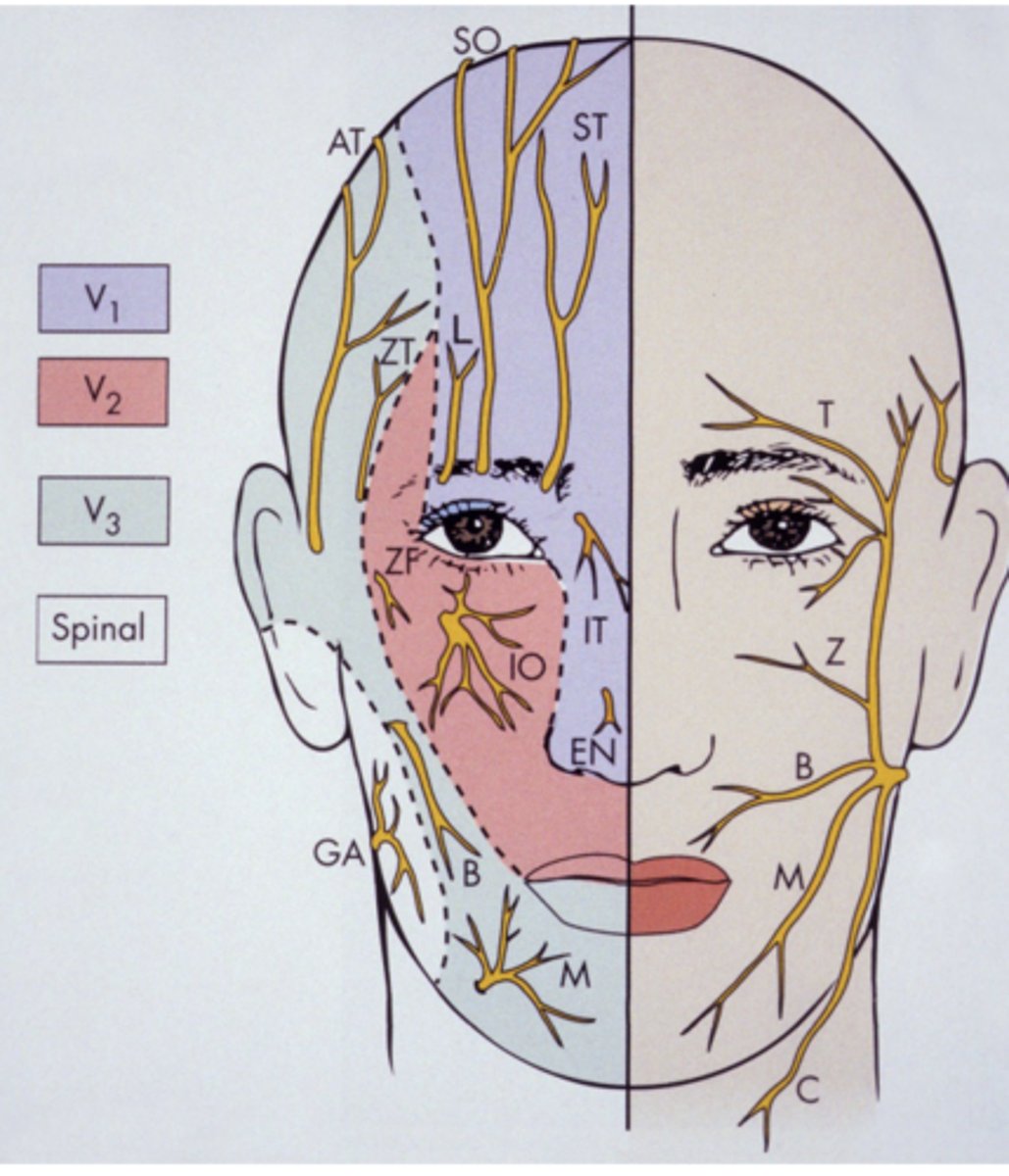
Mixed
V1 = Sensory
V2 = Sensory
V3 = Sensory, motor
Trigeminal
Moves eye laterally
Innervates lateral rectus muscle
Abducens
5 branches:
- Temporal
- Zygomatic
- Buccal
- Marginal Mandibular
- Cervical
Innervates muscles of facial expression
- The chorda tympani branch provides taste sensation to the anterior 2/3 of the tongue. Travels along with the lingual nerve
Facial:
Provides sensation of hearing and equilibrium
Innervates cochlear and semicircular canals of inner ear
Auditory:
Provides taste and sensation to posterior 1/3 of tongue
Carries parasympathetic fibers to parotid gland
Elevates pharynx during swallowing
Innervates stylopharyngeus muscle
Glossopharyngeal:
Provides parasympathetic innervation to influence breathing, heart rate, digestion, etc
Innervates thoracic and abdominal organs, heart, lungs, upper digestive tract
Also innervates skeletal muscle of pharynx and larynx which assists in swallowing
Vagus:
Innervates SCM, trapezius, larynx muscles
The spinal portion of the nerve innervates SCM/Trapezius which influences shoulder and neck movement
The Accessory cranial fibers innervate intrinsic laryngeal muscles of the larynx which controls the vocal cords
Accessory:
Innervates hyoglossus, styloglossus, genioglossus, geniohyoid, and intrinsic muscles of tongue
Hypoglossal:
Smaller than sensory root
Exits cranium through foramen oval with V3, the mandibular division of the sensory root of trigeminal nerve
It then enters the infratemporal fossa
Trigeminal Nerve motor root
1. Masticatory
- masseter
- temporalis
- medial pterygoid
- lateral pterygoid
2. Mylohyoid
3. Anterior belly of digastric
4. Tensor tympani
5. Tensor veli palatine
Trigeminal nerve- motor root innervates the following muscles
Ophthalmic division V1:
- Exits skull via superior orbital fissure into the orbit
Maxillary division V2:
- Exits skull via foramen rotundum into the pterygopalatine fossa
Mandibular division V3:
- Exits skull via Foramen oval into infratemporal fossa along with motor root of the trigeminal nerve
Trigeminal nerve- sensory root
Pure sensory
Smallest division
Divdes into 3 branches
- nasociliary nerve
- frontal nerve
- lacrimal nerve
Supplies eyeball, conjunctiva lacrimal gland, mucous membrane of nose and paranasal sinuses and skin of forehead, eyelids, and nose
Ophthalmic division V1:
Supplies mucous membrane of nasal septum and lateral wall of nasal cavity
Ethmoidal and sphenoid sinuses
Iris, cornea
Skin of lacrimal sac
Skin of nose
Ophthalmic Division V1- Nasociliary Nerve Branch
Largest branch of ophthalmic division
Divides into supratrochlear and supraorbital N.
Supplies conjunctiva and skin of upper eyelid, skin of forehead, anterior scalp and mucus membrane of frontal sinus
Ophthalmic Division V1- Frontal nerve branch
Smallest branch of ophthalmic division
Supplies skin and conjunctiva of lateral aspect of upper eyelid, forehead, and lacrimal gland
Ophthalmic Division V1- Lacrimal Nerve Branch
Pure sensory
Intermediate in size
Supplies:
1. Dura mater within cranium
2. Skin of
- Middle portion of face
- Lateral forehead
- Cheek bone
- Lower eyelid
- Side of nose
- Upper lip
3. Mucous membrane
- Nasopharynx
- Maxillary sinus
- Soft palate
- Tonsil
- Hard palate
4. Maxillary teeth and periodontium
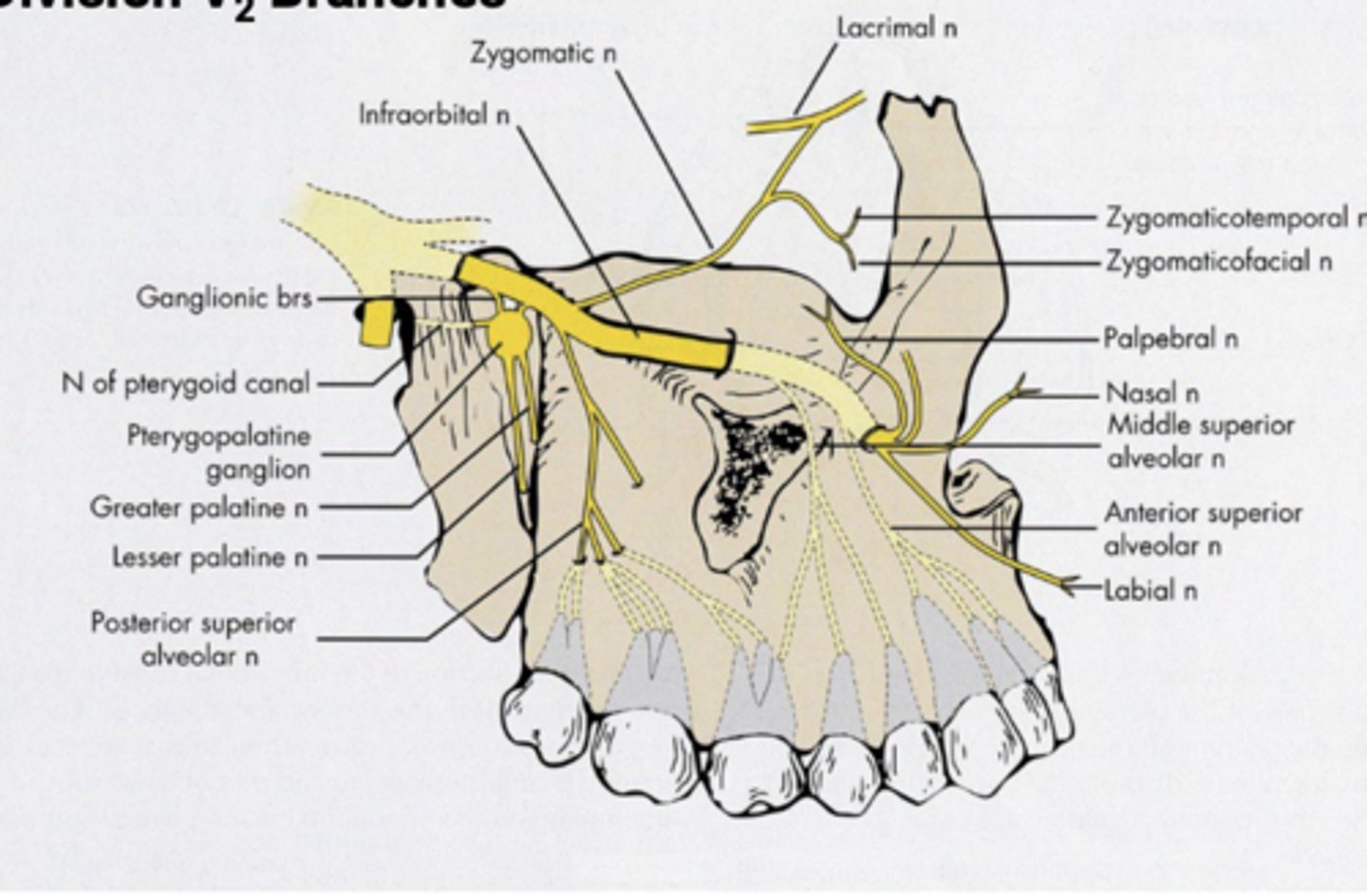
Maxillary division of V2
Branches within the cranium
- middle meningeal nerve
Branches within the pterygopalatine fossa
- zygomatic nerve
-- zygomaticotemporal nerve
-- zygomaticofacial nerve
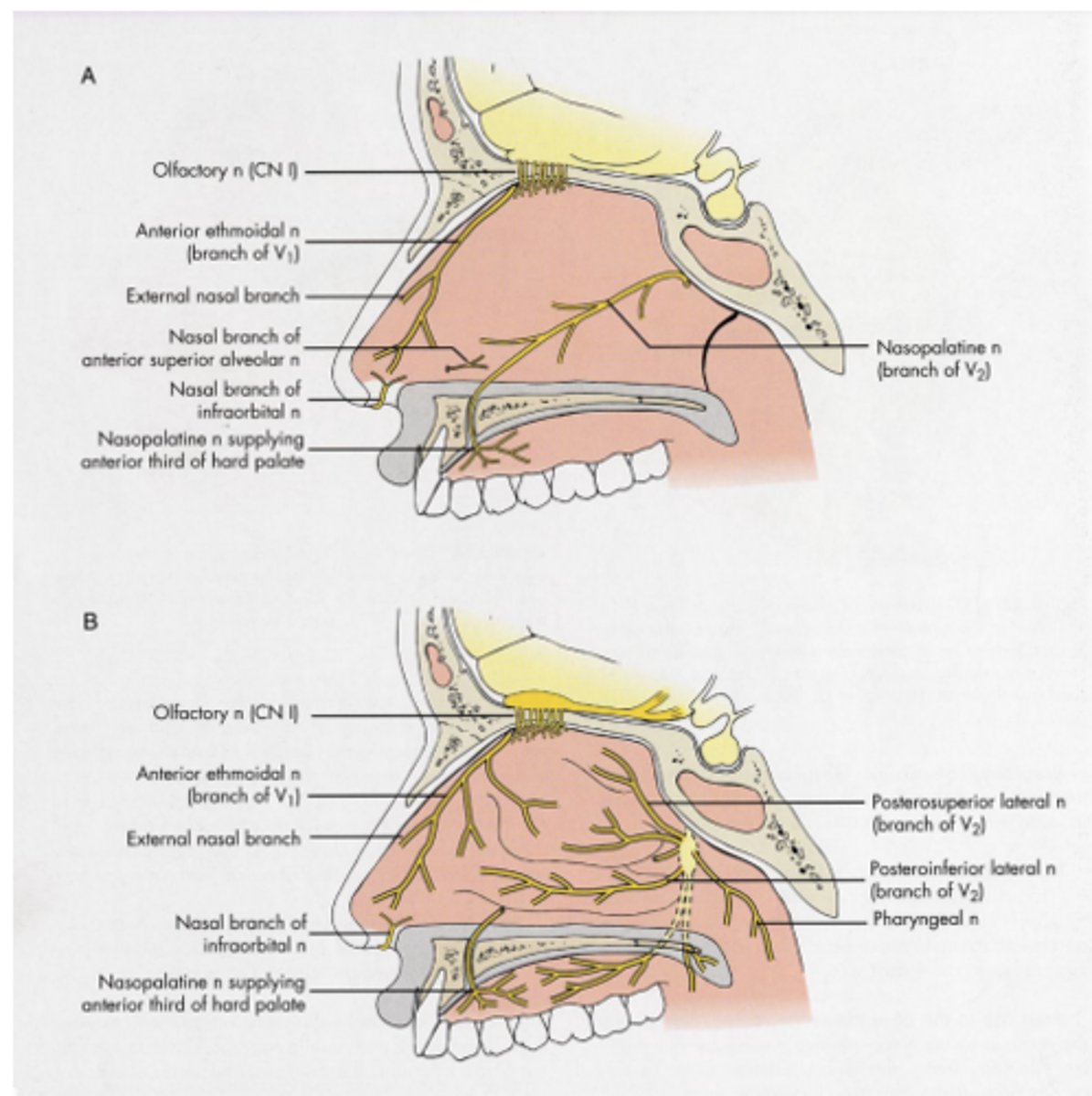
- Pterygopalatine nerve
-- Orbital brancehs
-- Nasal branches
---- Nasopalatine nerve
---- PSLN
---- PILN
-- Palatine branches
-- Greater (anterior) palatine nerve
-- Lesser (middle and posterior) palatine nerves
-- Pharyngeal branch
- Posterior superior alveolar nerve
Maxillary division of (V2) Branches
Zygomaticotemporal N.
- Innervates skin of lateral forehead
Zygomaticofacial N.
- Innervates skin over cheekbone
Maxillary division of V2- Zygomatic Nerve
Orbital branch
- Innervates periosteum of orbit
Nasal branches
- Nasopalatine nerve: innervates anterior septum, floor of nose, anterior 1/3 of hard palate
- Posterior superior lateral nasal
- Posteror inferior lateral nasal
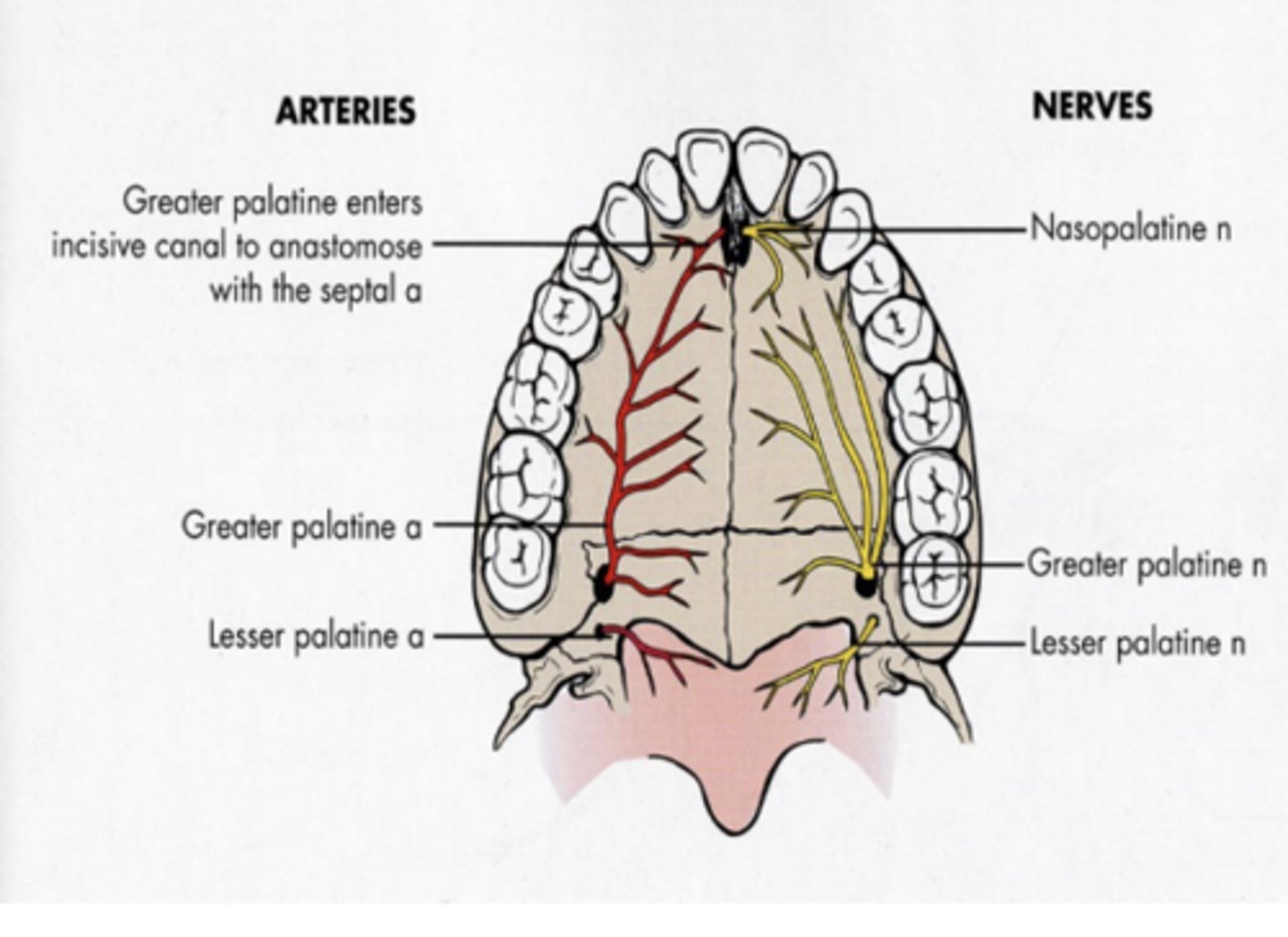
Palatine branches
- Greater palatine N: innervates posterior 2/3 of hard palate
- Lesser palatine N: innervates soft palate and tonsil
Pterygopalatine nerves
- Pharyngeal branch: innervates nasopharynx
Maxillary division of V2- Pterygopalatine nerves
Innervates
1. Mucous membrane of posterior maxillary sinus
2. Buccal gingiva of maxillary molars
3. Periodontium and pulp of maxillary molars except mesiobuccal root of 1st molar in 28% of cases
Maxillary division of V2- Posterior superior alveolar nerve
a. middle superior alveolar nerve
b. anterior superior alveolar nerve
Maxillary division of V2- Branches within the infraorbital canal
Present in about 46-72% of patients
Provides pulpal and periodontium innervation to the maxillary premolars and perhaps the mesiobuccal root of the maxillary 1st molar. Also innervates the buccal gingiva in the premolar area
Middle superior alveolar nerve
Provides pulpal, periodontium, and labial gingival innervation to the maxillary central incisor, lateral incisor, and canine
note:
- MSA nerve is NOT present the ASA nerve also typically innervates the premolars
Anterior superior alveolar nerve
a. Inferior palpebral branches
- Innervates skin of lower eyelids
b. external nasal branches
- Innervates skin of lateral aspect of nose
c. superior labial branches
- Innervates skin and mucous membrane of upper lip
Maxillary division of V2- Branches of the face
Nasal branches
- nasopalatine nerve
Palatine branches
- greater palatine nerve
- Lesser palatine nerves
Posterior Superior Alveolar nerve
Branches of Maxillary Division with Special Significance in Dentistry: Pterygopalatine nerves
Middle superior alveolar N
Anterior superior alveolar N
Branches of Maxillary Division with Special Significance in Dentistry: Infraorbital canal
Incisive papilla
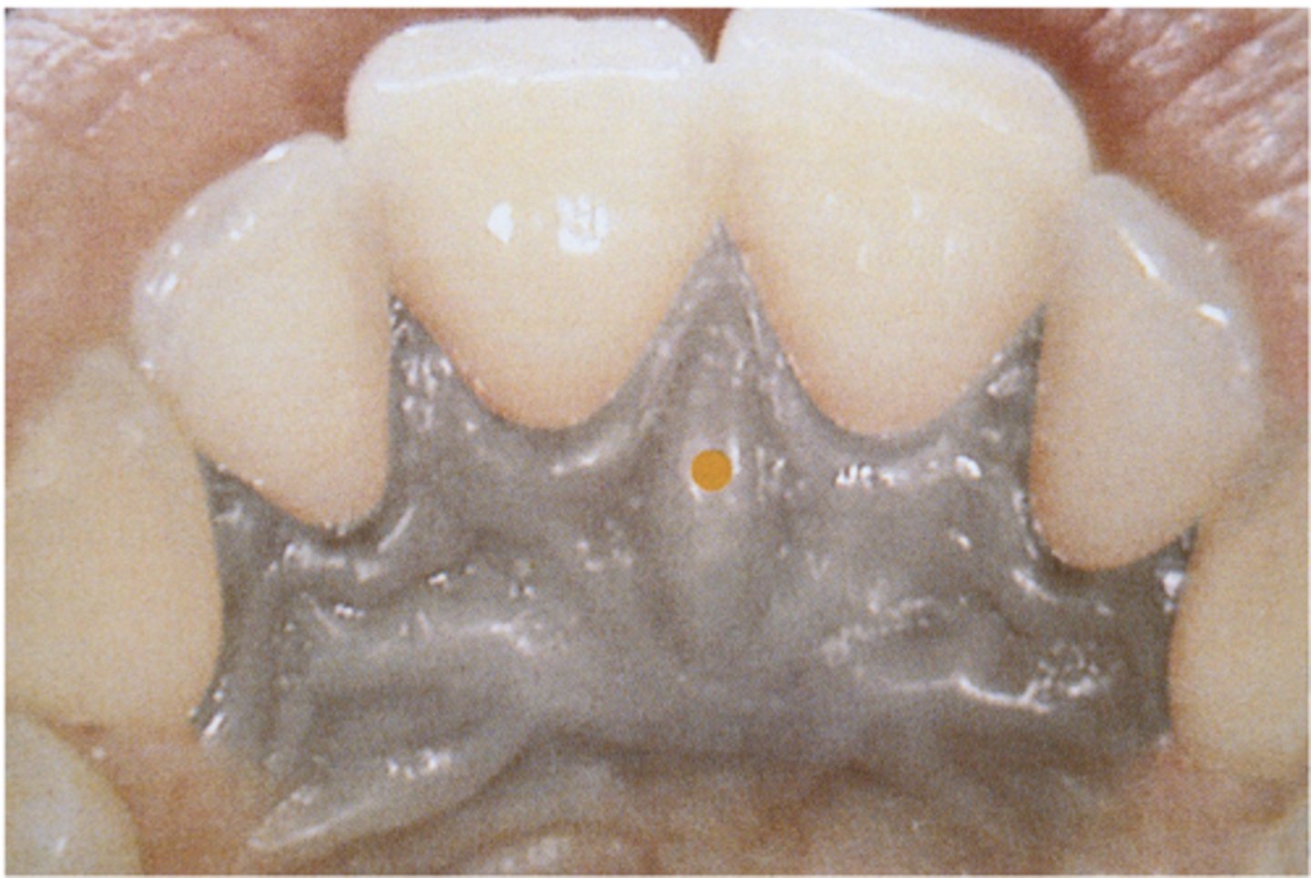
Greater palatine foramen- nerve innervates posterior 2/3 of hard palate
Nasopalatine foramen- nerve innervates anterior 1/3 of hard palate
Lesser palatine foramen- nerve innervates soft palate and tonsils
Palatine nerves
Mixed nerve
Largest division of trigeminal nerve
Branches in thee areas:
- Branches from the undivided nerve
- Branches from anterior division
- Branches from the posterior division
Mandibular Division of V3
Skin of:
- Temporal region
- Auricular region
- External auditory meatus
- Cheek
- Lower lip
- Chin
Mucous membrane of:
- Cheek
- Tongue (anterior 2/3rds)
- Mastoid cells
Mandibular teeth and periodontium
Bone and mandible
Temporomandibular joint
Parotid gland
Mandibular Division of V3: Sensory innervation
Masticatory muscles
Mylohyoid
Anterior belly of digastric
Tensor tympani
Tensor veli palatini
Mandibular Division of V3: Motor innervation
Nervus spinosus:
- Dura mater
- Mastoid air cells
Nerve to medial pterygoid muscle
- Small br. to tensor veli palatini
- tensor tympani
Mandibular Division of V3: Undivided nerve
Nerve to lateral pterygoid muscle
Nerve to masseter muscle
Nerve to temporalis muscle
Buccal Nerve (buccinator nerve or long buccal nerve)
Mandibular Division of V3: Divided nerve (anterior division)
sensory
Buccal nerve provides ______ innervation to skin of cheek, and mucous membrane of cheek and buccal gingiva of mandibular molars
buccinator muscle, facial nerve innervates buccinator muscle
Buccal nerve does not innervate the
Auriculotemporal nerve
Lingual nerve
Mylohyoid nerve
Inferior alveolar nerve: dental br
Incisive branch: dental branches
Mental nerve
Mandibular Division of V3: Divided nerve (posterior division)
Skin over zygomatic area, cheek, and mandible
Parotid gland
Skin of external auditory meatus and tympanic membrane
Posterior portion of temporomandibular joint
Skin over temporal region
Auriculotemporal nerve-innervates
Pure sensory
Runs anterior and medial to inferior alveolar nerve. Located at the side of the base of the tongue just below and behind mandibular 3rd molar
innervates
- anterior two-thirds of tongue (general sensation)
- Mucous membrane of floor of mouth
- Lingual gingiva of mandible
NOTE:
- Chorda tympani (a branch of the facial nerve) provides senastion of taste to anterior two-thirds of tongue
Lingual nerve
Mixed nerve
Branches from inferior alveolar Nerve before IAN enters mandibular canal
Runs downward and forward in mylohyoid groove on medial surface of mandible
Innervates
- mylohyoid muscle (motor)
- Anterior belly digastric (motor)
- Skin of mental protuberance (sensory)
- Sensory innervation of mandibular incisors
- Sensory innervation to mandibular molars
Mylohyoid nerve
Supplies pulpal innervation to mandibular molars and second premolars and sensory innervation to buccal periodontium of these same teeth
IAN
Supplies pulpal innervation and sensory innervation to buccal periodontium of mandibular 1st premolar, canine, and incisors
Incisive nerve
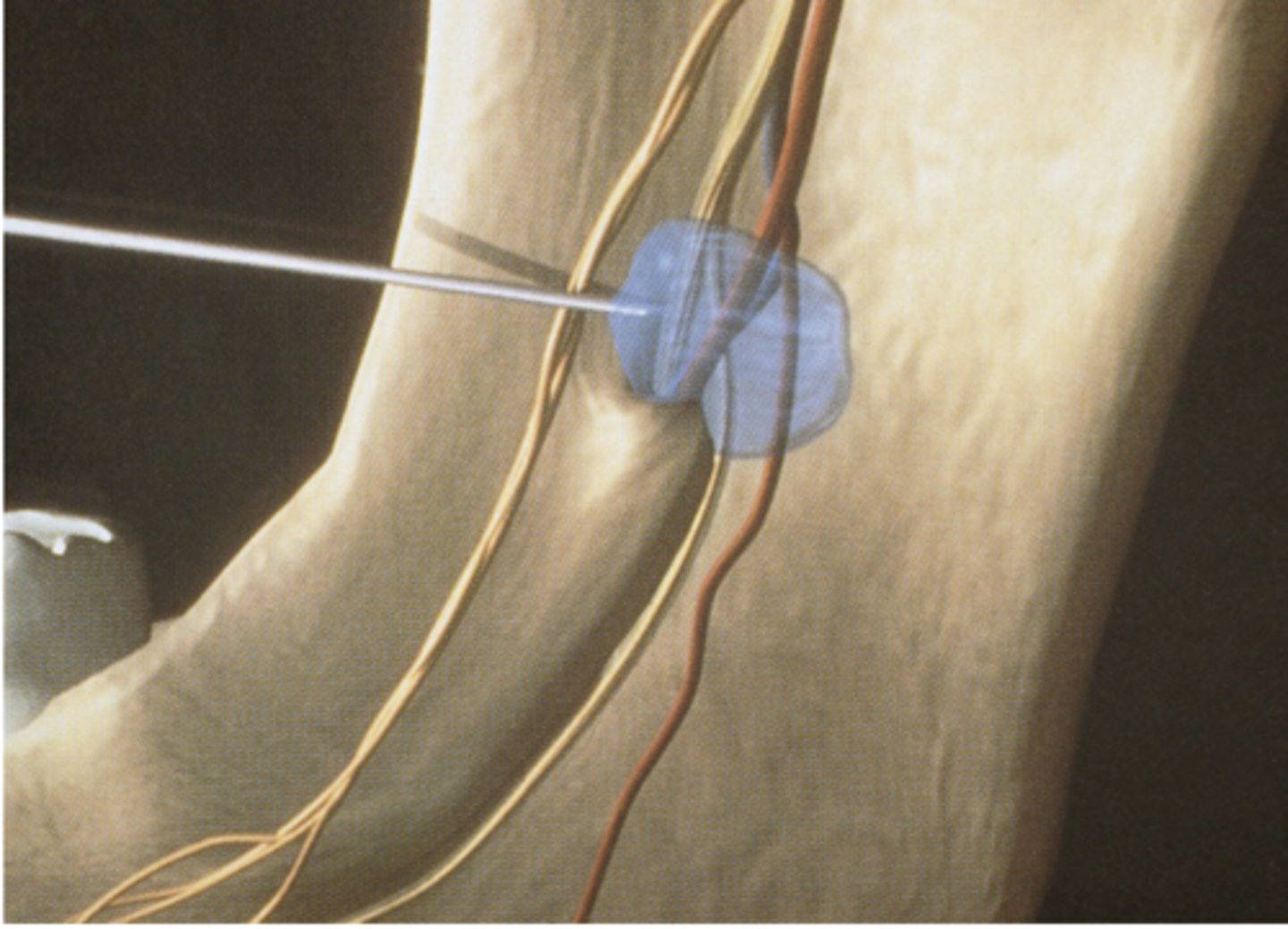
Mandibular foramen- target area for inferior alveolar nerve block
Deposition of local anesthetic just superior to mandibualr foramen
Exits inferior alveolar canal through the mental foramen
Divides into three branches that innervate the skin of chin and lower lip and mucous membrane of lower lip and labial gingiva of mandibular premolars and anterior teeth
Mental nerve
- Root eminences
- Canine eminence
- Canine fossa
- Infraorbital foramen
- Pyriform rim
- Anterior nasal spine
- Nasal septum
- Nasal bones
- Zygoma
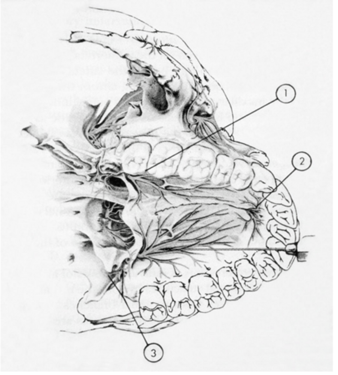
Important bony landmark in maxilla (anterior surface)
- Pterygomaxillary fissure and fossa
- Foramina for posterior superior alveolar nerve
- Maxillary tuberosity
- Lateral pterygoid plate
- Medial pterygoid plate
Important bony landmark in maxilla (Inferior Temporal surface)
- Palatal processes of maxilla (anterior 3/4ths of hard palate)
- Alveolar process
- Greater (anterior) palatine foramen
- Greater palatine groove
- Incisive foramen (descending palatine artery nasopalatine nerve)
- Premaxilla
- Horizontal process of palatine boen (posterior fourth of hard palate)
- Lesser palatine foramen
- Posterior nasal spine
Important bony landmark in maxilla (palatal surface)
- Condylar head
- Condylar neck
- Coronoid process
- Coronoid notch
- Ramus
- Body of mandible (thick, dense bone)
- Mental foramen
- Alveolar process (less dense bone in anterior)
- Mental protuberance
Important bony landmark in Mandible (Anterior view)
- Condylar head
- Condylar neck
- Sigmoid notch
- Coronoid process
- Coronoid notch (concavity of anterior ramus)
- Ramus
- Body of mandible
- External oblique line
- Mental foramen
- Mental protuberance
- Antegonial notch
- Antelingula
Important bony landmark in Mandible (Lateral View)
- Condylar head
- Condylar neck
- Coronoid process
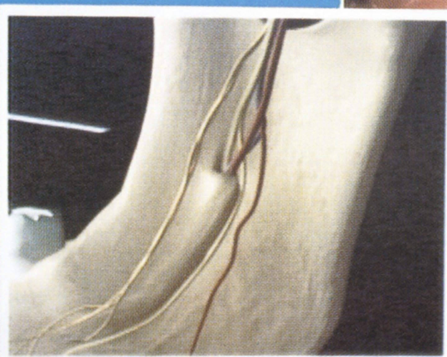
- Mandibular foramen
- Lingula
- Mylohyoid line
- Lingual foramina
- Genial tubercles
Important bony landmark in Mandible (Lingual view)
halfway between superior and inferior borders and 65%-75% of the distance from the anterior border of the ramus to its posterior border
May range from 1 to 19 mm or more above the level of the occlusal plane
The mandibular foramen is usually located
20-25mm
The thickness of soft tissue between needle penetration and bone of the ramus at the level of the mandibular foramen averages about