Skill Station 3 - ECG Interpretation
1/37
There's no tags or description
Looks like no tags are added yet.
Name | Mastery | Learn | Test | Matching | Spaced | Call with Kai |
|---|
No analytics yet
Send a link to your students to track their progress
38 Terms
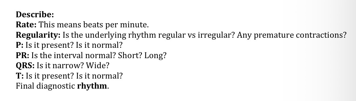
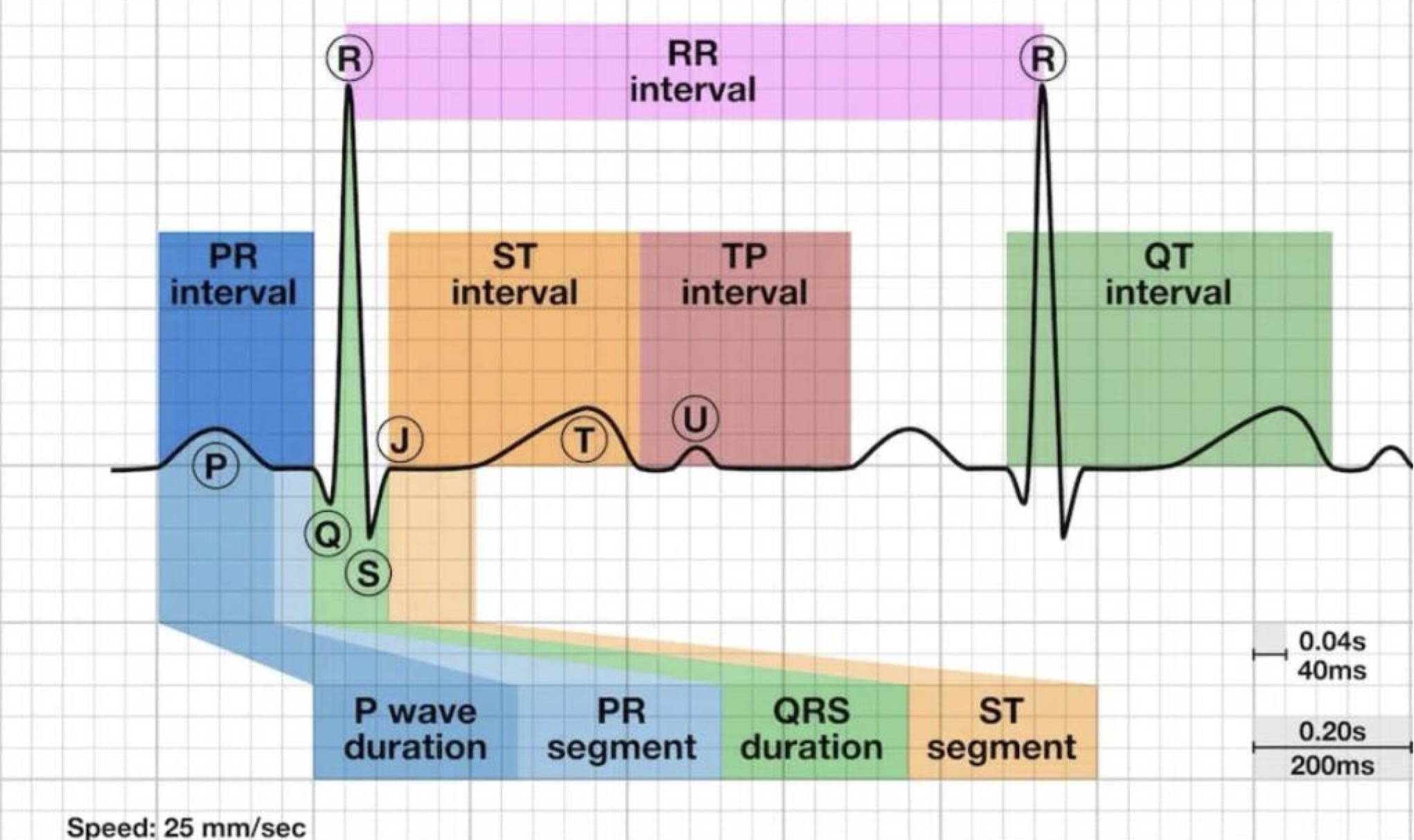
What is the 6 step rule to ECG interpretation?
rate
regularity
P wave
PR interval
QRS complex
T wave
Why is it important to interpret rate?
confirms whether the heart’s pacing is within normal range (60-100 bpm), bradycardic (under 60 bpm), or tachycardic (over 100 bpm)
Why is it important to measure regularity?
determine if the heart is beating at a regular or irregular rhythm
identification of premature contractions
What are the properties of a normal P wave?
less than 0.12 seconds (120 ms or 3 small squares)
amplitude is than 2.5 mm (0.25 mV) in the limb leads
smooth, rounded, and typical monophasic in most leads
may be biphasic in lead V1
frontal plane axis between 0-75 degrees
upright (positive) in leads I, II, and aVF
consistently inverted (negative) in aVR

What are the properties of a normal PR interval?
120-200 ms in duration (three to five small squares)
What are the properties of a normal QRS complex?
70-100 ms in duration

What is important to note about QRS length and its correlation to beat origin?
narrow complexes (QRS < 100 ms) are supraventricular in origin
broad complexes (QRS > 100 ms) may be either ventricular in origin or due to aberrant conduction of supraventricular complexes (e.g., due to bundle branch block, hyperkalaemia, or sodium-channel blockade)
What are the properties of a normal T wave?
asymmetrical
initial slope is more gradual followed by a steeper downslope
smooth
rounded
usually upright
0.10-0.25 secs
scales proportionately with the QT interval
amplitude is < 5 mm in limb leads
amplitude is < 10 mm in precordial leads

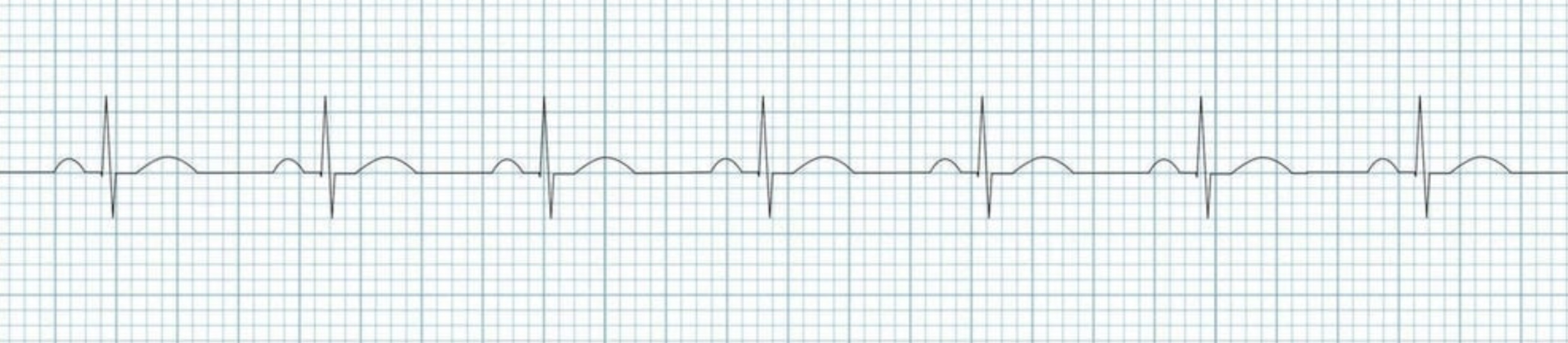
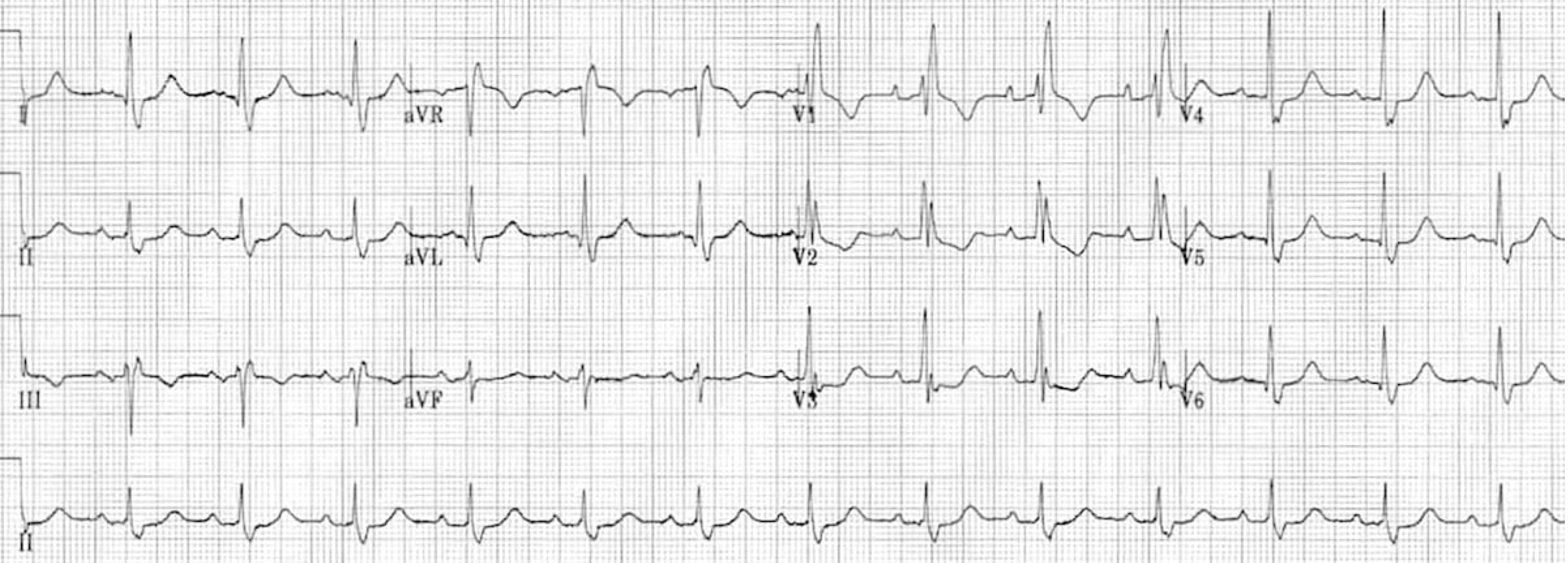
What rhythm/abnormality is seen below?
sinus rhythm
What are the diagnostic criteria for normal sinus rhythm?
regular rhythm (60-100 bpm)
or age-appropriate in children
each QRS complex is preceded by a normal P waves without progressive
normal P wave axis
upright in leads I and II
inverted in aVR
PR interval remains constant
QRS complexes are < 100 ms wide
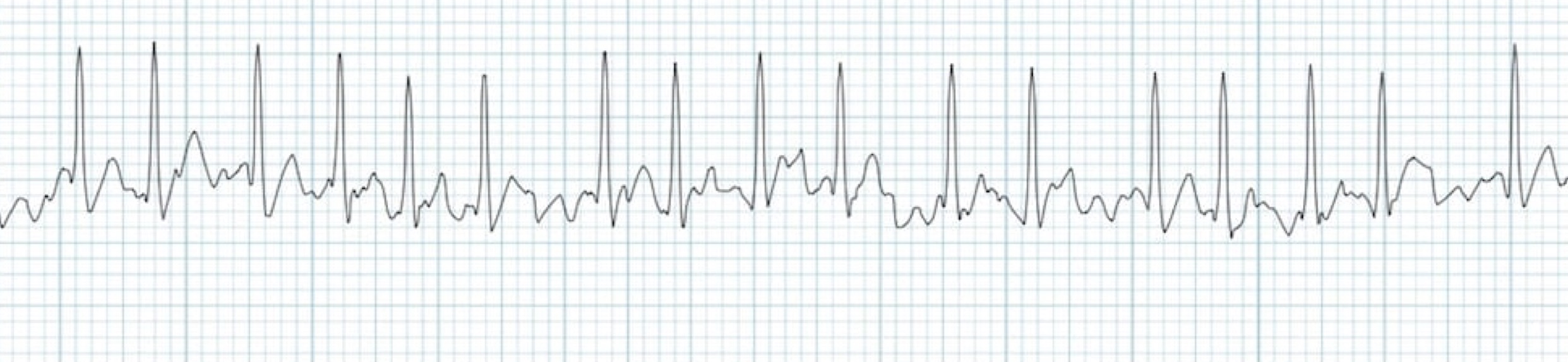
What rhythm/abnormality is seen below?
atrial fibrillation
What are is diagnostic criteria for atrial fibrillation?
irregularly irregular rhythm
P waves are absent
lack of isoelectric baseline
variable ventricular rate
QRS complex usually < 120 ms
unless pre-existing BBB, accessory pathway, or rate-related aberrant conduction
fibrillatory waves may be present (mimic P waves leading to misdiagnosis)
fine (amplitude < 0.5 mm)
coarse (amplitude > 0.5 mm)
What rhythm/abnormality is seen below?
atrial flutter
What is the diagnostic criteria for atrial flutter?
narrow complex tachycardia
regular atrial activity at ~300 bpm
loss of the isoelectric baseline
“saw-tooth” pattern of inverted flutter waves in leads II, III, and aVF
upright flutter waves in V1
may resemble P waves
ventricular rate depends on AV conduction rate
fixed
2:1 block = 150 bpm
3:1 block = 100 bpm
4:1 block = 75 bpm
variable
ventricular rate is irregular and may mimic atrial fibrillation
may be a pattern of alternating 2:1, 3:1, and 4:1 conduction ratios
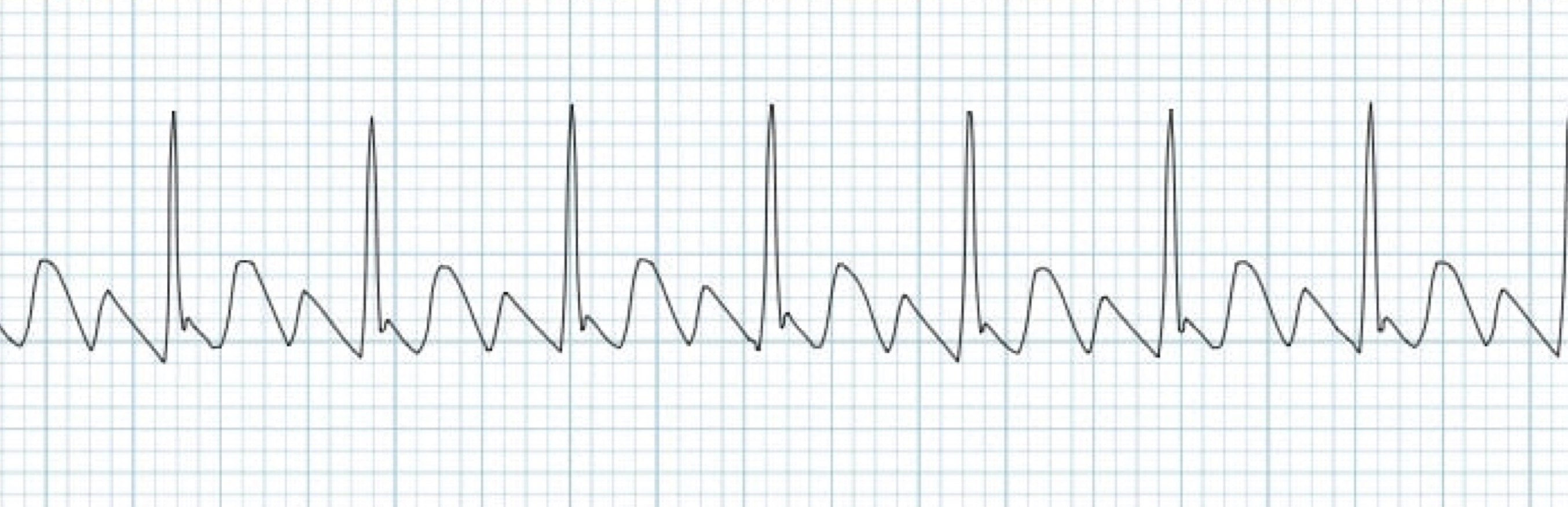
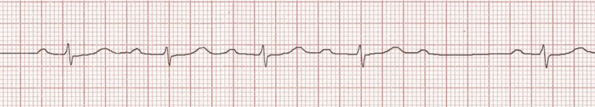
What rhythm/abnormality is seen below?
pre-ventricular complex (PVC) - bigeminy
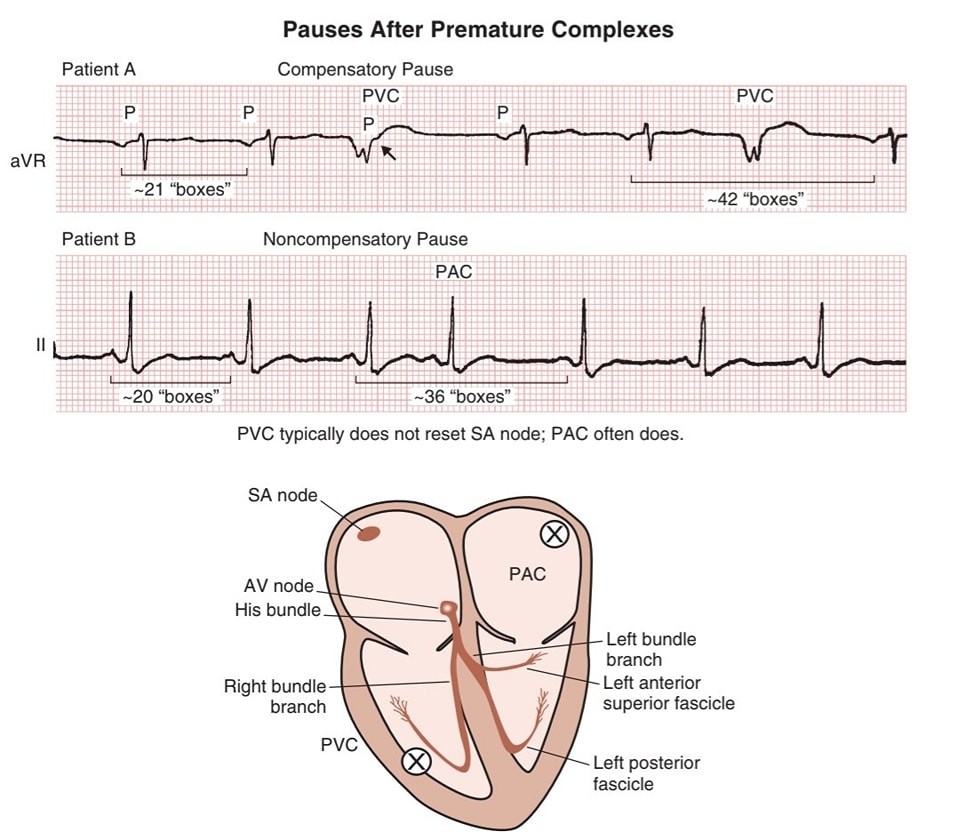
What are the diagnostic criteria for PVCs?
broad QRS complex with abnormal morphology
premature
discordant ST segment and T wave changes
usually followed by a full compensatory pause
retrograde capture of the atria may or may not occur
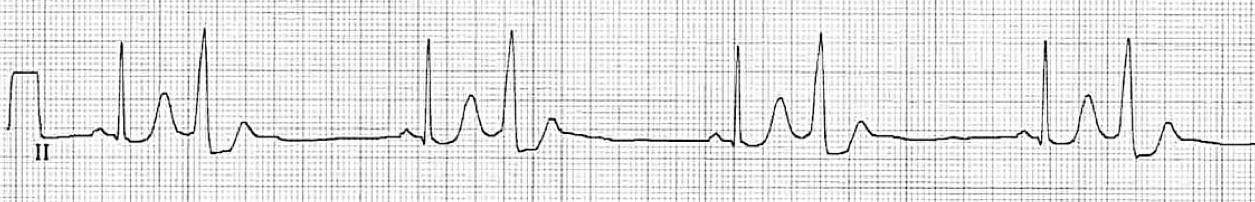
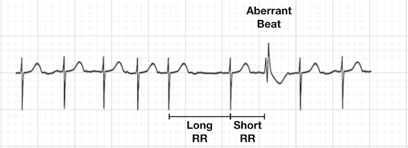
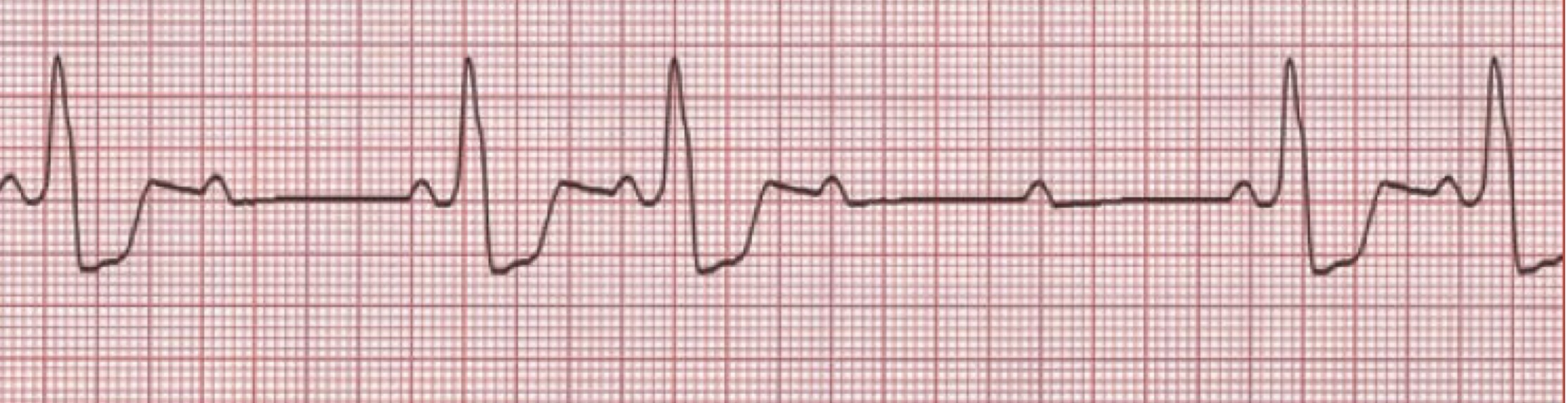
What rhythm/abnormality is seen below?
pre-atrial complex (PAC)
What are the diagnostic criteria for PACs?
abnormal P wave usually followed by a normal QRS complex
post-extrasystolic pauses may be present
may or may not be conducted aberrantly or not conducted at all
either way, P wave will still be visible
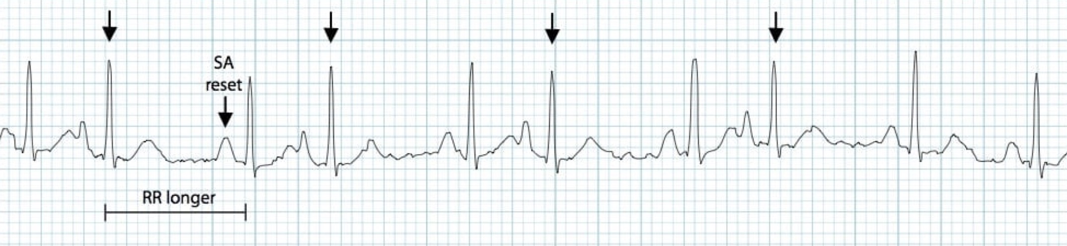
What are post-extrasystolic pauses?
brief pauses that result after a premature/extra beat depolarizes the SA node = SA node resets with a longer than normal interval before the next sinus beat arrives
What rhythm/abnormality is seen below?
right bundle branch block (RBBB)
What is aberrant conduction?
temporary, abnormal travel of an electrical impulse through the heart’s ventricles
occurs when an impulse arrives before the heart’s conduction pathways (specifically the bundle branches) have fully recovered from a previous beat
causes a delay/block that results in a widened QRS complex
What are the diagnostic criteria for RBBBs?
QRS duration > 120 ms
RSR’ pattern in V1-3 (“M” shaped QRS complex)
wide, slurred S wave in lateral leads (I, aVL, V5-6)
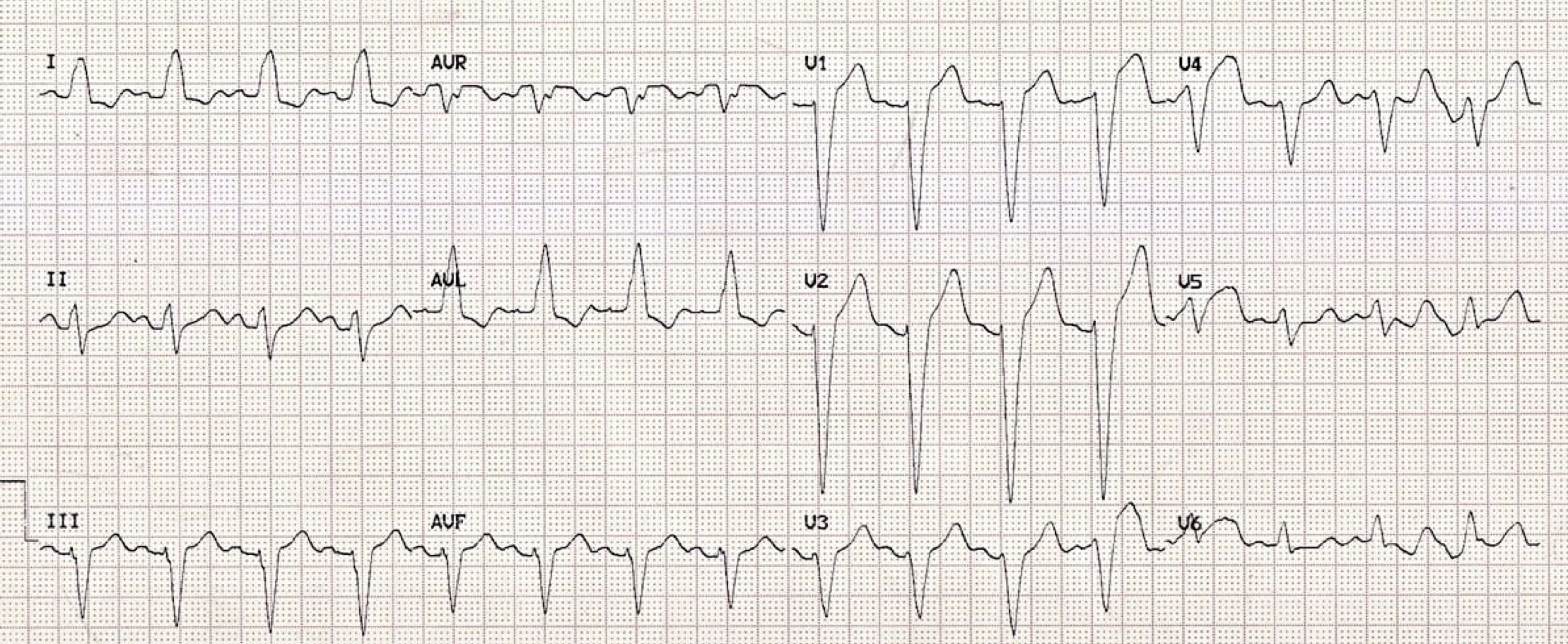
What rhythm/abnormality is seen below?
left bundle branch block (LBBB)
What are the diagnostic criteria for LBBBs?
QRS duration > 120 ms
dominant S wave in V1
broad monophasic R wave in lateral leads (I, aVL, V5-6)
absence of Q waves in lateral leads
prolonged R wave peak time > 60 ms in leads V5-6
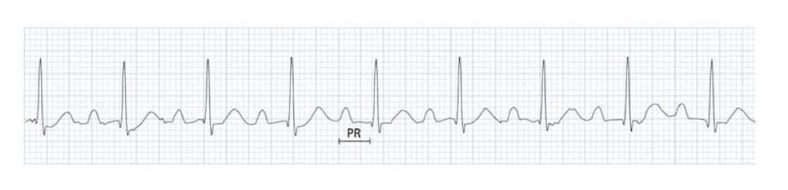
What rhythm/abnormality is seen below?
1st degree AV block
What are the diagnostic criteria for 1st degree AV block?
PR interval > 200 ms (five small squares) caused by delay without interruption in conduction from atria to ventricles
What rhythm/abnormality is seen below?
2nd degree AV block (Mobitz type I)
What are the diagnostic criteria for 2nd degree AV block (Mobitz type I)?
progressive prolongation of the PR interval culminating in a non-conducted P waves without progressive
PR interval is longest immediately before dropped beat
PR interval is shortest immediately after dropped beat
What rhythm/abnormality is seen below?
2nd degree AV block (Mobitz type II)
What are the diagnostic criteria for 2nd degree AV block (Mobitz type II)?
2nd degree AV block with intermittent non-conducted P waves without progressive prolongation of the PR interval
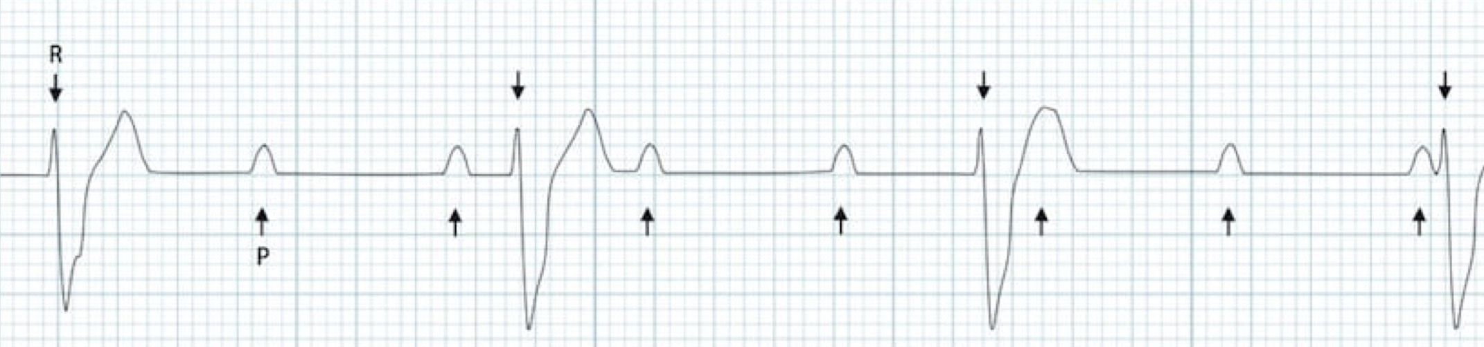
What rhythm/abnormality is seen below?
3rd degree AV block (complete heart block)
What are the diagnostic criteria for 3rd degree AV block (complete heart block)?
severe bradycardia due to absences of AV conduction
complete AV dissociation with independent atrial and ventricular rates
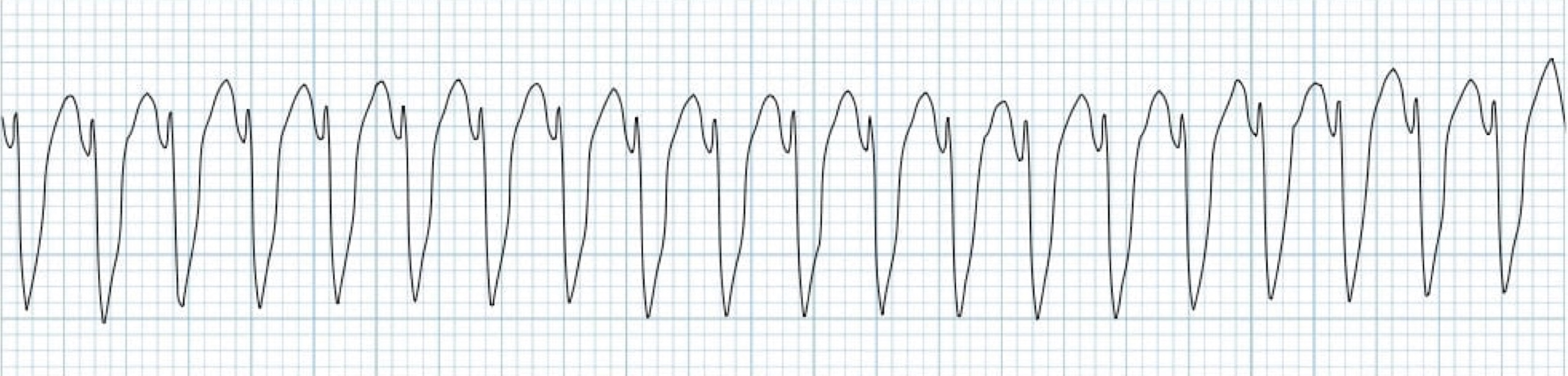
What rhythm/abnormality is seen below?
ventricular tachycardia
What are the diagnostic criteria for ventricular tachycardia?
regular broad complex tachycardia
uniform QRS complexes within each lead
with the exception of fusion/capture beats
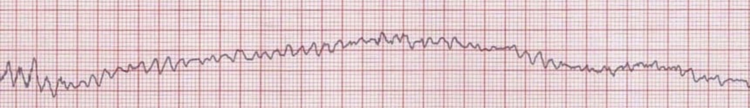
What rhythm/abnormality is seen below?
ventricular fibrillation
What are the diagnostic criteria for ventricular fibrillation?
chaotic irregular deflections of varying amplitude
no identifiable P waves, QRS complexes, or T waves
rate of 150-500 bpm
amplitude decreases with duration
coarse v-fib eventually turns into fine v-fib
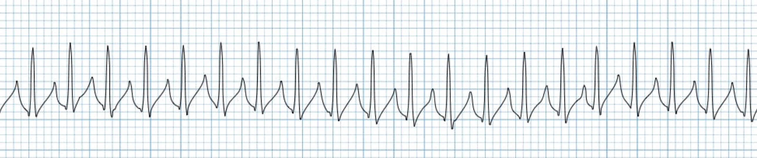
What rhythm/abnormality is seen below?
supraventricular tachycardia
What are the diagnostic criteria for supraventricular tachycardia?
any tachydysrhythmia arising from above the level of the Bundle of His
encompasses regular atrial, irregular atrial, and regular atrioventricular tachycardias