Anterior and medial thigh and femoral triangle
1/136
There's no tags or description
Looks like no tags are added yet.
Name | Mastery | Learn | Test | Matching | Spaced | Call with Kai |
|---|
No analytics yet
Send a link to your students to track their progress
137 Terms
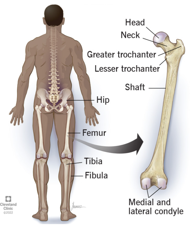
Major bony features of the femur
Femoral head - the "ball" in the proximal end of the femur. It is connected to the acetabulum by the ligament of the head of the femur (LHF) which inserts at the fovea capitis.
Femoral neck - between the head and shaft
Greater trochanter - a large, irregular and quadrilateral eminence (bony projection). Because of its location on the lateral aspect of the femur, muscles attaching there will abduct the hip.
Lesser trochanter - a conical and posteromedial eminence (bony projection). It serves as the principal insertion site of the iliopsoas muscle.
Linea aspera - a prominent, rough longitudinal ridge on the posterior surface of the femoral shaft, crucial for muscle attachment.
Medial and lateral condyles (condylar surfaces) - on the distal end of the femur, they articulate with the tibia at the tibial plateau. The lateral condyle is larger and more prominent than the medial condyle.
Patellar surface - on the anterior surface of the distal end of the femur, articulates with the posterior surface of the patella.
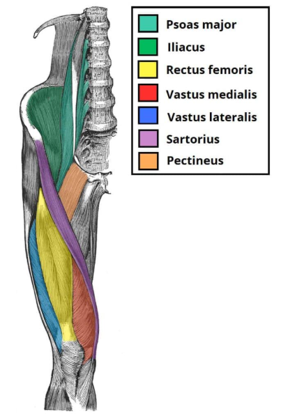
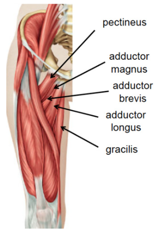
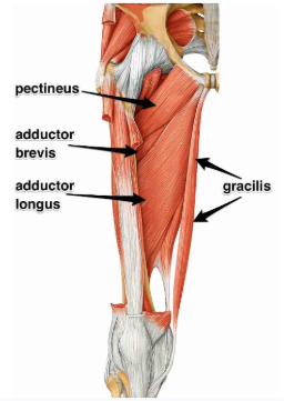
Which muscles make up the floor of the femoral triangle?
adductor longus, pectineus, psoas muscles and iliacus muscles
Major muscles in the anterior thigh + their actions
flexion and extension of knee, flexion and rotation/adduction of hip
Identify these muscles
adductor brevis, longus and magnus
Function of gracilis
flexes and adducts the hip joint, while also flexing and medially rotating the knee
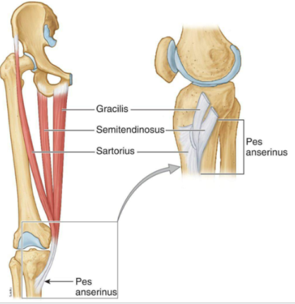
Pes anserus
conjoined tendons of semitendinosus, sartorius, and gracilis muscles.
Function of pectineus
At the hip joint, it acts by flexing (up to 45°) and adducting the thigh. Because it's so close to the hip bone, it also stabilises the pelvis.
Function of obturator externus
main action is to laterally rotate the thigh and abduction when the hip is flexed, also helps to stabilise the hip joint, maintaining the head of the femur in place.
Where does adductor brevis lie relative to pectineus?
deep to pectineus which is superior and anterior to it
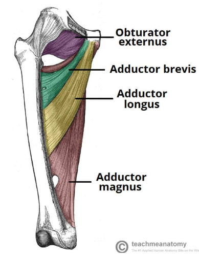

Where does adductor longus lie relative to adductor brevis and adductor magnus?
lies inferior to adductor brevis, superficial to adductor magnus
Which part of the adductor magnus acts like an adductor? Considering its insertion on the shaft of the fmur, what action does it carry out?
pubofemoral portion
only adducts the hip

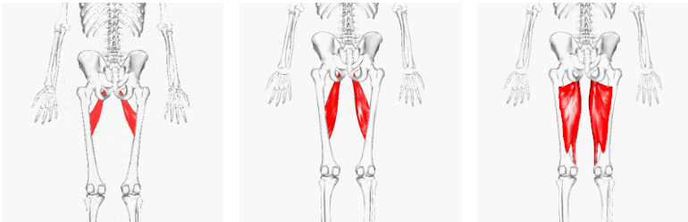
Major muscles in the medial thigh + actions
lateral rotation and abduction of thigh + adduction of hip
Major nerves that enter the lower limb from the trunk + describe the groups of muscles each one supplies
sciatic - posterior, obturator - medial, femoral - anterior
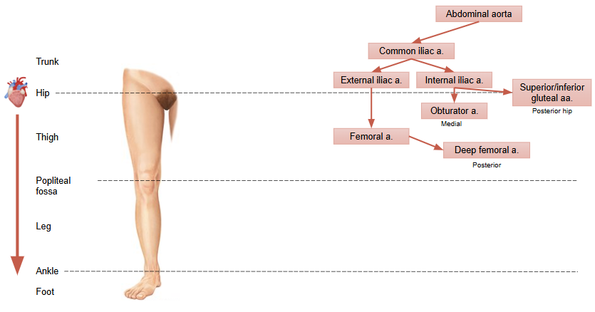
Major blood vessels that enter the lower limb from the trunk + describe the groups of muscles each one supplies/drains in the thigh
external iliac arteries becoming the femoral and deep femoral arteries + obturator and superior/inferior gluteal arteries + same veins are deep and great saphenous is superficial
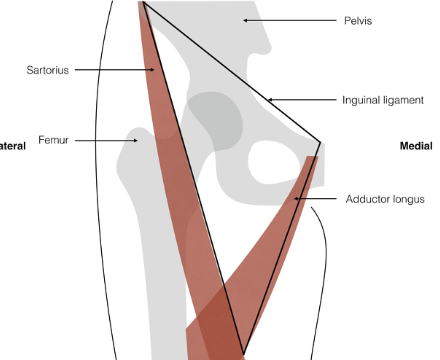
Identify the femoral triangle, its boundaries and contents
Borders:
Lateral border – sartorius m.
Medial border – adductor longus m.
Superior border – inguinal ligament
Roof – fascia lata, skin
Floor – 4 muscles (adductor longus, pectineus, psoas major and iliacus)
Femoral nerve – supplies anterior thigh
Femoral branch of the genitofemoral nerve – sensory to skin over femoral triangle
Femoral artery – continuation of external iliac, gives off deep femoral a.
Femoral vein – continuation of popliteal v., receives the deep femoral and great saphenous vv.
Femoral canal – potential space containing fat and lymph nodes
What is the bone of the thigh?
femur
What are the 3 components of the thigh?
anterior, medial and posterior
Which bone does the femur form a functional unit with allowing weight-bearing and locomotion?
hip bone
What are the 2 axes of the femur?
anatomical (weight baring) vs mechanical (locomotion)
What is the angle of inclination of the femur?
up to 150° at birth; 120°–130° in adults
What are the 2 movements specific to the femur?
femoral anteversion and retroversion
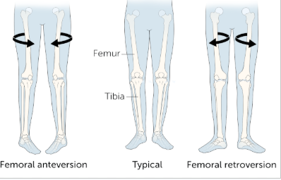
Is the angle of inclination of the femur more pronounced in men or women? Why?
women because larger hips
Which nerve passes just under the inguinal ligament?
femoral nerve
Why are the muscles of the anterior thigh termed quadriceps?
four heads of the thigh, all share one insertion point and meet at the quadriceps tendon which crosses into the tibia
3 originate on the femur, rectus femoris - straight muscle on top of the femur - most superficial and only one to come from and move the hip bone - flexes the hip
Innervation to posterior thigh
sciatic nerve
Innervation to medial thigh
obturator nerve
Innervation to anterior thigh
femoral nerve
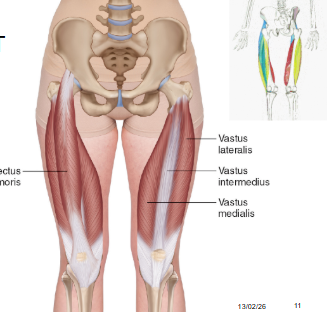
Which 6 muscles make up the anterior thigh?
iliopsoas, sartorius, rectus femoris, vastus intermedius, medialis and lateralis
Which 3 muscles of the anterior compartment of the thigh are responsible for the extension of the knee and share a part of their name?
vastus intermedius, medialis and lateralis (biggest so also stabilises the knee avoiding patellar from escaping and kneecap moving laterally)
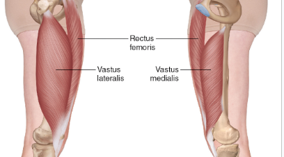
Movement of rectus femoris of anterior compartment of thigh
extension of knee and flexion of hip
Which artery and nerve supply the anterior thigh muscles?
femoral artery and nerve
What makes up the iliopsoas muscle?
psoas major and iliacus
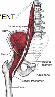
What is iliopsoas antagonistic to?
main flexor of hip so antagonist is main extensor ie gluteus maximus
Which artery supplies the iliopsoas?
iliolumbar, obturator, external iliac and femoral aa. - really long so multiple blood supplies
Innervation of iliopsoas
lumbar plexus and femoral nerve
Which muscle is part of both the anterior and medial compartments of the thigh?
pectineus

Blood supply and innervation of the pectineus
obturator artery (medial) and femoral nerve (anterior)
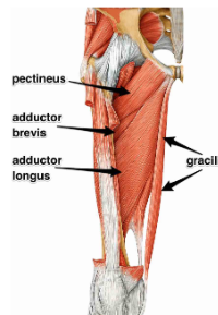
Movement of the pectineus
Flexion (up to 45°) and adduction of the hip, stabilises the pelvis

Is the pectineus supplied and innervated as the anterior or medial thigh compartment?
blood supply is medial (obturator), innervation is anterior (femoral)
Obturator externus blood supply and innervation
obturator artery and nerve
Movement of obturator externus
Lateral rotation of the thigh, abduction of the thigh when hip flexed
Where is the obturator externus found relative to the obturator foramen and obturator internus?
comes from obturator foramen and deep to obturator internus from posterior view - both muscles sandwich the obturator foramen
Which muscles makes up the medial compartment and lateral rotator group of the thigh?
obturator externus

Blood supply to medial compartment of the thigh
deep femoral artery
Innervation to medial compartment of the thigh
obturator nerve
Muscles that make up the medial compartment of the thigh
adductor brevis, adductor longus, adductor magnus, gracilis
Movement adductor brevis
Adduction and flexion of hip, stabilises pelvis
Movement adductor longus
Adduction and flexion of hip, stabilises pelvis - most superficial and visible muscle
Movement adductor magnus
adduction of hip (medial) - has 2 portions and is huge, lies just deep to adductor longus
Blood supply and innervation of gracilis
deep femoral artery and obturator nerve
In which thigh compartment is the gracilis and what action does it allow?
medial - flexion and adduction of the hip; flexion and medial rotation of the knee

What movement do both portions of the adductor magnus allow?
adduction of hip
What movement does the ischiocondylar portion of the adductor magnus allow?
extension of hip
What movement does the pubofemoral portion of the adductor magnus allow?
flexion of the hip
Which portion of the adductor magnus is most similar to a hamstring?
ischiocondylar
What is the pes anserinus and what forms it?
conjoined goose shaped tendons of semitendinosus, sartorius, and gracilis muscles.
What is the femoral triangle?
Triangular space on medial aspect of anterior thigh that is a passage for femoral nerve + main blood vessels between pelvis and lower limb
What 4 structures does the femoral triangle allow the passage of?
1 nerve, 2 vessels and 1 space
What are the boundaries of the femoral triangle?
3 borders, roof and floor

What are the borders of the femoral triangle?
SAIL:
Lateral border – sartorius m.
Medial border – adductor longus m.
Superior border – inguinal ligament
What make up the roof of the femoral triangle?
fascia lata and skin
What make up the floor of the femoral triangle?
4 muscles - adductor longus, pectineus, psoas major and iliacus
What are the contents of the femoral triangle lateral to medial?
NAVEL - femoral nerve, femoral branch of the genitofemoral nerve, femoral artery, femoral vein, femoral canal
What is and what makes up the femoral sheath?
membrane that covers the femoral artery, vein and the femoral canal (fat and lymph nodes) and excludes the femoral nerve
Which 2 structures are in the femoral sheath but not the femoral canal?
femoral artery and vein
What does the term popliteal refer to?
knee
Why is the femoral triangle important?
Vital structures passing between trunk and lower limb
Only protected by skin and fascia
Inguinal lymph nodes (deep and superficial)
Femoral pulse
‘Easy’ access to femoral vessels
What is the main superficial vein of the lower limb? What does it extend from and to?
great saphenous vein
from hip to ankle
Describe the blood supply to the lower limb?
common iliac artery → external iliac artery → femoral artery → deep femoral artery (posterior thigh) and internal iliac artery → obturator artery (medial thigh) and superior/inferior gluteal artery (posterior hip)
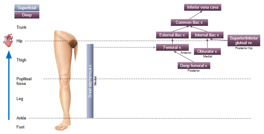
Describe the blood drainage of the thigh?
superficial: great saphenous vein
deep: deep femoral vein posterior drains into anterior femoral vein thwn external iliac vein and medial obturator vein drains into internal iliac vein - both iliac drain into common iliac vein finally into the IVC
Which 2 bones does the femur connect to?
femur to hip joint and tibia at knee joint
Summarise the anterior compartment of the thigh
contains the quadriceps (rectus femoris, vastus medialis, vastus lateralis and vastus intermedius), which extend the knee (rectus femoris also flexes the hip)
+ sartorius, a long muscle with multiple actions
compartment is innervated by the femoral nerve
Summarise the medial compartment of the thigh
contains the adductors (brevis, longus and magnus), which adduct the hip (the ischiocondylar portion of adductor magnus muscle acts as a hamstring!) + gracilis + pectineus. They are all innervated by the obturator nerve except pectineus (femoral n.).
Summarise the femoral triangle
triangular space on the medial aspect of the anterior thigh. It serves as a passage for femoral nerve + main blood vessels between pelvis and lower limb
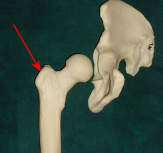
This picture shows the right hip bone and femur. A muscle that originates from the ilium and inserts at the arrow would act to?
abduct the hip
Which nerve passes through the gluteal region between piriformis and superior gemellus muscles?
sciatic
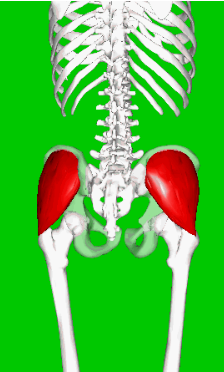
Which is the most powerful extensor of the hip?
gluteus maximus
Which of the following is correct about the hip joint?
has an incomplete bony articular surface, the acetabulum
while standing, it transfers the weight from the trunk to the lower limb mainly through the ischial bone
has a capsule which is strengthened anteriorly by the ischiofemoral ligament
has the pubofemoral ligament inside its capsule
has an incomplete bony articular surface, the acetabulum
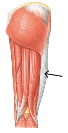
Which of the following statements correctly described the structure indicated by the arrow?
its a cartilaginous reinforcement of the fascia lata, the deep fascia of the thigh
its a fibrous reinforcement of the fascia lata on the medial side of the thigh
it is a reinforcement of the fascia lata that runs between the lumbar area to the lateral knee
its a fibrous reinforcement of the fascia lata and helps stabilise the knee
its a fibrous reinforcement of the fascia lata and helps stabilise the knee
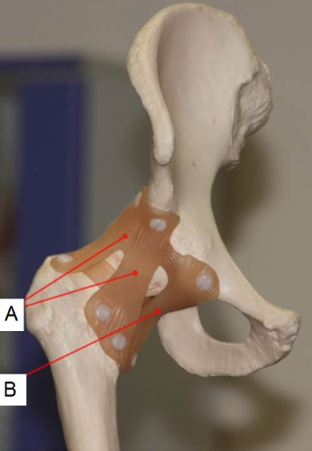
Which of the following is correct about the structures showed in the image?
Structure A is the ischiofemoral ligament, and structure B is the pubofemoral ligament
Structure A is the iliofemoral ligament, and structure B is the ischiofemoral ligament
Structure A is the iliofemoral ligament, and structure B is the pubofemoral ligament
Structure A is the iliofemoral ligament, and structure B is the transverse acetabular ligament
Structure A is the iliofemoral ligament, and structure B is the pubofemoral ligament
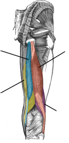
Regarding the muscles in the picture, which of the following statements is correct?
they are innervated by the femoral nerve
they act on the knee, extending it
they act on the hip, extending it
they are supplied by the superior gluteal and the deep femoral arteries
they act on the hip, extending it
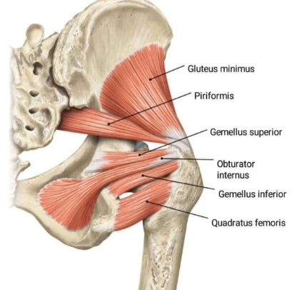
Which statement concerning piriformis muscle is correct?
it is an abductor of the thigh in hip flexion
it is innervated by the sciatic nerve
passes anterior to the femoral head
lies deep to the sciatic nerve
it is an abductor of the thigh in hip flexion
Which statement concerning this structure is correct?
it is innervated by the inferior gluteal nerve
it is completely covered by another muscle
it acts to abduct and medially rotate the hip
it is one of the lateral rotators of the hip
it acts to abduct and medially rotate the hip
Which of the following is not a movement of the hip joint?
flexion
lateral rotation
circumduction
opposition
opposition
Name the three bones that fuse to form the acetabulum and identify which one
contributes the largest portion to the socket.
The ilium, ischium, and pubis fuse to form the acetabulum, with the ilium contributing the largest portion (approximately two-fifths).
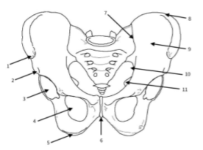
features of the pelvic girdle
1. Anterior superior iliac spine (ASIS)
2. Anterior inferior iliac spine (AFIS)
3. Acetabulum / hip joint
4. Obturator foramen
5. Ischial tuberosity / ischium
6. Pubic symphysis
7. Sacroiliac joint
8. Iliac crest
9. Iliac fossa
10. Greater sciatic notch
11. Ischial spine
Which bony landmark acts as a weight-bearing point when a person is in a seated
position?
ischial tuberosity
Despite its mobility, why is the hip joint less prone to dislocation than the shoulder joint?
The hip joint has a deeper socket (acetabulum) and stronger ligaments compared to the
shoulder, prioritising stability over mobility. The muscles around the hip joint help with
stabilisation of the pelvis but that’s not their main function (like the rotator cuff in the
shoulder)
Which ligament is continuous with the acetabular labrum? Is it intracapsular or capsular?
transverse acetabular ligament is the structure continuous with the acetabular
labrum. While the labrum is a fibrocartilaginous rim that attaches to the bony margin of
the acetabulum, the transverse acetabular ligament bridges the acetabular notch at the
bottom of the socket, completing the circle of the labrum and providing it with a
continuous base. It is a capsular ligament.
What is the main function of the ligament to the head of the femur?
also known as the ligamentum teres, changes significantly with age. In infants and children, its primary role is to serve as a protected conduit for the acetabular branch of the obturator artery, which provides a critical blood supply to the developing head of the femur. In adults, this artery usually becomes narrow or obliterated, and the ligament functions instead as a secondary stabiliser for the hip joint. It becomes taut during specific movements (particularly adduction, flexion, and external rotation) acting as a "sling" or "check" to prevent the femoral head from subluxating or dislocating at these extremes of motion. Additionally, it contains nerve endings that contribute to proprioception, helping the body sense the position and movement of the hip joint
The pubofemoral ligament primarily limits which specific movement of the hip joint?
Abduction; it also limits extension and external rotation to a lesser degree
During childbirth, the fetal head must pass through the pelvic outlet. Which pelvic joint
must undergo significant hormonal "softening" to allow for increased diameter?
The pubic symphysis. The hormone relaxin increases the laxity of the dense connective
tissues in this joint, allowing the pelvic outlet to expand.
Which ligament forms a "Y" shape and is primarily responsible for limiting excessive
external rotation and extension?
iliofemoral ligament
How do the iliofemoral, pubofemoral, and ischiofemoral ligaments work together to
stabilise the hip? Think about the direction of the fibres and the arrangement around the
joint.
function as a synergistic unit by fusing into each other, converting the hip joint into a stable and close-packed unit. These extracapsular ligaments are arranged in a spiral orientation around the femoral neck, meaning they naturally loosen during flexion to allow for a wide range of mobility but become progressively tighter as the hip extends. The iliofemoral ligament, characterised by the "Y-shaped", is one of the strongest in the body and acts as the
primary restraint against hyperextension by reinforcing the anterior aspect of the joint
capsule. Working alongside it, the pubofemoral ligament reinforces the anterior and
inferior capsule, specifically tightening to limit excessive abduction and external
rotation. On the posterior side, the ischiofemoral ligament completes the triad by
spiraling over the superior aspect of the femoral neck to attach anteriorly, which allows it
to resist excessive internal rotation and further secure the femoral head within the
acetabulum during extension. Together, these three ligaments ensure that as a person
stands upright, the femoral head is mechanically compressed into the socket, providing
maximum passive stability with minimal muscular effort
Why is the pelvis considered both a weight-bearing and a protective structure?
The pelvis transmits body weight from the axial skeleton to the lower limbs through the hip joints, while also protecting pelvic viscera such as the bladder, reproductive organs, and rectum. Its thick, curved bones and ring-like structure distribute mechanical stress
efficiently while forming a rigid protective basin.
Which anatomical landmark is used to identify the exit point of the sciatic nerve from the
pelvis?
The piriformis muscle, as the sciatic nerve typically exits the pelvis into the
posterior thigh below it
Which nerves innervate the gluteal group of the gluteal region?
The gluteal muscles are primarily innervated by two key nerves from the sacral plexus: the superior gluteal nerve, which supplies the gluteus medius, gluteus minimus, and tensor fasciae latae, and the inferior gluteal nerve, which provides motor innervation to the large gluteus maximus muscle
Which nerve innervates the posterior aspect of the hip joint?
Nerve to quadratus femoris.
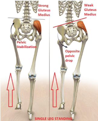
If the superior gluteal nerve gets damaged, what movement would be impaired?
hip abduction and medial (internal) rotation of the thigh. This occurs because the nerve provides the sole motor innervation to the gluteus medius, gluteus minimus, and the tensor fasciae latae muscles. Beyond simple movement, the most critical functional loss is the inability to stabilise the pelvis during walking. Under normal conditions, these muscles contract to keep the pelvis level when the opposite foot is lifted off the ground; when they are paralysed, the pelvis drops
toward the unsupported side, a clinical manifestation known as the Trendelenburg sign
(see the question below!).
Ask a classmate to stand on both feet, then lift one foot. Watch their pelvis from posterior
for 30 seconds, focusing on the side of the lifted (unsupported) leg. If their pelvis stays
level or rises slightly on the unsupported side, that is a negative Trendelenburg sign
(normal). However, if the pelvis drops on the side of the lifted (unsupported) leg, that is a
positive Trendelenburg sign (pathological). Which muscles would be weakened in a
person with a positive Trendelenburg sign? Why?
hip abductors, primarily the gluteus medius and gluteus minimus muscles on the supported (stance) side, innervated by the superior gluteal nerve and are critical for maintaining pelvic stability during unilateral weight-bearing activities. The reason the pelvis drops on the unsupported side is due to a failure of the "hip abductor mechanism." To prevent the pelvis from tilting downward toward the unsupported leg under the force of gravity, the gluteus medius and minimus on the weight-bearing side must contract isometrically (contracting without changing length, like holding a plank). This contraction pulls the ilium (origin) toward the greater trochanter of the femur (insertion), effectively "levelling" the pelvis. If these muscles are weak, paralysed, or inhibited by pain, they can not generate sufficient tension to counteract the weight of the swinging limb and trunk, resulting in the characteristic contralateral pelvic drop.