ANATOMY OF THE SPINAL CORD, ASCENDING, DESCENDING, AND INTERSEGMENTAL TRACTS (PART 1)
1/98
Earn XP
Description and Tags
SKIBIDI DOB DOB DOB YES YES
Name | Mastery | Learn | Test | Matching | Spaced | Call with Kai | Chat |
|---|
No analytics yet
Send a link to your students to track their progress
99 Terms
Support skull, pectoral girdle, upper limbs, and thoracic cage and through the pelvic girdle, transmit body weight to the lower limbs
VERTEBRAL COLUMN
What structures make the vertebral column flexible?
Intervertebral Discs (IV DISCS)
Enumerate all types of vertebra and how many are there per region.
7 cervical
12 thoracic
5 lumbar
5 sacral
4 coccygeal (fused lower 3)
PARTS OF THE VERTEBRA:
most prominent, rounded anterior
Vertebral body
posterior
Arch / Vertebral Arch
Form the side of the arch
Pedicles
Extends from the pedicles; fuses to form spinous process
Lamina / Flattened lamina
How many processes are there in a vertebra?
7 : 1 spinous, 2 transverse, 4 articular
VERTEBRAL LIGAMENTS
Above the spinous process; between tips of spines
Supraspinous Ligament
Key word: Tip to tip action
Connects adjacent spine
Interspinous Ligament
Key word: Shaft on shaft action
supraspinous & interspinous ligament on the neck region
Ligamentum nuchae
Level of the ligamentum nuchae
C1-6
Connects laminae
Ligamentum Flavum
ligaments on anterior portion of vertebral column
Anterior longitudinal ligament
ligaments on posterior portion of vertebral column
Posterior Longitudinal Ligament
Anterior and posterior longitudinal ligament extends from _____ to ______ (Hint: Skeleton parts)
Skull to sacrum
Which ligament is stronger and wider of the two longitudinal ligaments?
Anterior longitudinal ligament (Flexion>Extension)
INTERVERTEBRAL DISC PARTS

Peripheral part
Fibrocartilage
Strongly attached to vertebral bodies and the anterior and posterior longitudinal ligaments
Annulus FibroSUS

Central Part; Ovoid
semifluid, gelatinous material
Nucleus PulpoSUS

Upper and lower surfaces of adjacent vertebral bodies are covered with thin plates of _________ cartilage
Hyaline
TRUE OR FALSE: IV discs are the thickest in the thoracic region where movement is most restricted
FALSE
IV discs are thickest where movement is greatest (cervical and thoracic)
SPINAL CORD
From _______ where it is continuous with the ______
Foramen magnum
Medulla oblongata
Terminates at what level for adults
Lower border of L1 vertebra
Terminates at what level for newborns
L3
Cone shaped termination of the spinal cord
Conus medullaris
Fusiformly enlarged at the areas that give off _____and _____plexus
Brachial and lumbosacral
Spinal nerves exiting at levels lower than L1
Cauda Equina
SPINAL CHORD MENINGES

pial folds that stretch from the surface of cord to dural sheath midway between the dorsal and ventral roots
Dura is NOT adherent to SC
_________ anchor spinal cord to vertebral body
Denticulate ligaments
Wrapped by ___ adherent to the SC
Covered by ___ until nerve roots
Pia
Enumerate all spinal chord segments
31 segments:
8 cervical
12 thoracic
5 Lumbar
5 Sacral
1 coccygeal
Dorsal and ventral roots join in the ___________ to form spinal nerve
Intervertebral foramen
True or false: spinal chord is inner gay outer whack
FALSE
Inner gray and outer white
PRACTICE NAMING SPINAL NERVES:
The next slides will give you a pair of vertebrae, name the spinal nerve in between them.
Above the atlas (C1), below foramen magnum
C1 spinal nerve
C4 and C5
C5 spinal nerve
T1 and C7
C8 spinal nerve
C2 and C3
C3 spinal nerve
L5 and L6
BUNYE
L5 and S1 = L5 spinal nerve
L4 and L5
L4 spinal nerve
PRACTICE NAMING SPINAL CHORD SEGMENTS
The next slides will give a spinal vertebra, name the corresponding spinal chord segment.
C1
C2 spinal segment
T6
T8 spinal segment
T9
T12 spinal segment
T10
L1-2 spinal segments
T11
L3-4 spinal segments
T12
L5 spinal segment
L1
Sacral and coccygeal spinal segments
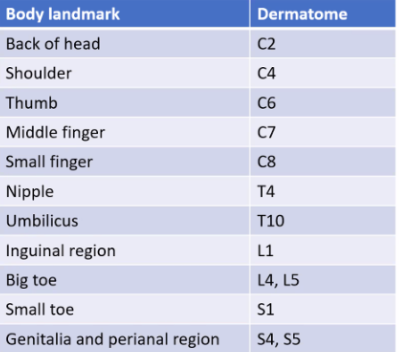
DERMATOMES (Memorize them bozo)
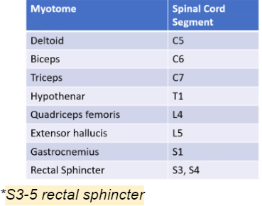
MYOTOMES
Groups of muscles innervated by a single spinal cord segment
Spinal Cord Meninges

PAD: Pia, Arachnoid, Dura
Arranged from inner to outer
Layer of pia closely adherent to the surface of the spinal cord
INTIMA PIA
Layer of pia that carries blood vessels
Denticulate Ligaments - Lateral flattened bands of _____ tissue
Epipia
Denticulate ligaments are epipial tissue
Between pia and arachnoid
Contains CSF
Subarachnoid Space
Layer that’s closely adherent to Dura
Arachnoid
Potential space; there should be no space but there are bridging veins; susceptible to rupture =___________
Subdural Space
Rupture of subdural space = Subdural hematoma
DURA
Attached to SC only at the level of the __________
Otherwise it attaches to the __________
Ensheath the ventral and dorsal roots DRG, and proximal portions of the spinal nerves
Closest to vertebra
Foramen magnum
Epidural Space
With adipose tissue and venous plexus
Prone to hematoma
Exists below level of foramen magnum
REAL space
Epidural Space
SPINAL CORD FISSURES
deep longitudinal midline fissure anteriorly
Anterior Median Fissure
shallow furrow on posterior surface
Posterior Median Septum
site of entrance of dorsal roots
Posterolateral Sulcus
site of exit of ventral roots
Anterolateral Sulcus
Separates fasciculus gracilis and fasciculus cuneatus
Dorsal/Posterior intermediate septum
TRUE or FALSE : The dorsal intermediate septum only exists at the level of T6 above
TRUE:
The dorsal intermediate septum separates the fasciculus gracilis and cuneatus but below T6 there is only gracilis.
Horns that are only present in levels T1-L3 (Outflow of sympathetic nerve fibers)
Lateral Horns
middle of anterior and posterior gray horns
Gray Commisure
contains CSF
Divides into gray commissure into anterior and posterior commissure
Central Canal
GRAY MATTER
H-shaped pillar with anterior and posterior gray horns
United by gray commissure containing the central canal
Amount of gray matter is related to amount of muscles innervated (Greatest at ________ and _________)
__________-cell bodies that receive axons of DRG; sensory (Horn)
____________-seen in thoracic and upper lumbar segments; preganglionic SY nerve fibers that exit via ventral root
Fibers from _______ join motor fibers and come out of ______ root
_________-motor; largest at cervical and lumbosacral
Cervical and Lumbosacral
Dorsal Horn
Lateral Horn / intermediolateral horn
Lateral horn joins motor fibers and comes out the ventral root
Ventral horn
ANTERIOR GRAY COLUMN NERVE CELL GROUPS
Large and multipolar
Axons pass in the anterior roots of spinal nerves
Innervate skeletal muscles
α Efferents / α motor neurons
Smaller and multipolar
Axons pass in the anterior roots of spinal nerves
Innervate intrafusal muscle fibers of neuromuscular spindles
𝜸 Efferents / 𝜸 motor neurons
Group that is present in most segments
Innervate skeletal muscle of neck, trunk, intercostal, and abdominal muscles
Medial Group
Smallest group, present in cervical and lumbosacral segments
Central Group
Which nerve roots form the phrenic nucleus for the diaphragm?
C3-5
Which nerve roots form the accessory nucleus (SCM and trapezius; axons form spinal part of accessory nerve)?
Upper 5 or 6 cervical segments (C1-5 / C1-6)
Which nerve roots form the lumbosacral nucleus?
L2-S1
Group that is present in cervical and lumbosacral segments
Innervate skeletal muscles
Lateral Group
POSTERIOR GRAY COLUMN NERVE CELL GROUPS

In the apex of posterior gray column
Composed of golgi type II neurons
Receives afferent fiber for pain, temperature, and touch from SRG (Sensory root ganglia / Dorsal root ganglia)
Substantia Gelatinosa Group
Anterior to SG
Main bulk of cells in posterior gray column
Receives fibers associated with proprioception, 2 point discrimination and vibration
Nucleus Pulposus group (2nd red arrow)
At the base of PGC (Posterior gray column)
C8-L3-4
Associated with proprioceptive endings (neuromuscular spindles and tendon spindles
Nucleus Dorsalis (Clarke column)
situated lateral to nucleus dorsalis
T1-L3
Receives visceral afferent information
Visceral Afferent Nucleus
LATERAL GRAY COLUMN NERVE CELL GROUPS
situated lateral to nucleus dorsalis
T1-L3
Receives visceral afferent information
Gives rise to preganglionic sympathetic fibers (focus on dis)
Intermediolateral Group
S2-S4
Give rise to preganglionic parasympathetic fibers (focus on dis)
Dorsal Outpouching of Ventral Horn
GRAY COMMISSURE AND CENTRAL CANAL
Take a 5 min break and rehydrate
connects anterior and posterior gray columns
GRAY COMMISSURE
center of gray commissure
continuous with central canal of the 4th ventricle superiorly
Expands terminal ventricle in the conus medullaris inferiorly and terminates within the root of filum terminale
Filled with CSF
CENTRAL CANAL
Central canal is lined with what type of cells?
ciliated columnar epithelial cells or EPENDYMA
posterior to canal
Posterior gray commisure
anterior to the canal
Anterior Gray commisure
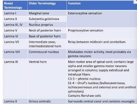
ZONES OF LAMINA
Summary:
1-6 Dorsal Horn
7- Lateral or Intermediate Zone
8-9 Ventral Horn
10 - Surrounding Central canal
Break muna medyo marami na ung slides
part 2 coming soon