Mosby's Dental Radiation Health and Safety DANB Review
1/225
There's no tags or description
Looks like no tags are added yet.
Name | Mastery | Learn | Test | Matching | Spaced | Call with Kai |
|---|
No analytics yet
Send a link to your students to track their progress
226 Terms
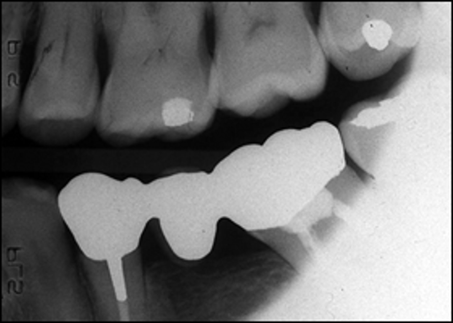
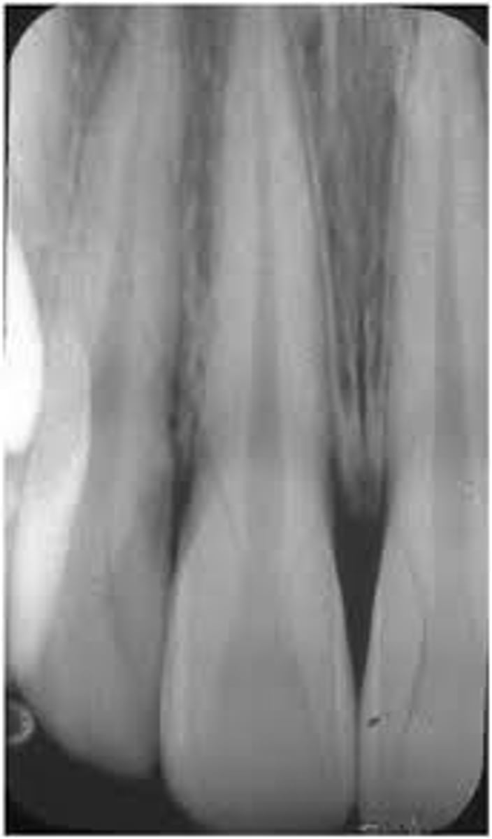
What is wrong with this image?
The image is cone cut
What causes black images on film or phosphor plates?
Exposure to light, unexposed sensors in some programs, or extreme overexposure.
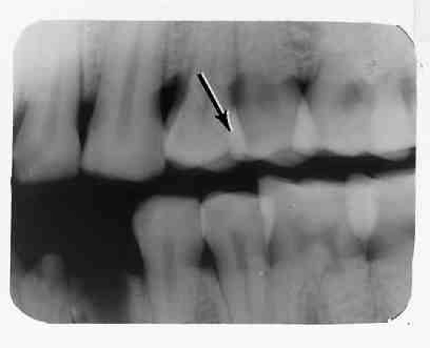
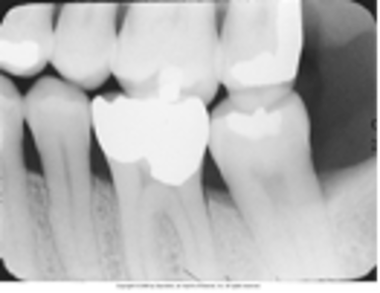
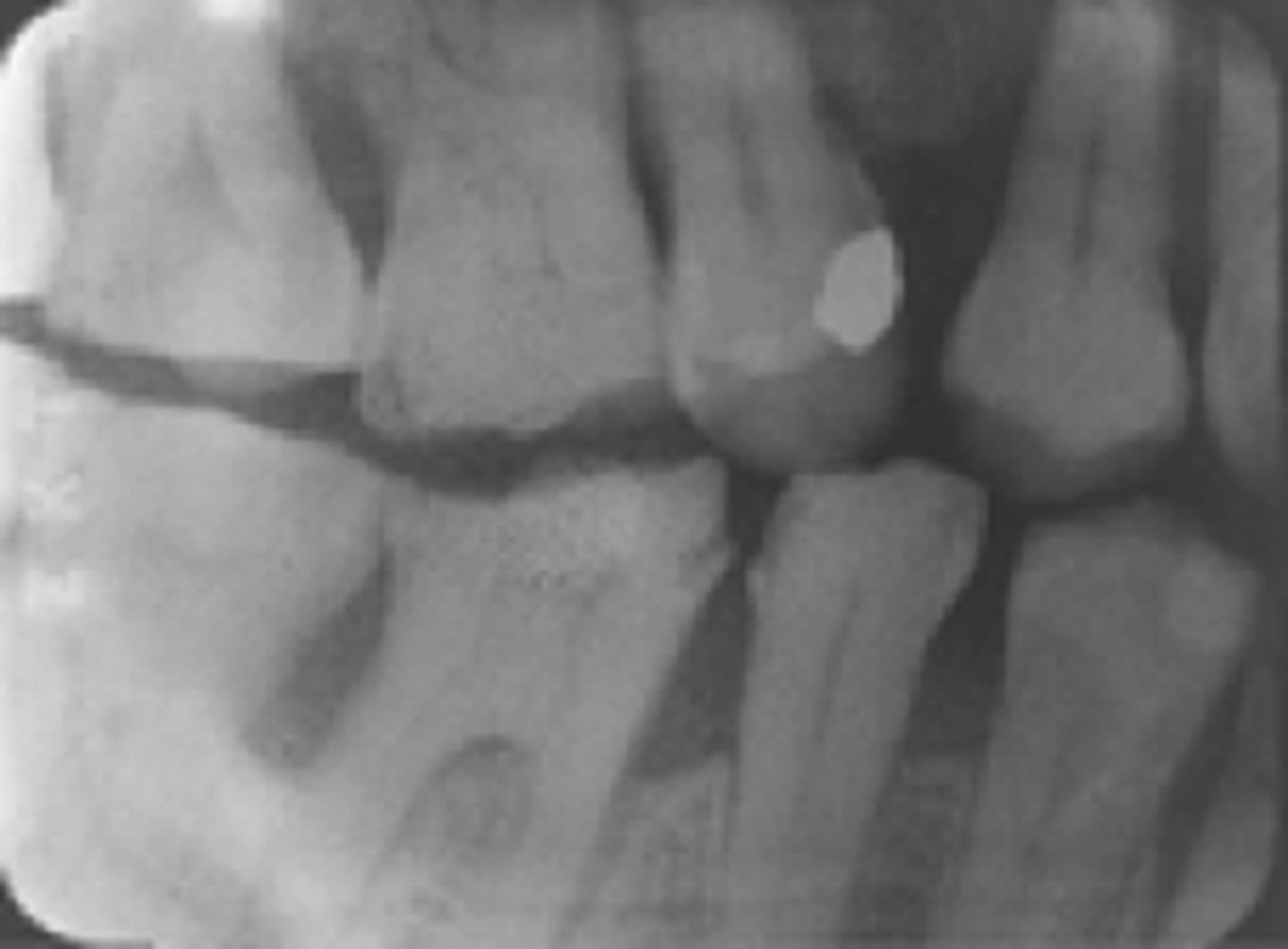
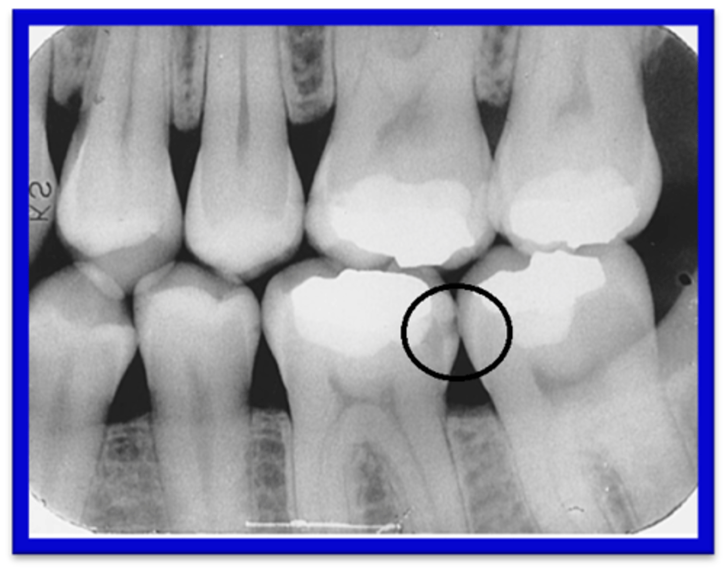
What causes overlap of interproximal spaces on an image?
Incorrect horizontal angulation.
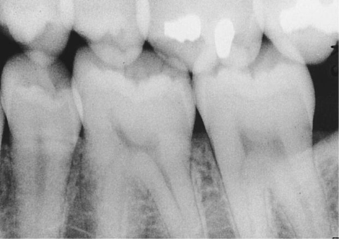
What causes one arch to be more visible than the other in a bitewing image?
Incorrect vertical angulation.
What causes too many teeth and distorted teeth on an image?
Double exposure of the receptor.
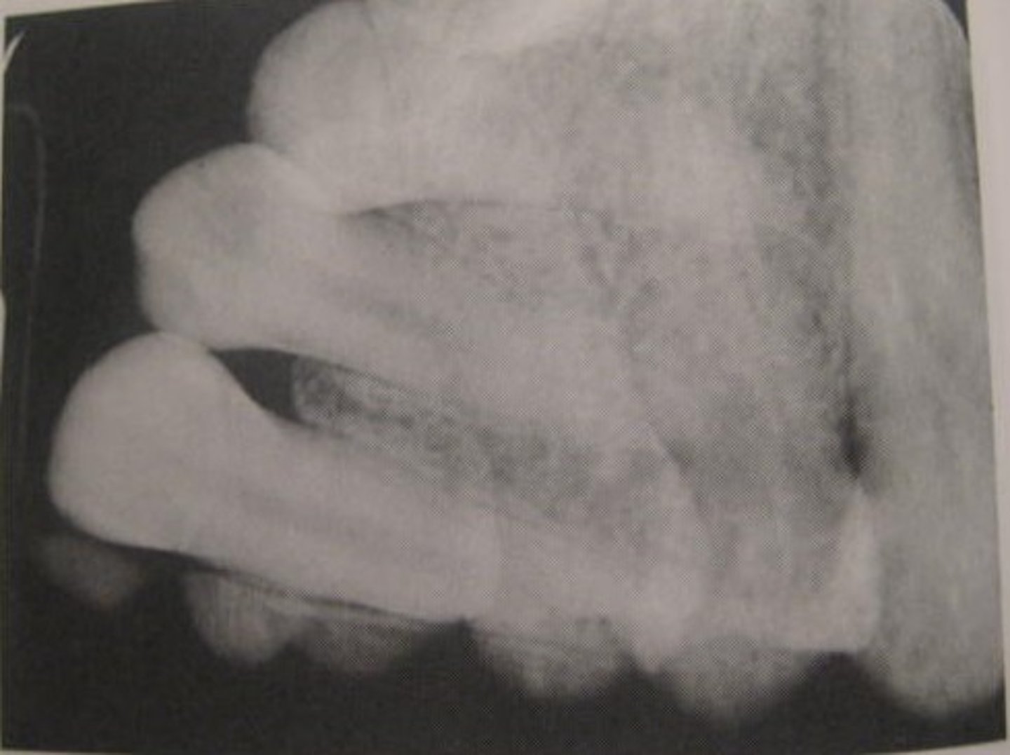
What causes a blurred radiographic image?
Patient or tube head movement.
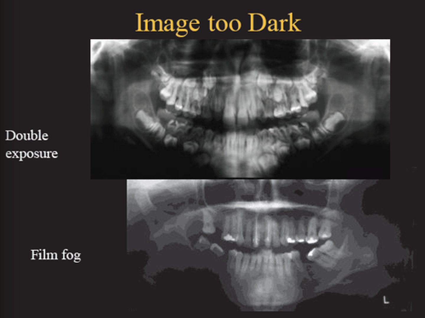
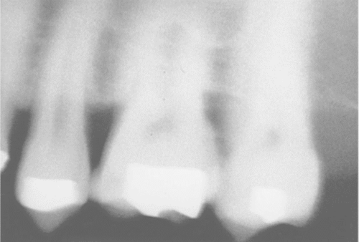
What causes an image to be too dark?
Overexposure due to high time, mA, or kV settings.
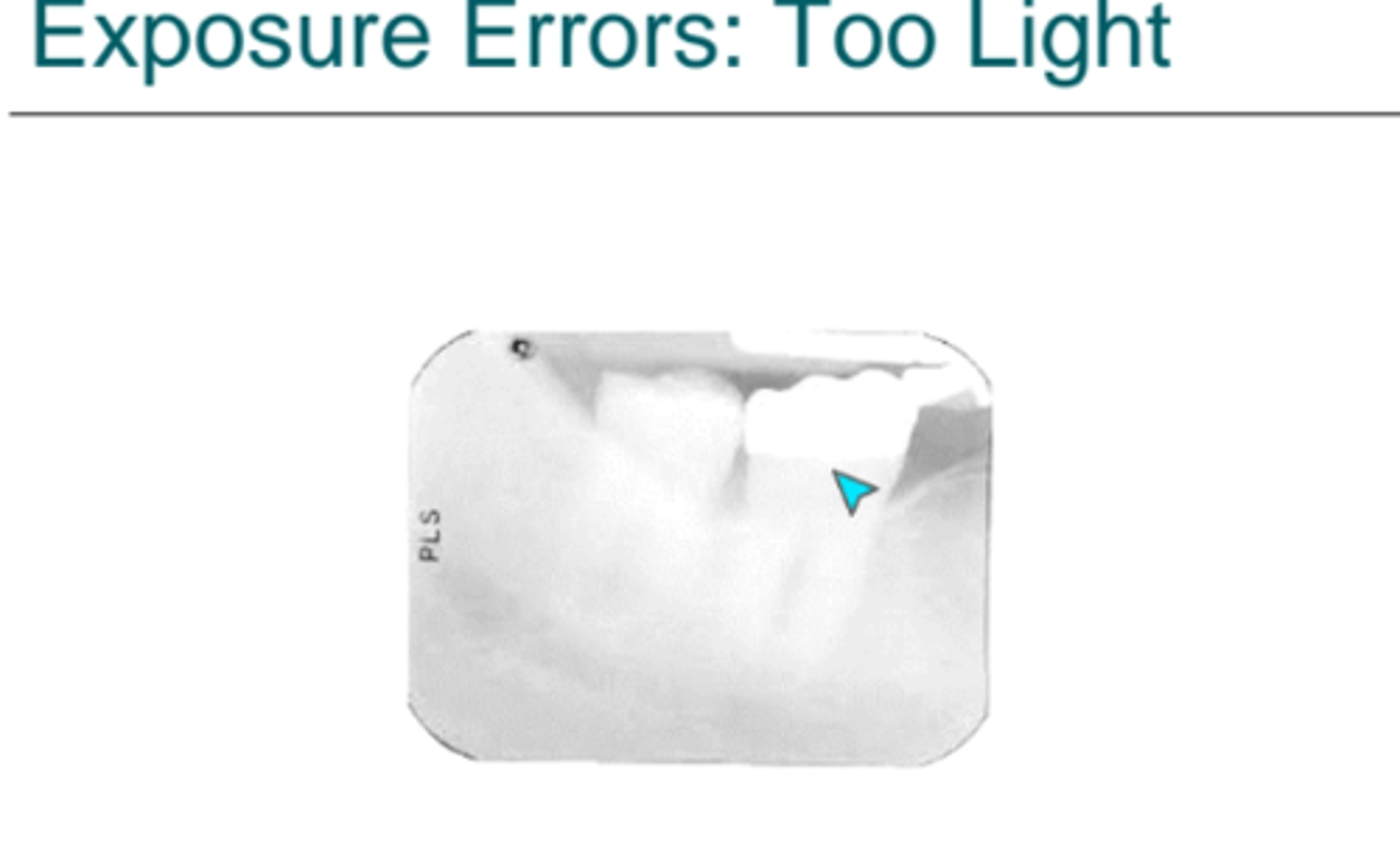
What causes an image to be too light?
Underexposure due to low time, mA, or kV settings.
What does a negative vertical angulation of the PID cause in a bitewing image?
Overlap of occlusal surfaces or more of the mandibular arch visible than the maxillary arch.
What causes elongation (longer crowns and roots) on an image?
Insufficient vertical angulation.
What causes foreshortening (shorter crowns and roots) on an image?
Excessive vertical angulation.

How does an indirect digital system work?
An exposed plate is placed in a scanner and converted to a digital image read by a computer.
How are intraoral sensors identified for the correct side?
An identification letter 'a' is used instead of an identification dot.
What is an intraoral sensor?
A small, bulky, and rigid detector placed intraorally to capture a radiographic image stored in a computer.
What are the two types of extraoral film?
Screen film and nonscreen film.
Which type of screen film is more efficient: blue or green?
Green screen film is more efficient and requires less exposure.
Why is nonscreen film not recommended for dental radiography?
It requires more exposure time than screen film.
What is duplicating film used for?
To make an identical copy of an intraoral or extraoral radiograph.
What is film speed?
A measure of a film's sensitivity to radiation.
What does kilovoltage (kV) control?
The force or penetrating ability of electrons, which determines beam quality, density, and contrast.
What does milliamperage (mA) regulate?
The quantity of electrons produced by regulating the cathode temperature.
What is radiographic density?
The overall darkness or blackness of an image, controlled by kV, mA, and time.
What controls the contrast of a radiographic image?
Only the kilovoltage (kV).
What is the difference between high and low contrast?
Low contrast (many shades of gray) occurs with increased kV; high contrast (few shades of gray) occurs with decreased kV.
What is the effect of decreasing kV on image contrast?
It increases contrast, producing an image with fewer shades of gray.
What is the quantity of x-rays regulated by?
mA and time.
What is the quality of the x-ray beam regulated by?
kV.
What is the function of the master switch on the control panel?
It turns on the x-ray machine.
What does the mA selector control?
The number of electrons produced.
What does the exposure button control?
The number of impulses per second that x-rays are emitted from the tube head.
What is the purpose of the step-down transformer?
To decrease incoming voltage from 110 or 220 volts to 3 to 5 volts for the filament.
What process releases electrons from the tungsten filament?
Thermionic emission.
What is the purpose of the step-up transformer?
To increase voltage to 65,000 to 100,000 volts to propel electrons to the anode.
What percentage of kinetic energy from electrons striking the target becomes x-ray photons?
1% (the rest becomes heat).
What are Bremsstrahlung x-rays?
X-rays formed when high-speed electrons slow down or stop upon hitting the nucleus of a tungsten atom.
What is the minimum kV required to create characteristic x-rays?
70 kV.
What is the purpose of the filter in the x-ray tube head?
To remove low-energy, longer wavelength x-rays.
What is the purpose of the collimator?
To restrict the size of the x-ray beam.
What is primary radiation?
The useful beam that comes from the target of the x-ray tube.
How should contaminated developer or fixer solutions be handled?
They must be disposed of, and the tank must be cleaned and refilled with fresh chemicals.
What is the recommended storage temperature for x-ray chemicals?
60°-70°F.
What are the characteristics of developer solution?
It is alkaline, caustic, and can burn tissue.
What should be done if skin contacts developer or fixer?
Wash the skin immediately.
Which maxillary teeth should be placed last to avoid a gag reflex?
Maxillary molars.
What is a technique to help a patient with a severe gag reflex?
Use a topical anesthetic.
What is the appearance of metallic restorations on a dental image?
Completely radiopaque (white).
How do dental caries appear on a dental image?
Dark or radiolucent.
What is the function of the PID?
To aim the x-rays at the film in the patient's mouth.
What does the extension arm house?
The wire between the tube head and the control panel.
What is the function of the molybdenum cup?
It contains the electron cloud until the exposure button is depressed.
How often does the American Academy of Oral and Maxillofacial Radiology recommend inspecting X-ray machines?
Annually
What is the appearance of fresh, properly processed film?
Clear with a slight blue tint
What is the appearance of expired film?
Fogged
How often should screens and cassettes be checked for light leaks and warping?
Monthly
How often should automatic and manual processing equipment be checked?
Daily
What is the purpose of a clearing test in film processing?
To test fixer strength
How often should processing solutions be replaced?
Every 3 to 4 weeks based on usage and test results
What is the purpose of a periapical examination?
To examine the entire tooth including the crown, root, and supporting tissues
What is the purpose of an interproximal (bitewing) examination?
To examine the crowns of maxillary and mandibular teeth, adjacent tooth surfaces, and crestal bone
What is the purpose of an occlusal examination?
To examine large areas of the maxilla or mandible to locate retained roots, supernumerary teeth, impacted teeth, foreign bodies, or salivary stones
What is the purpose of a panoramic examination?
To obtain an overall view of the maxilla and mandible to evaluate impacted teeth, eruption patterns, growth, and detect lesions or diseases
What is the purpose of lateral cephalometric images?
To evaluate facial growth and development, trauma, and developmental abnormalities
What structures are visualized in TMJ imaging?
The mandibular condyle, the glenoid fossa, and the articular eminence
How does CBCT (Cone Beam Computed Tomography) compare to traditional 2D imaging regarding radiation exposure?
It requires more radiation exposure than 2D intraoral and extraoral imaging
What is the SLOB rule used for?
Localization of objects (Same Lingual, Opposite Buccal)
What are the three component parts of an intraoral X-ray machine?
Tube head, extension arm, and control panel
What is the likely cause of an underdeveloped, light film?
Inadequate development time or developer solution that is too cool
What is the likely cause of an overdeveloped, dark film?
Excessive developing time or developer solution that is too hot
What causes reticulation of the emulsion on a film?
Sudden temperature change between the developer and the water bath
What modification is recommended for a patient with a narrow arch?
Use a smaller receptor
Where should the receptor be placed for a patient with a maxillary torus?
On the opposite side of the torus, between the torus and the tongue
What can be used to substitute missing teeth in edentulous patients?
Cotton rolls
What is the purpose of a cleaning film in processor maintenance?
To clean the rollers each day
What is the frequency for cleaning processor tanks?
Monthly
What is the primary use of skull imaging?
To examine the bones of the face and skull for orthodontics and oral surgery
What is the purpose of lateral jaw imaging?
To examine the posterior region of the mandible for impacted molars, fractures, or pathology
What causes dark or black spots on a processed film?
Developer coming in contact with the film before processing.
What causes white or light spots on a processed film?
Fixer coming in contact with the film before processing.
What causes yellow-brown stains on a processed film?
Exhausted developer or fixer, insufficient fixing time, or insufficient rinsing.
What is film mounting?
The process of placing dental radiographs into a holder and arranging them in anatomic order.
What information must be labeled on a film mount?
Patient's full name, date of exposure, dentist's name, and radiographer's name.
What does the labial mounting method assume?
The viewer is looking directly at the patient, where the patient's left side is the viewer's right side.
How is intensity of an x-ray beam defined?
The total energy and number of photons contained in the x-ray beam in a given area at a given time.
What factors affect the intensity of an x-ray beam?
kV, mA, exposure time, and distance.
What is the function of an aluminum filter in an x-ray machine?
It reduces beam intensity and removes long-wavelength, low-energy x-rays.
What does the inverse-square law state regarding x-ray intensity?
The intensity of the x-ray beam is inversely proportional to the square of the distance from the source.
What factors influence the density of a radiographic image?
kV, mA, exposure time, and subject thickness.
How does kV affect radiographic contrast?
Low kV produces high contrast (short-scale), while high kV produces low contrast (long-scale).
How does screen thickness affect the sharpness of an image using intensifying screens?
Thinner screens produce sharper images.
Why is a long (16 inch) PID used in the paralleling technique?
To prevent magnification caused by the receptor being away from the tooth.
What causes distortion in a radiographic image?
When the object and receptor are not parallel or the x-ray beam is not perpendicular to the object and receptor.
How often should darkrooms be checked for light leaks?
Every month.
How often should a safelight check be performed?
Every 6 months.
How often should viewboxes be checked?
Weekly.
What is the rule regarding the storage of lead aprons?
They should be checked for damage and never folded.
What did the 1968 Radiation Control for Health and Safety Act accomplish?
It standardized radiation equipment.
What did the 1981 Consumer-Patient Radiation Health and Safety Act accomplish?
It standardized education and certification for persons using radiation equipment.
In most states, how long should dental images be kept?
7 years.
What is the statute of limitations for dental negligence claims in most states?
3 years following reasonable discovery of damages.
What is the statute of limitations for children regarding dental negligence?
3 years following the legal age of adulthood.