Neurobiology test 3
1/234
There's no tags or description
Looks like no tags are added yet.
Name | Mastery | Learn | Test | Matching | Spaced | Call with Kai |
|---|
No analytics yet
Send a link to your students to track their progress
235 Terms
3 parts of ear
outer, middle, inner
From _____ _____ outward is considered the outer ear
Tympanic membrane
Sound progression
1. sound waves arrive at tympanic membrane
2. movement of the tympanic membrane causes displacement of the ossicles
3. movement of the stapes at the oval window establishes pressure waves in the perilymph of the scala vestibuli
______ wiggle when vibration occurs and moves towards the inner ear
ossicles (malleus, incus, stapes)
What does the tympanic membrane do?
transmits and amplifies sound
How much amplification occurs between the tympanic membrane and the oval window?
8-9x
What does the stapes do?
Moves like a piston to disrupt fluid in the cochlea
Round window is aka the ____ _____
release valve
along the way, the waves are acting with the _______ of ______-
organ of corti
external acoustic meatus
Canal leading to eardrum and middle ear
internal acoustic meatus
A passage for CN VIII from the inner ear to the brain.
What are the cochlea and vestibular apparatus embedded in?
bone
vestibular apparatus=
semicircular canal
-at 90 degrees to each other
-coordinates balance
stimulus of hearing
compression waves of air particles
frequency
pitch
amplitude
volume
in/out of inner ear
in-sound
out-action potential
branch of facial nerve runs thru
middle ear
tensor tympani
the muscle attached to the malleus; tensing the tensor tympani decreases vibration
- reduce transmission of loud noise
stapedius
the muscle attached to the stapes; tensing the stapedius decreases vibration
-reduce transmission of loud noise
The inside of the vestibule and cochlea are filled with
endolymph or perilymph
Vestibule
balance and equilibrium
maculae
sensory receptors for static equilibrium in the vestibule
-collection of hair cells detecting the center of gravity
-inside utricle or saccule
ampullae
a bulbous structure at the base of semicircular canals in the inner ear's vestibular system
-It houses sensory hair cells within a gelatinous structure called the cupula
-playing a crucial role in maintaining balance
cupulla
a gelatinous mass found in the ampulla of the semicircular canals; moves in response to the flow of the fluid in the canals
-motion sensor for rotational acceleration
saccule and utricle
2 chambers with endolymph in vestibule
-utricle connected to (u-shaped) semicircular canals
Cranial nerve 8
vestibulocochlear nerve
otoconia otoliths
-crystals in the macullae, can cause stereo
(Ear) synapsing occurs between _____ ____ and _____ ____
hair cells and afferent neurons
Where does sound land?
Superior aspect of the temporal lobe
Medial geniculate nucleus
Sound processing in the thalamus
lateral lemniscus
band of fibers carrying auditory information through the medulla and pons
corpora quadrigemina
located in the midbrain; contains reflex centers for vision and auditory reflexes
Auditory reflexes go through this
inferior colliculus
______ _____ is in the top of the nasal cavity
olfactory epithelium
-these run thru the cribriform plate
Inside nasal epithelium:
-olfactory receptors
- detect chemicals
-odorant molecule binds to receptor and activated G- protein and depolarizing event begins
How many genes are in the olfactory receptor class?
4-500
Visible structures on tongue
papilla
cranial nerves for taste
Facial (VII)
Glossopharyngeal (IX)
Vagus (X)
cranial nerves for smell
olfactory
vestibule of ear
head motion, gravity, and position, sending signals to the brain to maintain stability
dynamic equillibrium
maintains balance during rotational movement, using the semicircular canals
static equilibrium
maintains balance relative to gravity when still, using the utricle and saccule within the vestibule
semicircular canals
motion sensors, detecting rotational acceleration of the head in three-dimensional space
Everything has a layer of ___ mediating the taste
saliva
Taste pores have an opening where ______ make ______ available
-microvilli
-g-protein coupled receptors
All gustatory neurons have ______ cells
they are subject to damage so they need to be replaced readily
basal
2 classes of taste receptors
-g- protein couples
-direct ion channels
mediating sour and salty
Taste
CN 7 and 9 (tiny bit 10)
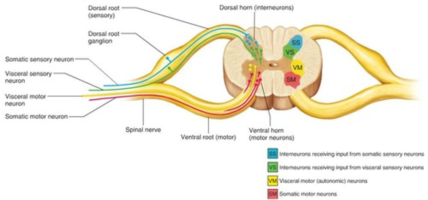
Functions of spinal cord
transmit, transmit, and integrate
transmitting info
-lots of pathways in the white matter
-most names start with where it's coming from and end with where they're going to (ascending or descending)
Ascending pathways
-dorsal white column
-dorsal spinocerebellar tract
-ventral spinocerebellar tract
-lateral spinothalamic tract
-ventral spinothalamic tract
Descending pathways
-ventral white commissure
-lateral reticulospinal tract
-lateral corticospinal tract
-rubrospinal tract
-medial reticulospinal tract
-ventral corticospinal tract
-vestibulospinal tract
-tectospinal tract
Cortical spinal tracts are most important
motor control
spinal thalamic
important sensory
rubrospinal
unique set of motor neurons
integration
-reflexes
-descending tracts have to come out at a spinal cord level
white matter
super highway
grey matter
off ramp to sideroads
somatic reflex
contraction of skeletal muscles
-receptor detection, signal transmission by a sensory neuron, central integration, signaling via a motor neuron, and effector muscle response
spinal cord is protected by
bone, meninges, CSF
meninges
pia mater
arachnoid mater
dura mater
Between the pia and white we have
CSF
A bulge is only on the _____ not the ______
dorsal root, ventral root
CSF is produced in
-choroid plexus (in each of the ventricles)
-support, protect, buffer
CSF functions
support, protect, buffer
Foramen magnum to L1 =
solid structure... spinal cord anatomy
-31 spinal nerve pairs
-31 spinal segments
-cervical= c1-c8
-thoracic nerves= t1-t12
-lumbar nerves= l1-l5
-sacral nerves=s1-s5
-coccygeal nerve=Co1
dermatomes
Skin areas innervated by specific spinal nerves
white matter structures of spinal cord
dorsal columns, lateral columns, ventral columns
grey matter structures of the spinal cord
posterior/dorsal horn, intermediate zone, anterior/ventral horn, and grey commissure with the central canal
Ventricles of brain
2 lateral ventricles, 3rd ventricle, 4th ventricle
-CSF flows thru these
Spinal enlargements
cervical and lumbar
___ ___ does not have an associated vertebrae bone
c8
_____ is fused in adulthood
Sacrum
Dermatomes
portion of skin innervated by which specific spinal nerve
4 main plexuses of ventral rami
cervical, brachial, lumbar, sacral
gray matter spinal cord organization
Ventral rami -> form _______----> nerve plexuses -----> 4 plexuses
interlaced bundles of nerve fibers
What happens when spinal nerves leave the CNS and PNS
branch into ventral/dorsal rami to innervate the body, while lower nerves form the cauda equina to exit lower down, changing from CNS-based, heavily myelinated tracts into peripheral nerves.
somatotopic
spatially mapped in the somatosensory cortex in correspondence to spatial events on the skin
anterolateral system
a somatosensory system that carries most of the pain information from the body to the brain
Cervical nerves
C1-C4/5 ish
-neck, thoracic cavity, diaphragmatic muscles
-phrenic= major nerve (c3-5)
cervical nerves overall function
motor control and sensation in the neck, shoulders, arms, and hands, while also controlling breathing and head movement
brachial
-C5 to T1
-gives way to axillary, muscular cutaneous, radial, ulnar
-roof and floor: deep investing fascia and prevertebral fascia
Thoracic nerves
T1-T12
-chest and abdominal muscles
ulnar nerve
C8-T1
-ring and pinky finger
-carries sensory info from the palm and the medial hand/ finger and sends motor commands to the wrist flexors/ intrinsic hand muscles
ulnar nerve function
controls fine motor movements of the hand (intrinsic muscles) and sensation in the pinky and half of the ring finger. It facilitates grip strength, finger adduction/abduction, and wrist flexion
musculocutaneous nerve
C5-C7
-anterior upper arm
-carries sensory info from shoulder region and motor commands to the deltoid muscle
musculocutaneous nerve function
flexion of arm at elbow, supination of forearm
median nerve
C5-C8, T1
-hand from ring finger to thumb
median nerve function
flexors of wrist and digits
radial nerve
C5-T1
-forearm - doesn't service hand
-carries sensory info from the posterior arm and motor commands to the triceps brachii, wrist extensors, and brachioradialis
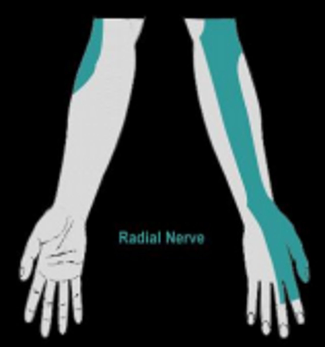
radial nerve function
drives elbow extension (triceps), wrist extension, finger extension, and thumb extension/abduction
axillary nerve
C5-C6
-deltoid and teres minor
-carries sensory info from the shoulder region and motor commands to the deltoid muscle
axillary nerve function
lifting the arm (deltoid), rotator cuff ( teres)
Lumbar nerves
L1-L5
-femoral nerve- (Pectineus, Illiacus, rectus femoris, vastus intermedius, vastus lateralis, vastus medialis) QUADS
-obturator nerve: any ADDUCTOR
-tibial nerve(every muscle below knee)
-common fibular nerve (every muscle below knee)
femoral nerve sensory/motor
-carries sensory info from much of the thigh, leg, and foot and sends motor commands to the quads
femoral nerve function
primary nerve for walking, running, and climbing stairs by operating the QUAD muscles
Obturator nerve
-L3-L5
-adductor brevis
-adductor longus
-adductor magnus
-gracilis
obturator nerve sensory/motor
-carries sensory info from much of the thigh
-sends motor command to adductor muscle