pract. 9 - operations of the thorax (sem 2)
1/21
There's no tags or description
Looks like no tags are added yet.
Name | Mastery | Learn | Test | Matching | Spaced | Call with Kai |
|---|
No analytics yet
Send a link to your students to track their progress
22 Terms
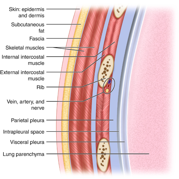
Layers of the Thorax
Skin
Subcutaneous fat
Superficial fascia
Deep fascia
Extrinsic muscles
Intrinsic muscles
Endothoracic fascia
Parietal pleura
Visceral pleura
Lung
Types of Thoracic Injuries
Crush injuries: lung, pleura, ribs
external pressure
Single rib fracture
Multiple rib fractures
Steering wheel injury: bilateral rib fractures, flail chest, sternal fracture
Stove-in chest / Flail chest
a section of the rib cage becomes detached from the rest of the chest wall
due to fractures in multiple ribs
Traumatic (open) pneumothorax
Hemothorax / Hemopneumothorax (commonly with fractured ribs)
Tension pneumothorax (most dangerous)
Pericardial or cardiac rupture, bronchial rupture
Associated injuries: liver, spleen, diaphragm, major vessels
Trauma Classification
Penetrating: parietal pleura is breached
Non-penetrating: parietal pleura remains intact
Thoracic Surgeries
Lobectomy
Surgical removal of a lobe of the lung
Pneumonectomy
Removal of an entire lung
Segmentectomy
Removal of a specific segment of a lung lobe
Wedge resection
Removal of a small, wedge-shaped portion of the lung that includes the lesion and a margin of healthy tissue.
ICD insertion
(Intercostal Drain / Chest Tube Insertion)
Insertion of a tube into the pleural space to remove:
Air (pneumothorax)
Blood (hemothorax)
Pus (empyema)
Fluid (pleural effusion)
thoracic incisions
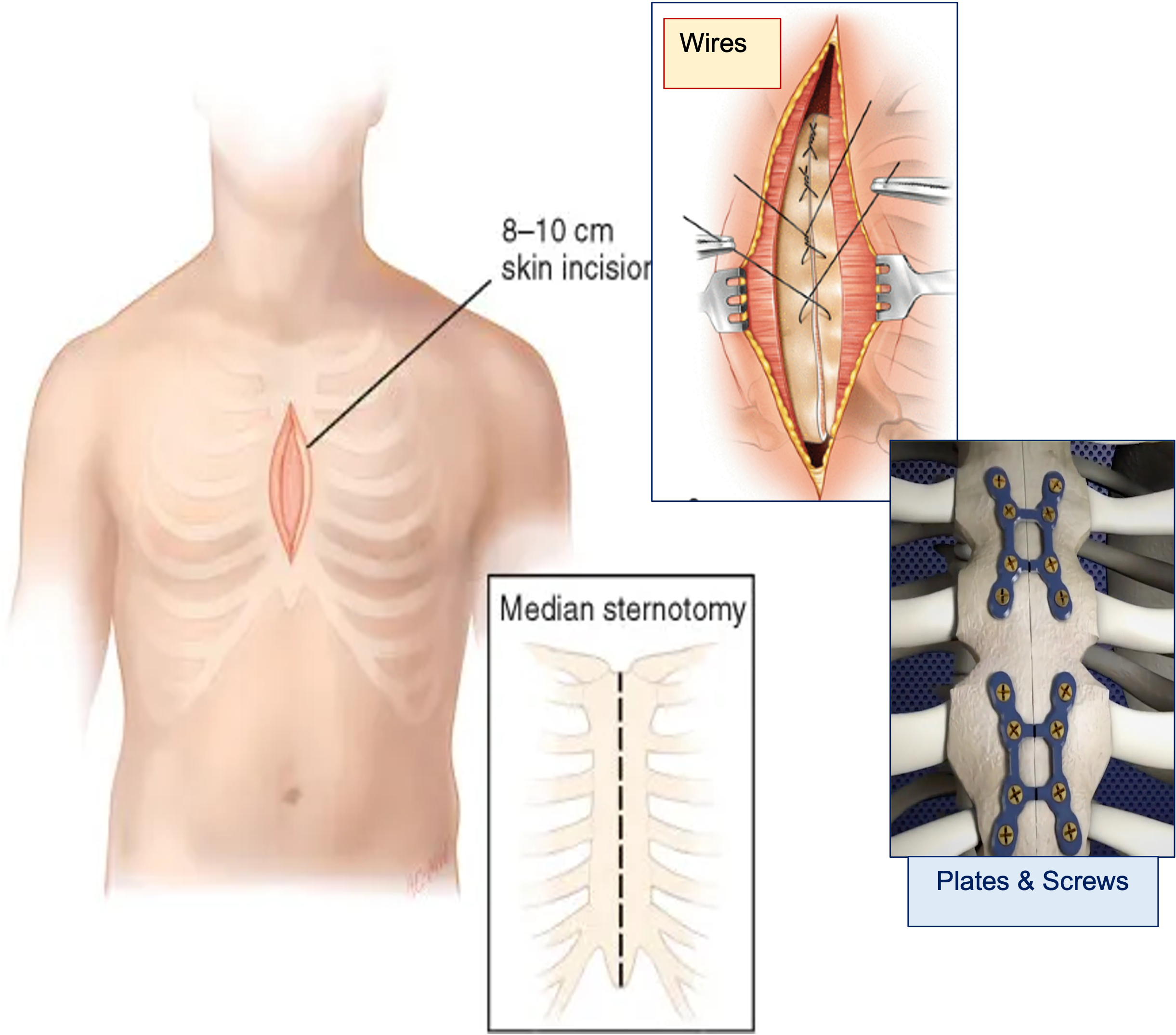
Median sternotomy
Posterolateral thoracotomy
Anterolateral thoracotomy
Clamshell incision
Median Sternotomy
Incision:
Vertical cut through the sternum
just below the neck to below the sternum
Access: Both lungs, heart and mediastinum
Commonly used for:
Cardiac surgeries
(e.g., CABG, valve replacement)
Bilateral lung procedures
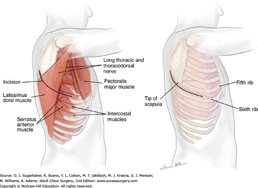
Posterolateral Thoracotomy
Incision: From back (posterior axillary line),
around to the front of the chest
Involves cutting through the latissimus dorsi and intercostal muscles
Access: One side of the thoracic cavity (excellent view of lung, oesophagus, aorta)
Used for:
Lobectomy
Pneumonectomy
Esophageal surgery

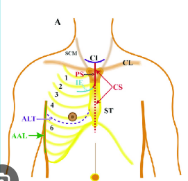
Anterolateral Thoracotomy
Incision: Along the anterior chest wall
(4th–6th intercostal space)
more toward the front
Access: Heart, anterior lungs, chest wall
Used for:
Emergency thoracotomies (e.g., trauma)
relieve the pressure- cardiac tamponade
clamp the blood vessel (aorta)- haemorrhage
Some cardiac procedures
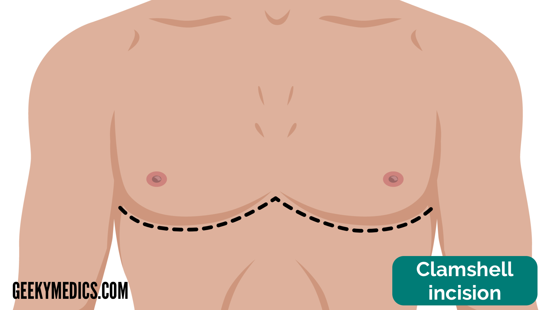
Clamshell Incision
Incision:
Bilateral transverse thoracotomy
across the chest (under the breasts),
joining both sides
→ opens chest like a clamshell
Access: Both lungs, entire mediastinum, heart, great vessels
Used for:
Bilateral lung transplantation
Extensive trauma
Mediastinal tumors
pathological effects of chest injuries
Immediate effects
Hypoxia
Hypercapnia
Acidosis
Hypovolemic Shock
Due to internal bleeding
Cardiac Arrhythmias
Bronchospasm
Reflex airway narrowing
In response to trauma or pain
→ worsens hypoxia
Late Effects
Empyema
Collection of pus in the pleural cavity
Fibrothorax
Thickening and fibrosis of the pleural space
Lung Abscess
Mediastinitis
Severe infection of the mediastinum
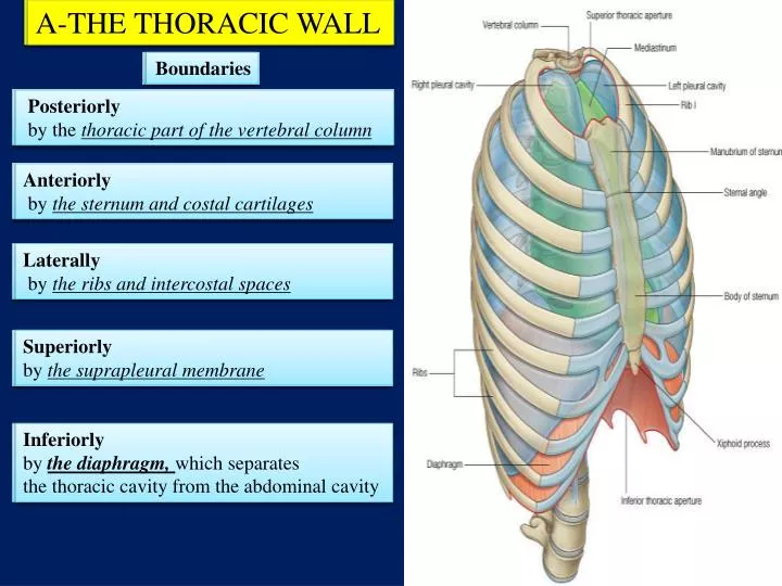
Thorax Boundaries (According to Stef)
Superior: suprapleural membranes
Posterior: vertebral bodies
Lateral: ribs & intercostal muscles
Anterior: sternum & costal cartilages
Inferior (floor): diaphragm
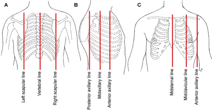
Anatomical lines
What are the 4 manipulations of the thorax? (stef question)
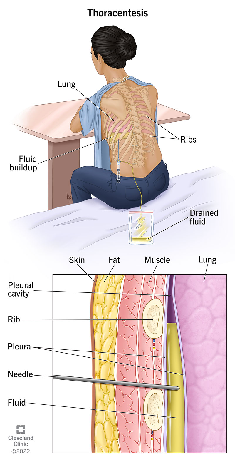
Puncture
Thoracocentesis
controlled puncture of the pleural space
using a needle or cannula.
To remove fluid or air from the pleural cavity.
VATS (video assisted thoracoscopic surgery)
using a thoracoscope (camera) and small incisions.
Purpose:
Lung biopsy
Pleural biopsy
Lobectomy or wedge resection
Decortication for empyema
Thoracotomy (incision)
A major open surgical incision into the thoracic cavity.
Purpose: Direct access to lungs, heart, oesophagus, and major vessels.
Operations- names only
Thoracocentesis
Pneumothorax
Pericardiocentesis
Rib resection
Pleurodesis
Empyema
Decortication
Thoracocentesis
(Pleural Puncture)
Purpose: Diagnostic or therapeutic aspiration of pleural fluid.
Position: Patient seated, leaning forward.
Site: 8th–9th intercostal space
mid/posterior axillary line.
Technique:
Local anesthesia (1%)
Needle inserted above upper border of rib below (to avoid neurovascular bundle)
Antibiotic instillation post-aspiration
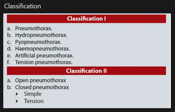
Pneumothorax
Definition: Entry of air into pleural cavity, collapsing lung.
Types:
Spontaneous
Traumatic
Tension → Most dangerous
Mechanism: Air enters during inspiration but can't exit
→ pressure ↑
→ mediastinal shift
→ ↓ venous return
→ heart failure
Emergency: Needs needle decompression
2nd ICS
midclavicular line
or chest tube
Pericardiocentesis
Purpose: Aspiration of fluid/blood from pericardial sac
Indication: Cardiac tamponade
Approach: Insert needle just below xiphoid,
angle 45°
directed upwards and leftwards
Needle: 16 or 18G
Rib Resection
Surgical removal of a rib
Purpose:
Access to thoracic organs
Rib tumour excision
Pleurodesis
part of the pleural space is artificially obliterated.
It involves the adhesion of the visceral and the costal pleura.
stick visceral to costal pleura
The mediastinal pleura is spared
Empyema
Definition: Pus in the pleural cavity
Caused by:
Lung abscess rupture
Pneumonia complications
Surgery, trauma
Phases:
Exudative: Thin pus
Fibropurulent: Thick, fibrinous
Organising: Fibroblast rind forms
Tx: Thoracocentesis + antibiotics ± surgery
Decortication
Removal of restrictive layer of fibrous tissue over the lung
Helps the lung tissue to re-expand
Terminology (stef question)
Pneumothorax- presence of atmospheric air in the thorax
Hemothorax- presence of blood in the thorax
Pyothorax- presence of pus in the thorax
Chylothorax- presence of lymphatic fluid in the thorax
Hydropneumothorax- presence of liquid and air in the thorax
Lobectomy- removal of a lung lobe
Pneumonectomy/ pulmonectomy- removal of the whole lung
Pneumonothorax- presence of pus and blood in the thorax