Embryology and Circulation of Fetal Heart
1/69
There's no tags or description
Looks like no tags are added yet.
Name | Mastery | Learn | Test | Matching | Spaced | Call with Kai |
|---|
No analytics yet
Send a link to your students to track their progress
70 Terms
Embryology
-cardiovascular system is 1st system to function in embryo
-most sensitive period in the first trimester for cardiac development is between 3.5-6.5 wks
-primitive circulatory system powered by a single chambered tube begins to beat 22 days after conception (day 35 GA/end of 5th wk)
3rd Wk
-development of the vascular system begins in the wall of the yolk sac
-by end of week circulation of blood has begun
6th Wk
-bulbous, hollow cardiovascular channel has established vascular communication w/ MAT circulation and main embryologic circulation
4th Wk
-paried endocardial heart tubes gradully fuse to form a single tubular heart
-fusion of the paired structures occurs from cranial to caudal end
-as heart elongates it bends upon itself
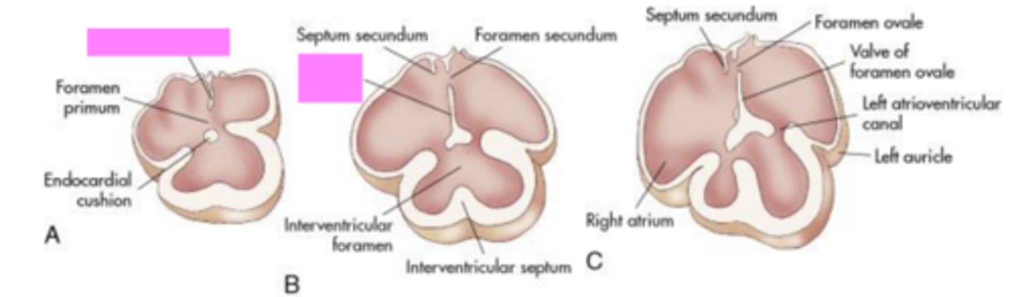
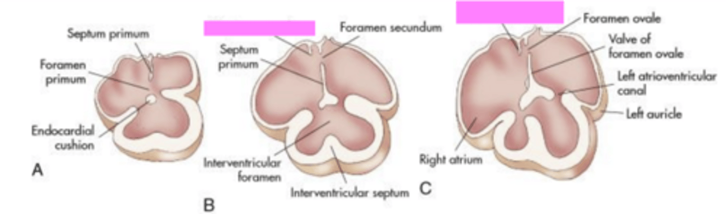
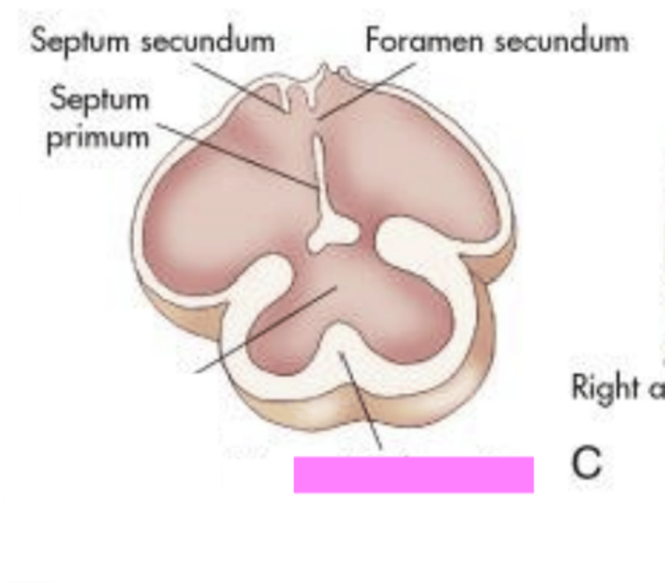
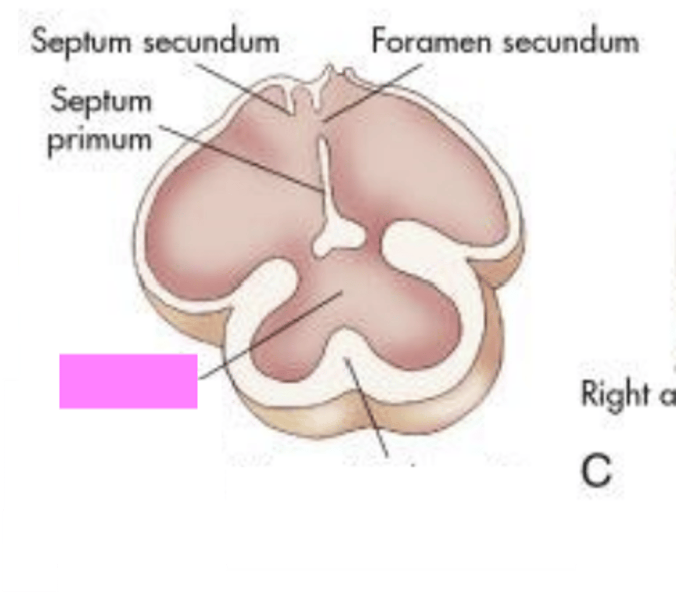
Septum Primum
grows from the roof of the single arterial chamber toward the centrally located endocardial cushions
Septum Secundum
grows adjacent to the septum primum
Atrial Septum
-formed by the septum primum and septum secundum
-partitions the right and left atria, leaving a gap that will eventually become the foramen ovale
Septum Primum
Septum Secundum
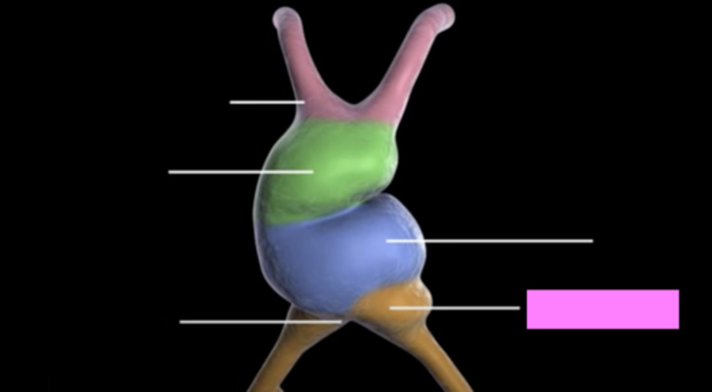
Truncus Arteriosus
-drumstick shaped
-sits cranial to the bilobed bulbus cordis
-partitions into the great arteries by the end of the 7th wk (aorta and pulmonary trunk)
Truncus Arteriosus
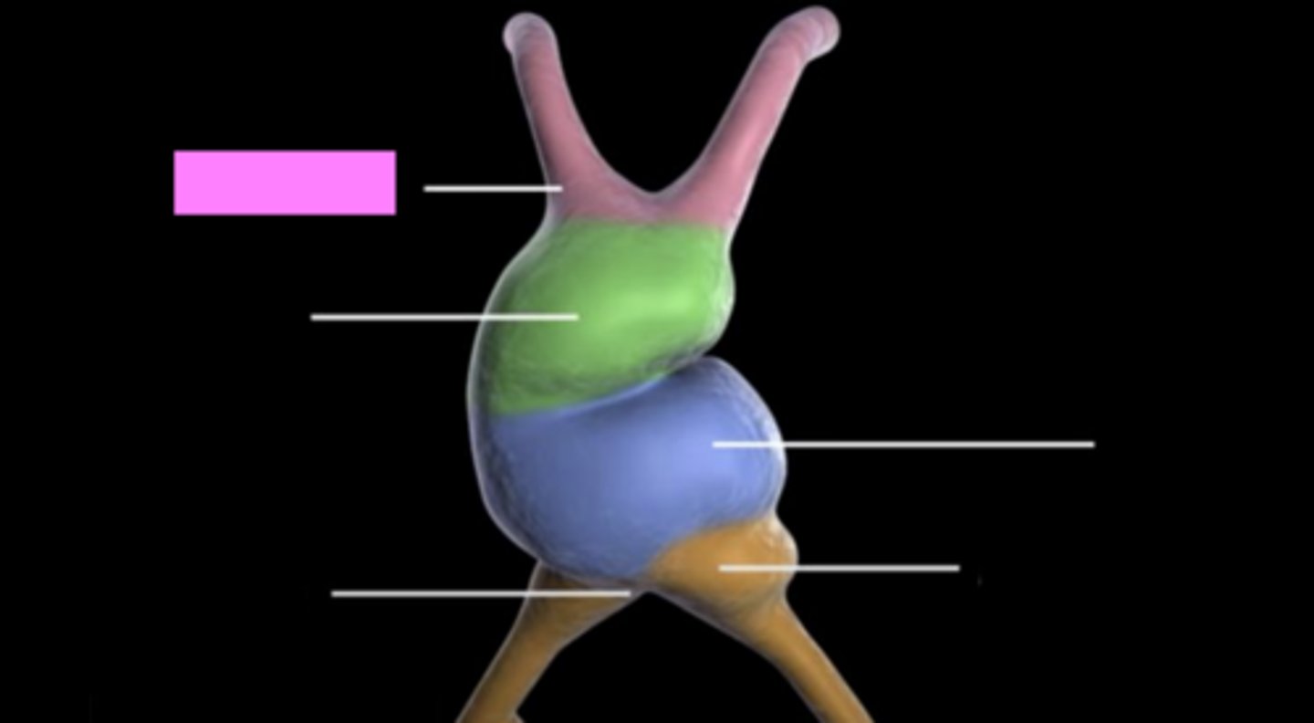
Bulbus Cordis
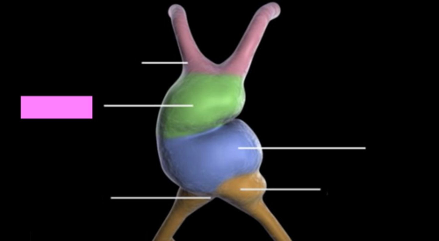
Ventricle
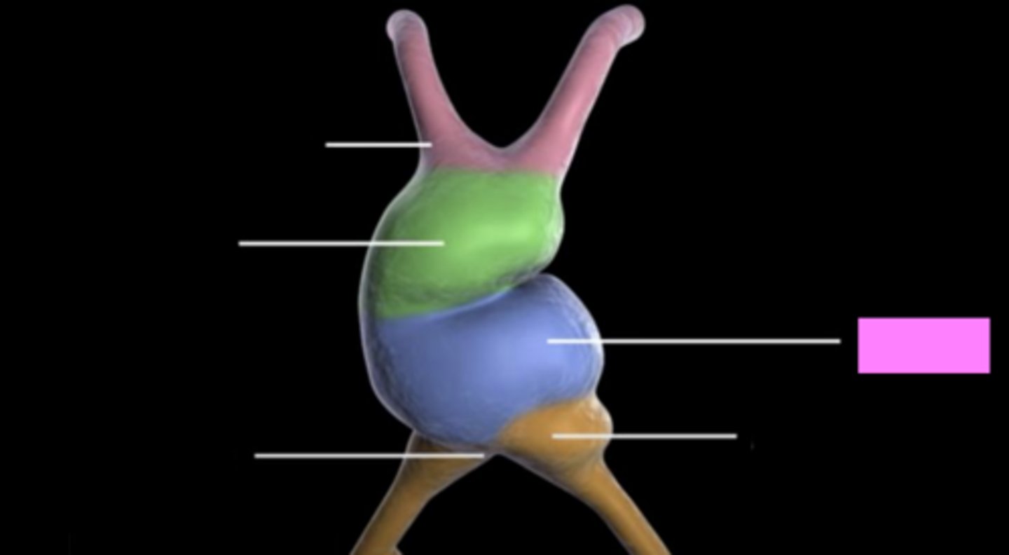
Sinus Venosus
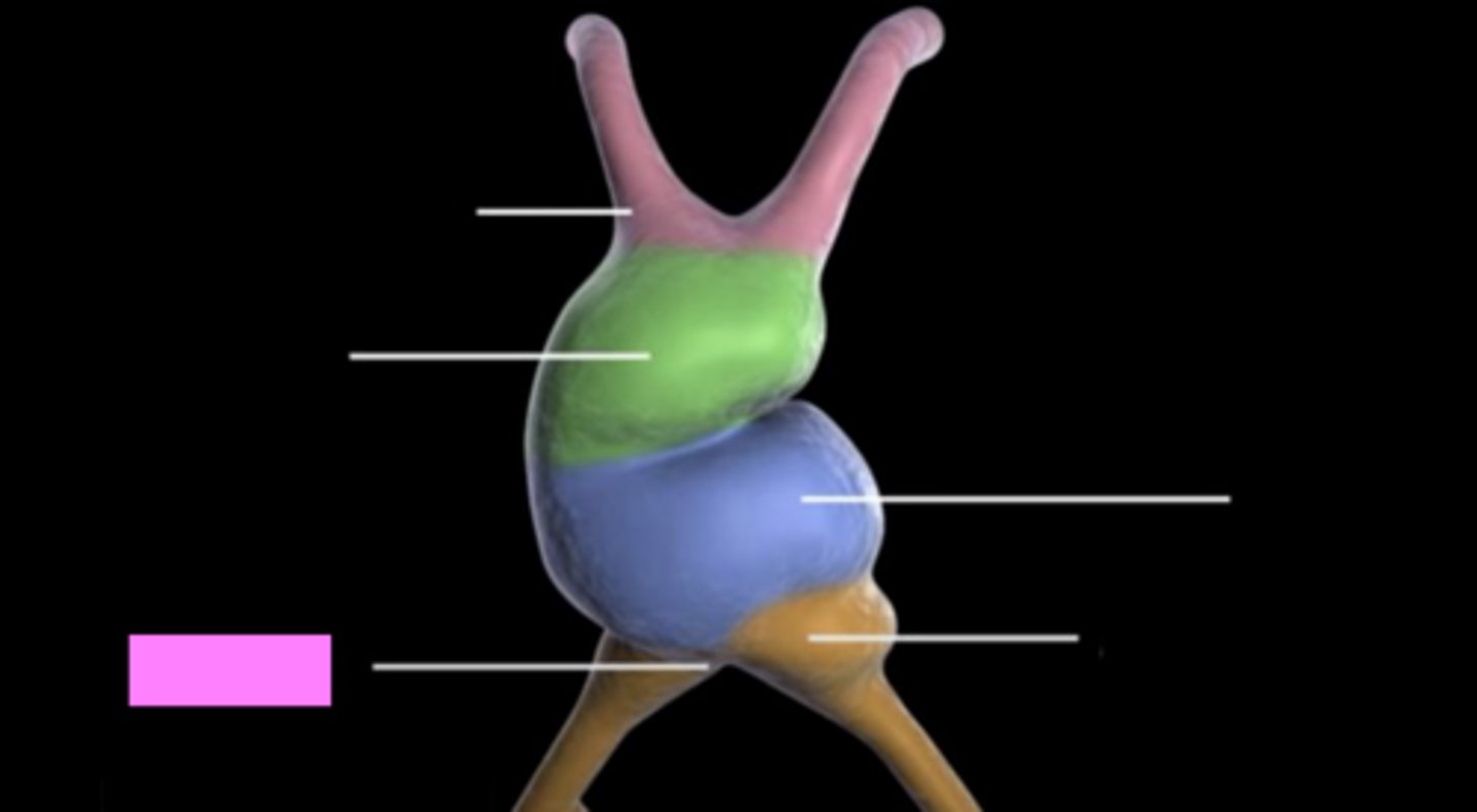
Primitive Atrium
Aorticopulmonary Septum
-ridges of tissue arise along the walls of the bulbus cordis and spiral into the aorticopulmonary septum over several days
-partitions the AO and pulmonary trunk; interference w/ this partitioning will result in conotruncal anomaly
Primordial Interventricular Septum
-arises from the apex (inferior)
-grows upward (toward the endocardial cushions) to separate the right and left ventricles
-leaves a space called interventricular foramen
Interventricular Foramen
permits cross circulation until end of the 9th wk
Primordial Interventricular Septum
Interventricular Foramen
Wk 4
-cardiovascular tube formation
-linear tube forms and begins beating as soon as its formed
5-6 Wks
-Looping
-linear tube bends into asymmetric right and left sides
-distinct chambers begin to form
6.8-9 Wks
-atrial septation
-atria septate by the septum primum and septum secondary
-endocardial cushions form
7-10 Wks
-outflow tract separation
-truncus arteriosis begins the develop aorta and pulmonary artery
7.4- 8.6 Wks
-ventricular septation
-right and left ventricles are formed by the growth of the interventricular septum
-embryonic development of herat is complete by approx 9 wks
Fetal Circulation
-primitive embryonic heart has evolved into a four chamber pump by 11 wks
-receives blood through a venous inflow system
-ejects blood via an arterial outflow system
-regulates flow within its chambers (series of valvews and temporary communication channels that seal off after birth)
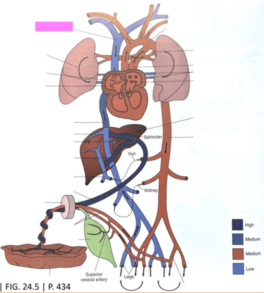
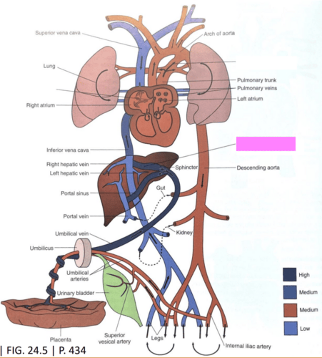
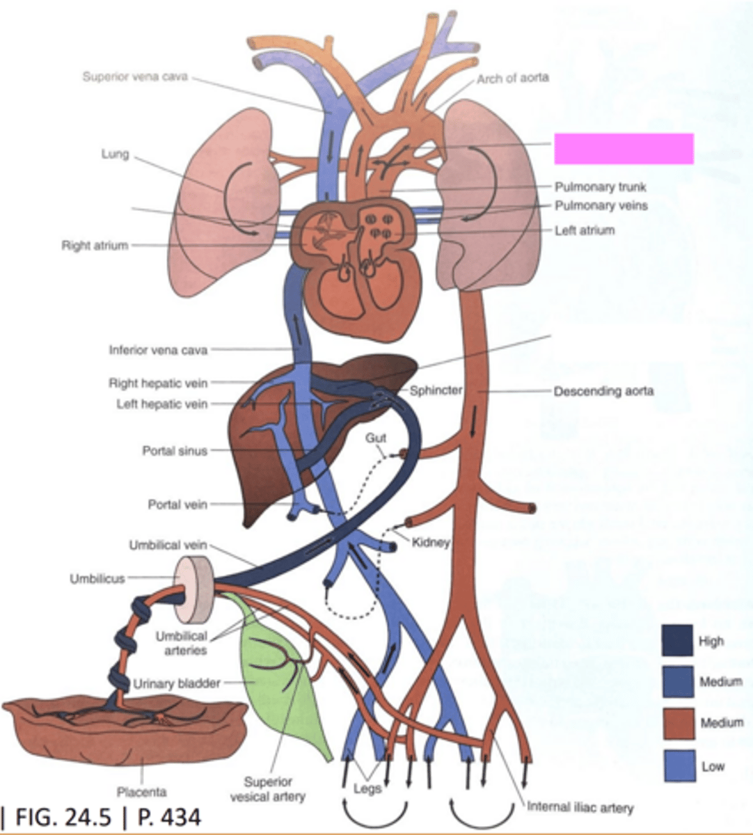
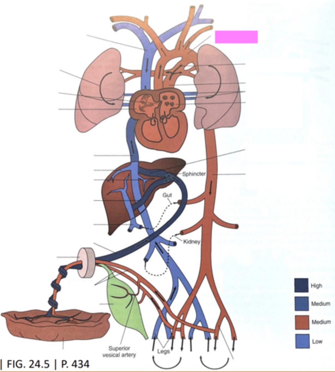
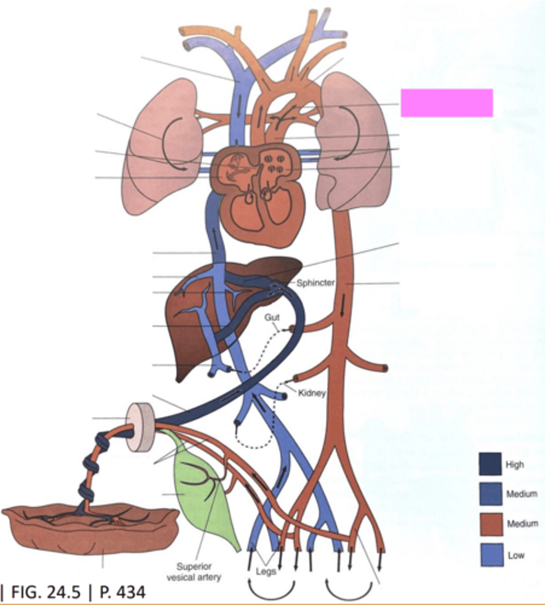
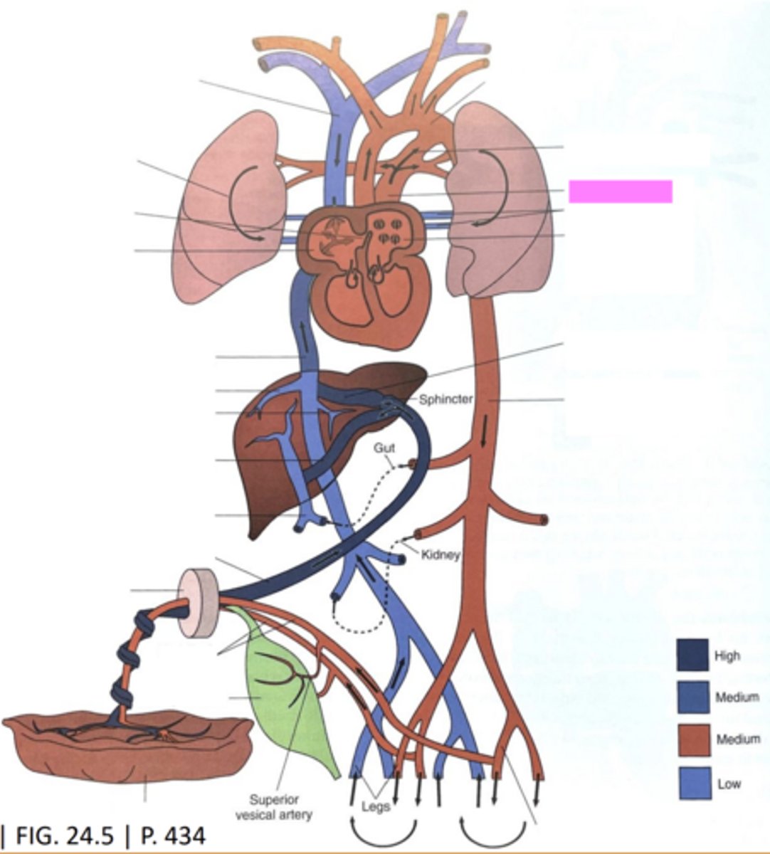
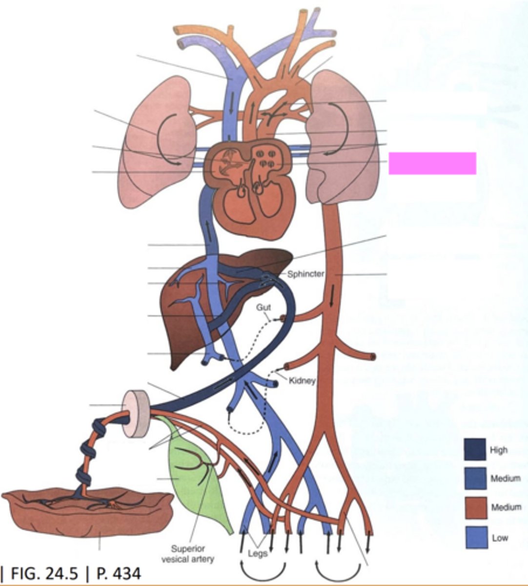
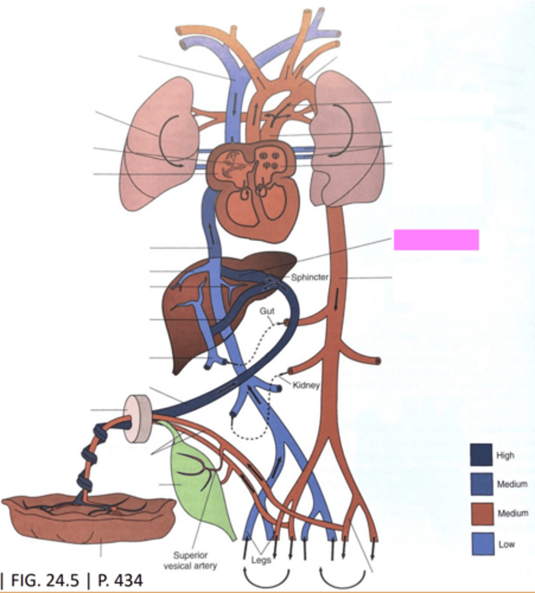
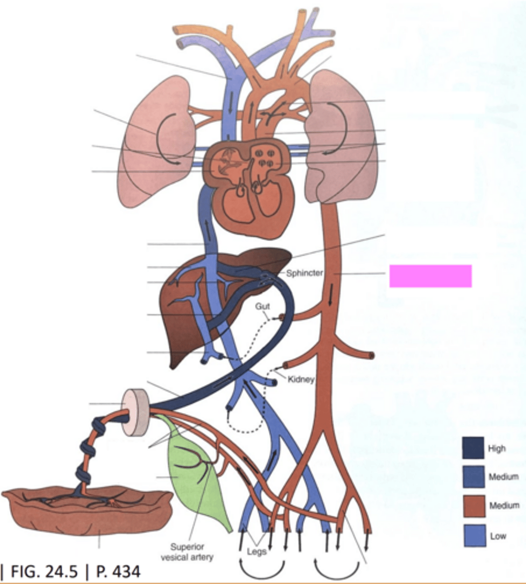
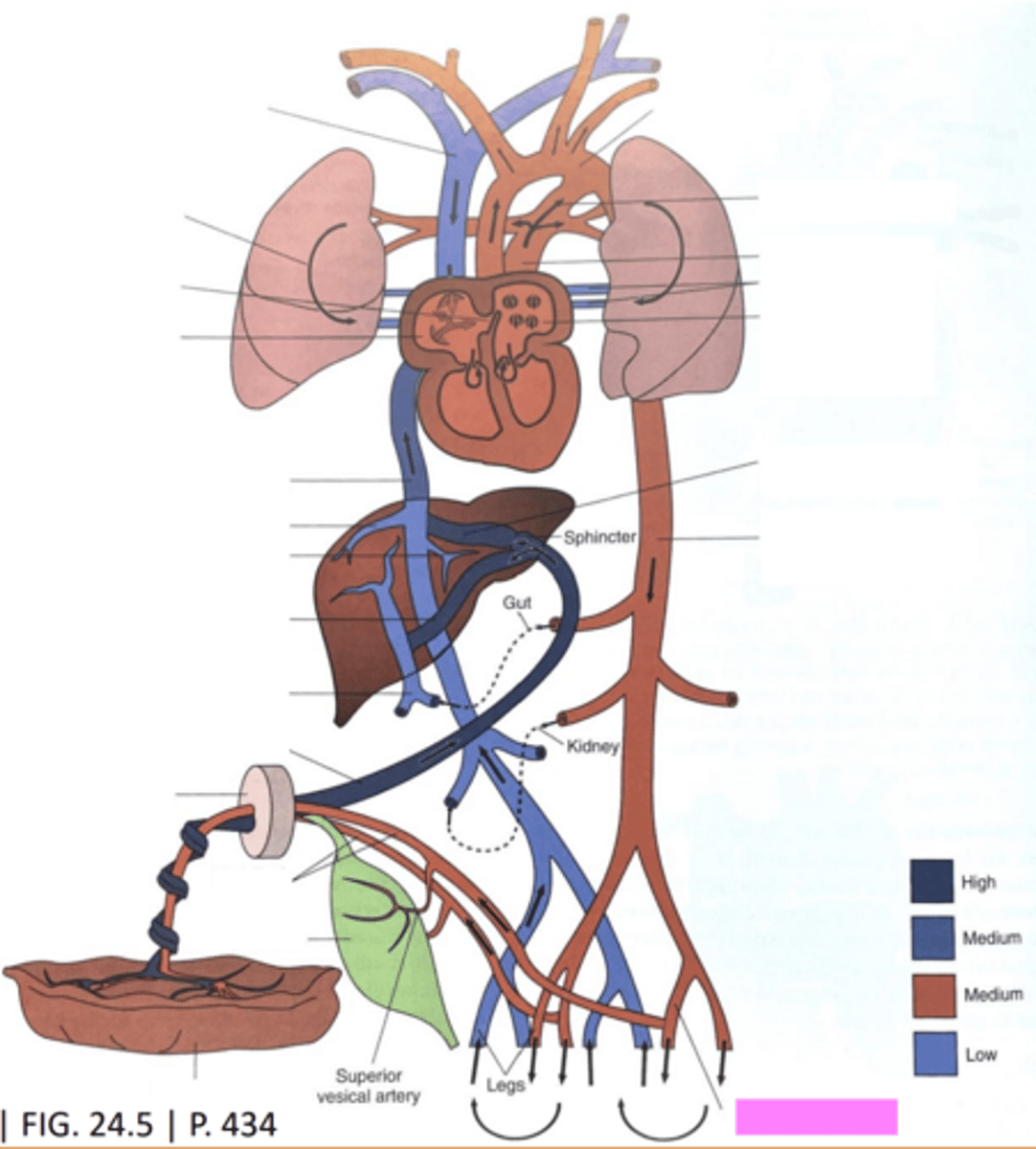
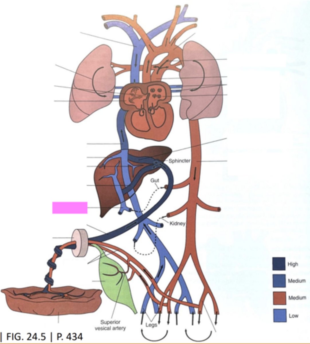
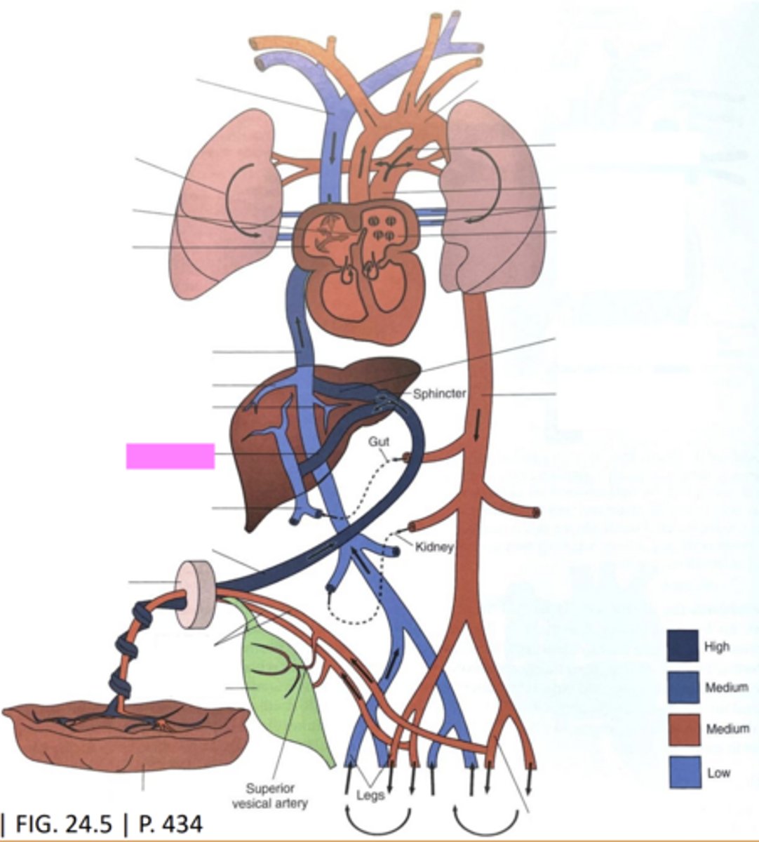
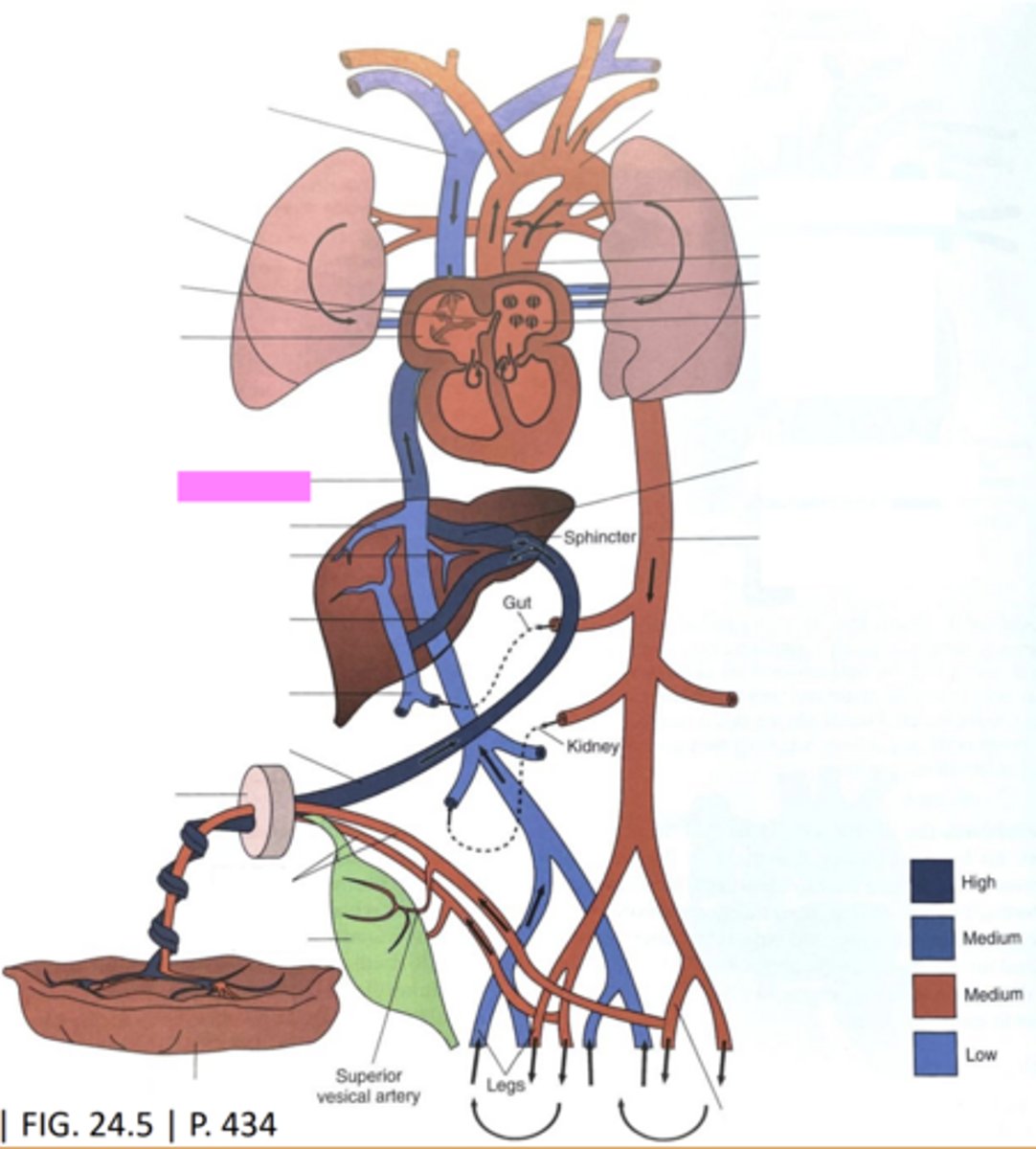
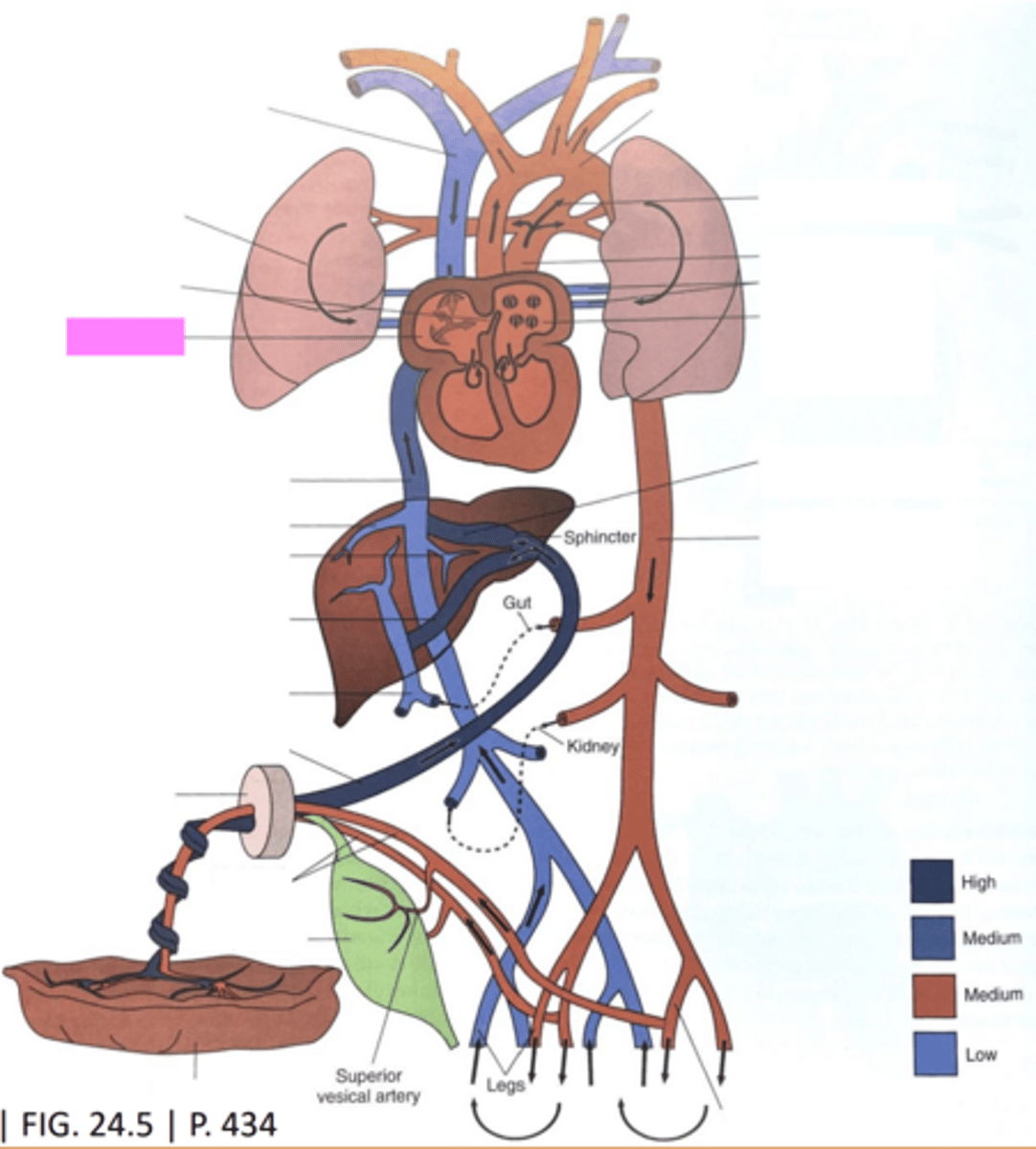
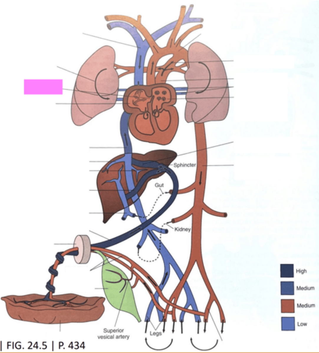
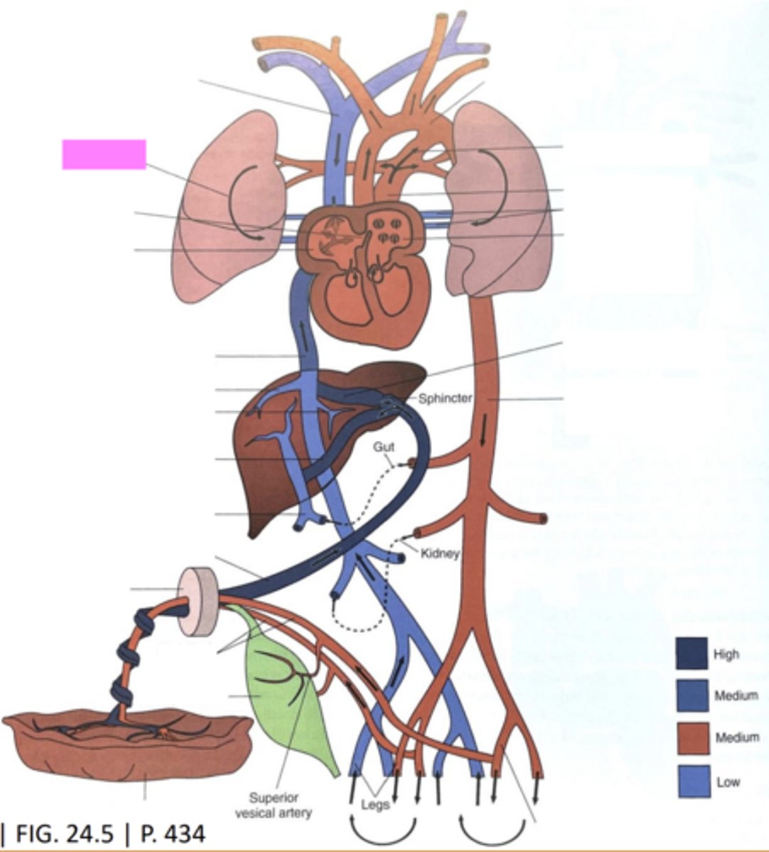
Fetal Cardiovascular Circulation
-parallel circulation in the fetus d/t arterial shunts
-in utero the lungs are not required to provide oxygen or remove carbon dioxide
Fetal Shunts
-allow blood to bypass lungs and liver and deliver oxygenated blood to brain
-foramen ovale
-ductus venosus
-ductus arteriosus
Ductus Venosus
-near the left portal vein
-allows some blood to bypass the liver
Foramen Ovale
-an opening btwn the right and left atria
-allows some blood to bypass the right ventricle
Ductus Arteriosus
-btwn the AO and pulmonary artery
-allows some blood to enter the aorta instead of the lungs
Ductus Venosus
Ductus Arteriosus
Foramen Ovale
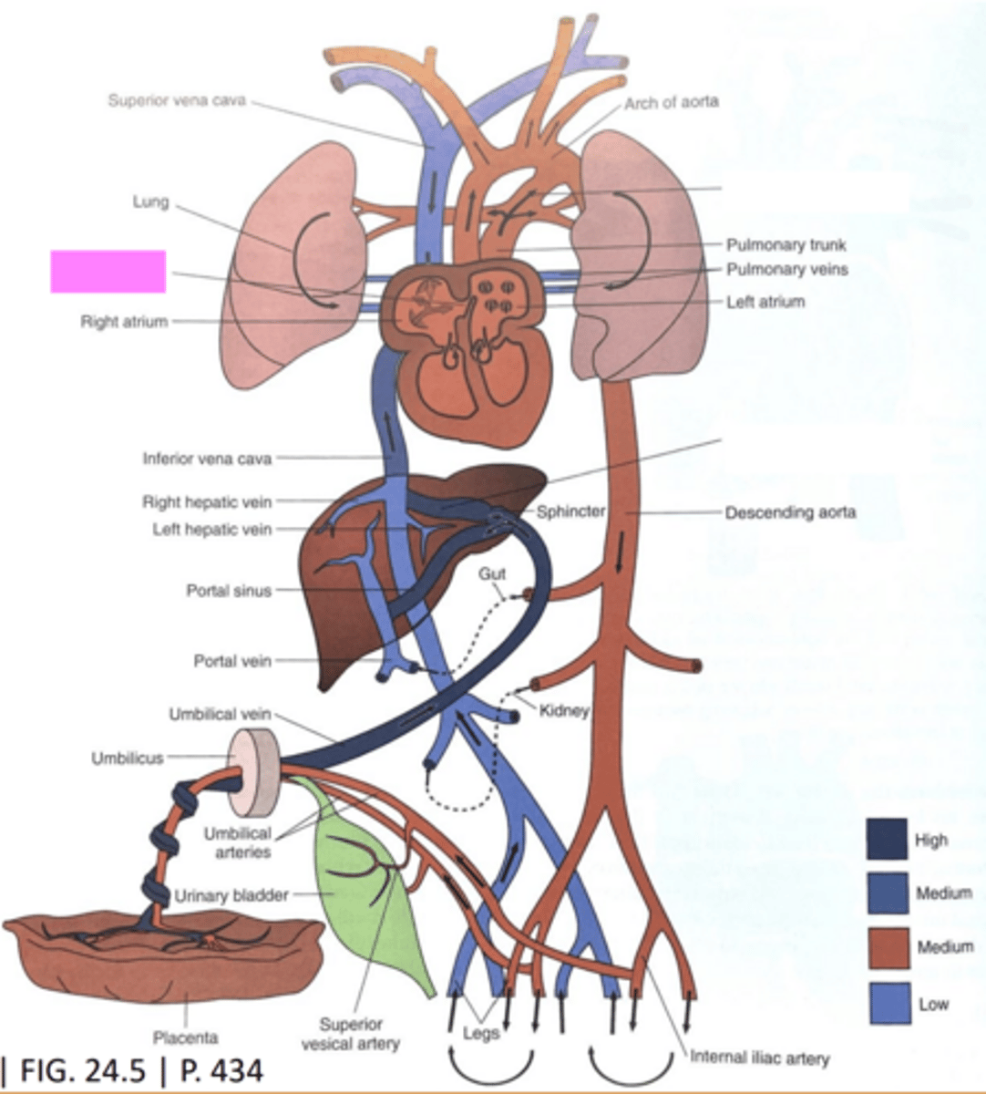
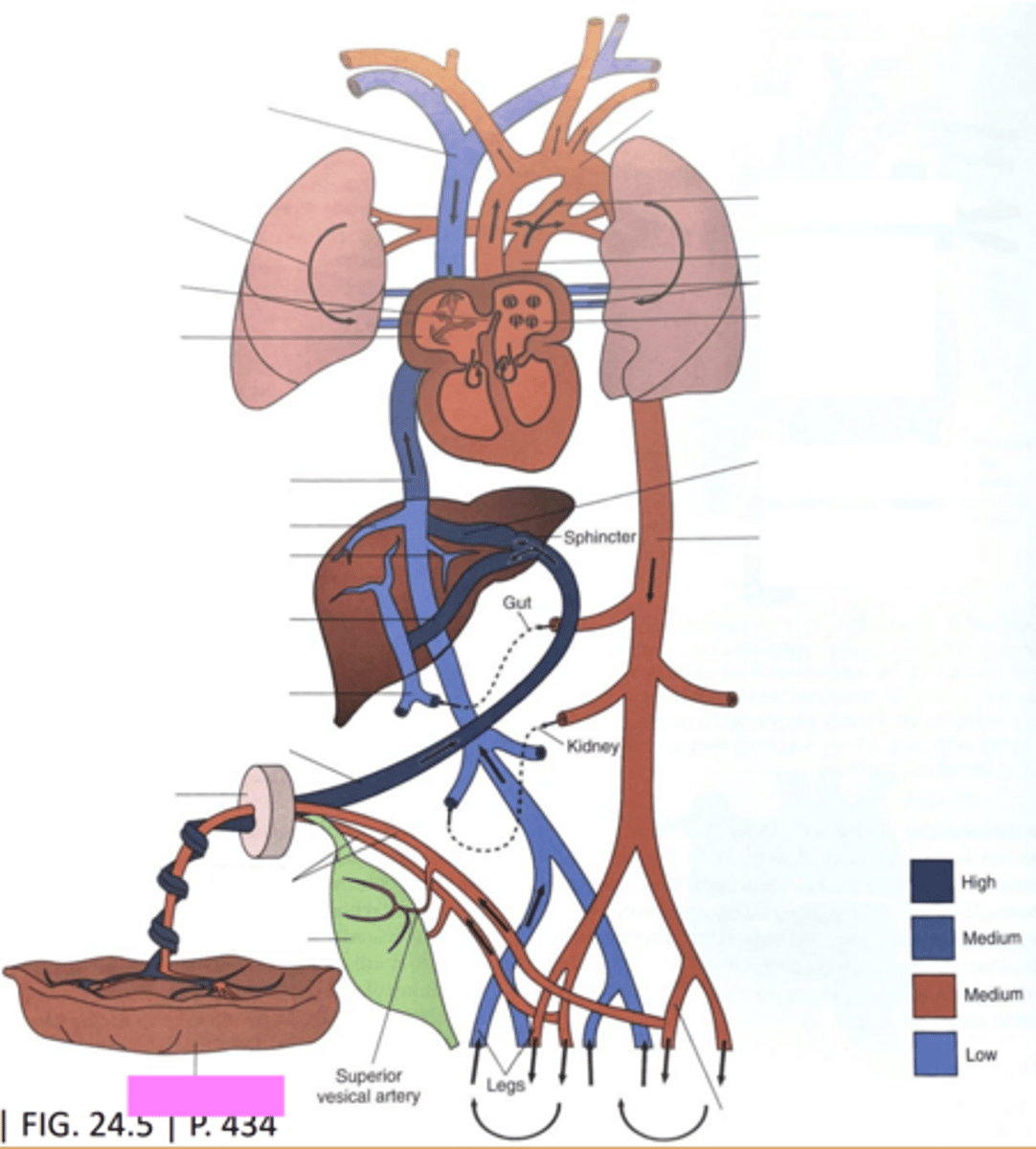
Maternal Placenta
serves as a transfer site of oxygen, carbon dioxide, and nutrition to and from fetus through the umbilical cord
Umbilical Arteries
carry unoxygenated blood from the fetus to the placenta
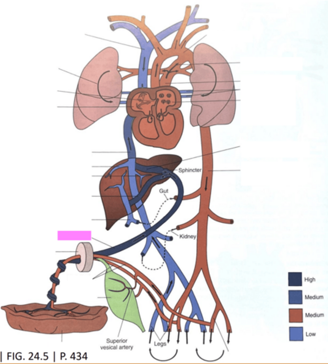
Umbilical Vein
-oxygen rich and nutrient rich blood is sent to the fetus from the placenta via the umbilical vein
-branches into the ductus venosus and left portal vein when it enters the liver
Ductus Venosus
-1st shunt in the fetal circulation
-vascular channel that connects umbilical vein to IVC
-bypasses the liver and joins the hepatic vein blood from the liver just before entering the IVC
Right Atrium
-blood now flows into right atrium from the IVC
-poorly oxygenated blood is also entering the right atrium via the SVC
-blood flows across the tricuspid valve and is directed toward the left atrium via the eustachian valve and right ventricle
Eustachian Valve
directs the ductus venosus blood across the foramen ovale into the left atrium
Foramen Ovale
-2nd shunt in the fetal circulation
-opening between right to left atria
Right Ventricle
blood entering the right ventricle is ejected through the pulmonary valve into the main pulmonary artery or pulmonary trunk toward the lungs
Ductus Arteriosus
-3rd shunt in the fetal circulation
-flow to fetal trunk and abdomen is supplied by ductus arteriosus
-blood exits the main pulmonary artery via the ductus arteriosus to enter the proximal portion of the descending AO
-diverts blood away from the lungs to protect them and help strengthen the right ventricle for future function after birth
Left Atrium
-blood enters the left atrium via the foramen ovale and pulmonary veins
-then flows across mitral valve into the left ventricle
Left Ventricle
blood exits the left ventricle through the aortic valve into the ascending AO
Ascending AO
-arises from the left ventricle
-delivers highly oxygenated blood to the heart muscle, brain, and upper extremities
-heart muscle > via the coronary arteries
-brain > via the carotid arteries
Descending AO
-blood from the ductus arteriosus mixes w/ blood from the AO
-blood continues down descending AO to supply the lower extremities and some will return to placenta for reoxygenation
-lower extremities > via iliac arteries
-placenta > via umbilical arteries
Postnatal Cardiac Circulation
-upon delivery, the umbilical placental circulation is interrupted
-the tasks of oxygen and carbon dioxide removal are now transferred from the placenta to the lungs
-fluid in the fetal airways is removed w/ the onset of breathing
-constriction of the ductus arteriosus occurs upon initial inflation of lungs
-the foramen ovale closes d/t increased left atrial pressure and decreased right atrial pressure (normally)
-the ductus venosus is open at the time of birth but as fibrin infiltrates it closes and becomes the ligamentum venosus
-serial circulation is present after birth once shunts close (1 side at a time)
Arch of Aorta
Ductus Arteriosus
Pulmonary Trunk
Pulmonary Veins

Left Atrium
Ductus Venosus
Descending AO
Internal Iliac Artery
Placenta
Urinary Bladder
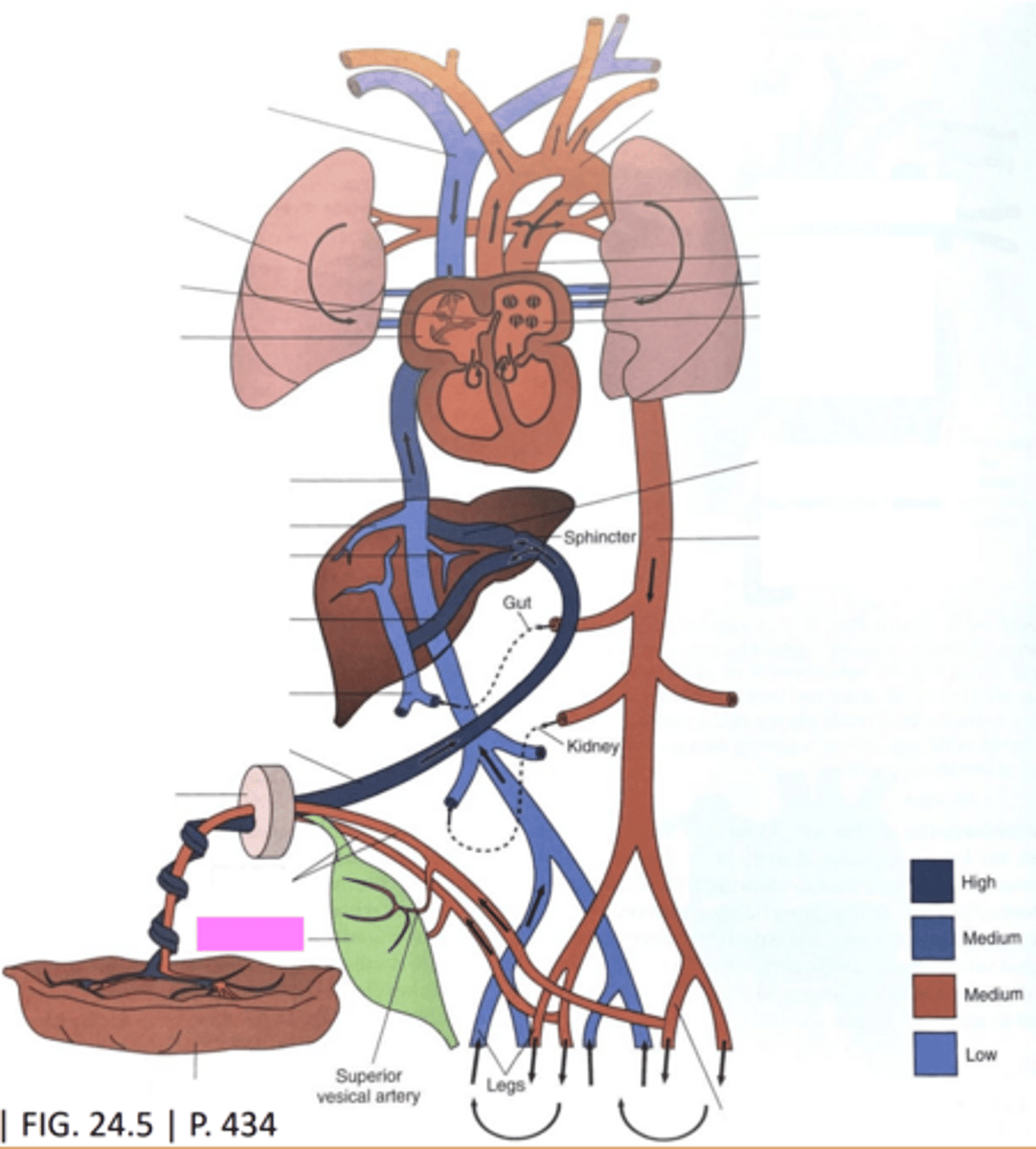
Umbilical Arteries

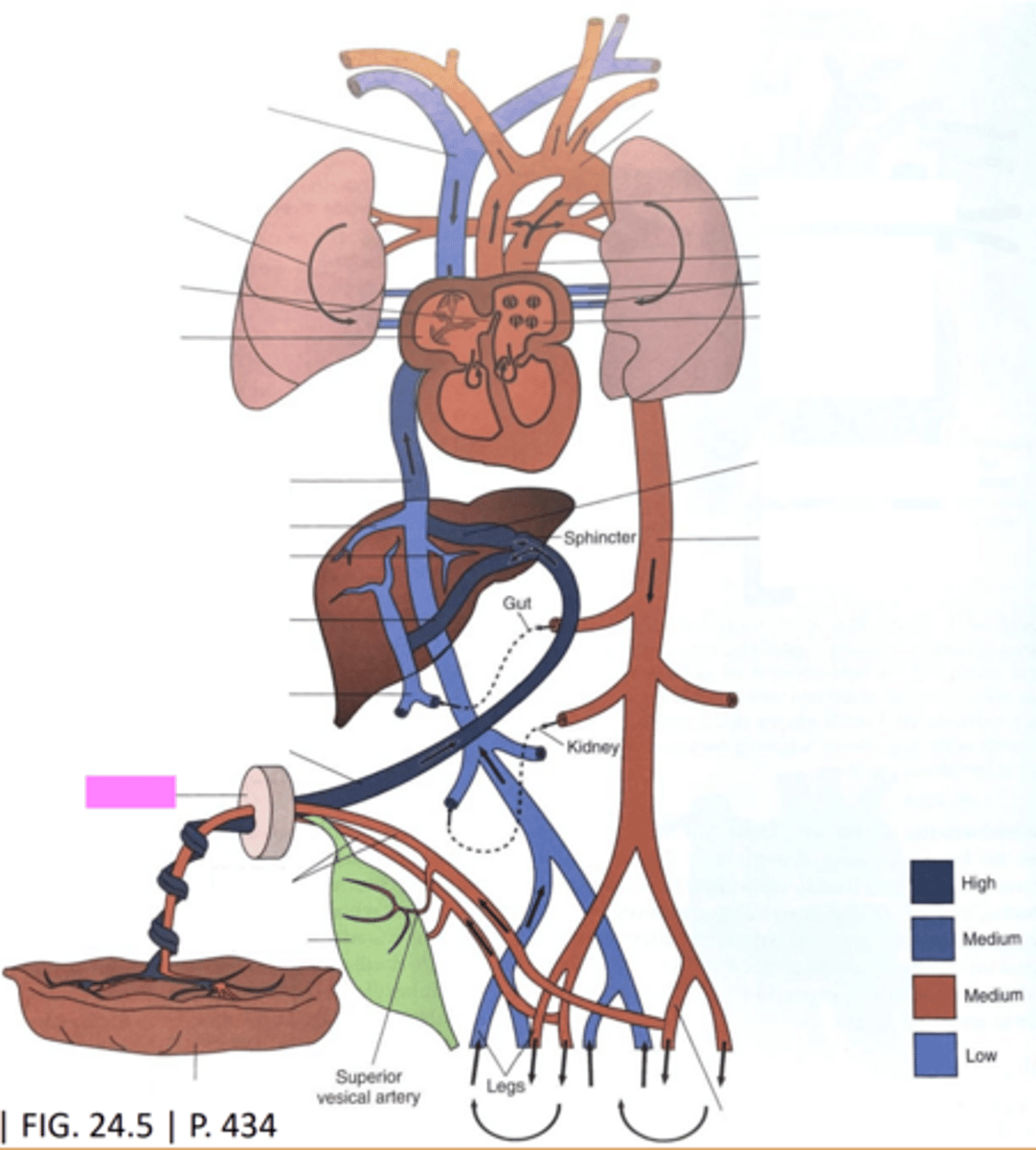
Umbilicus
Umbilical Vein
Portal Vein
Portal Sinus
Left Hepatic Vein

Right Hepatic Vein

Inferior Vena Cava
Right Atrium
Foramen Ovale
Lung
Superior Vena Cava