Lecture 2: Anterior and medial thigh and femoral triangle
1/106
Earn XP
Description and Tags
Identify the major bony features of the femur. Recognise the major muscles in the anterior and medial thigh, and explain their actions. Recognise and name the major nerves that enter the lower limb from the trunk and describe the groups of muscles each one supplies. Identify the major blood vessels that enter the lower limb from the trunk and describe the groups of muscles each one supplies/drains in the thigh. Identify the femoral triangle, recognising its boundaries and contents.
Name | Mastery | Learn | Test | Matching | Spaced | Call with Kai |
|---|
No analytics yet
Send a link to your students to track their progress
107 Terms
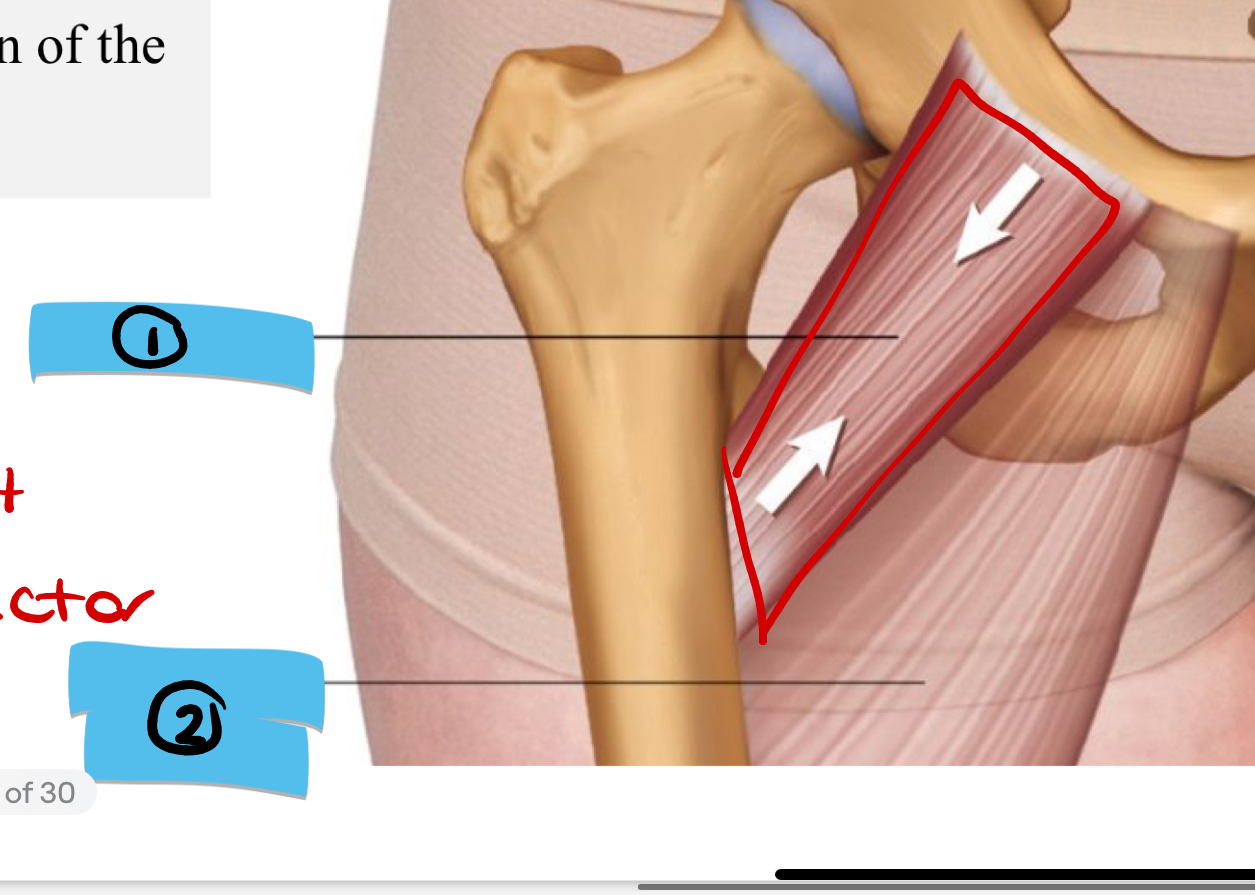
Label the missing structure on this diagram
Adductor Longus
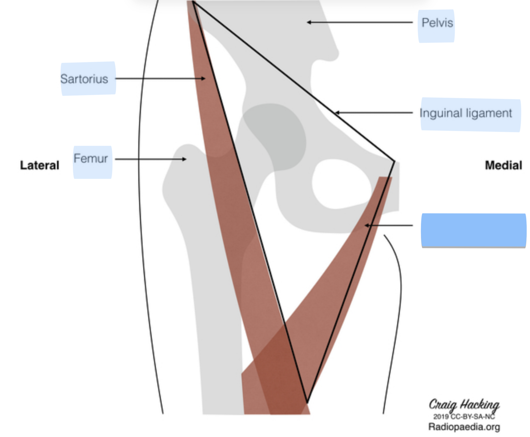
What are the boundaries of the femoral triangle? (lateral, medial, superior)
Sartorius muscle, Adductor longus muscle, Inguinal Ligament
What are the boundaries of the femoral triangle? (roof and floor)
Fascia Latte and Skin; 4 muscles (adductor longs, pectineus, posts major, iliacus muscle)
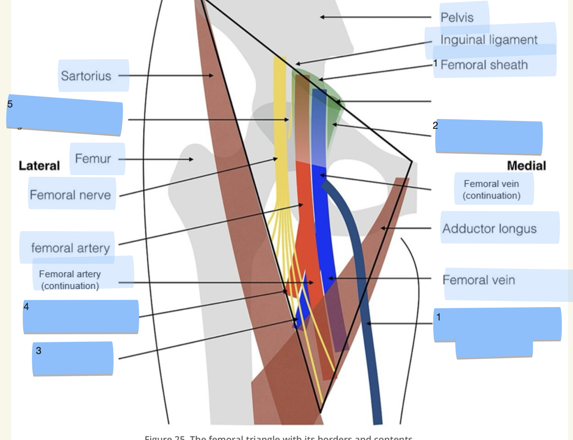
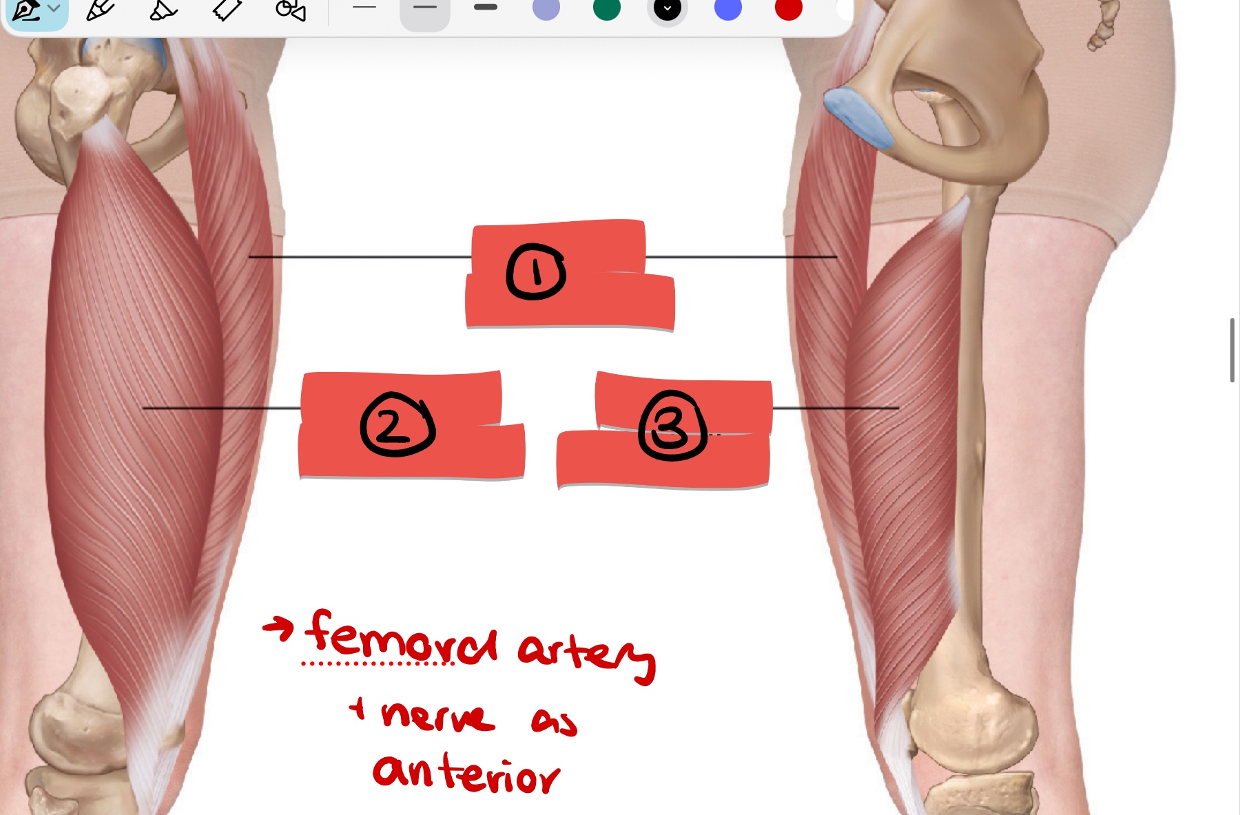
Label structures 1-3
1) Femoral Nerve 2) Femoral Artery 3) Femoral Vein
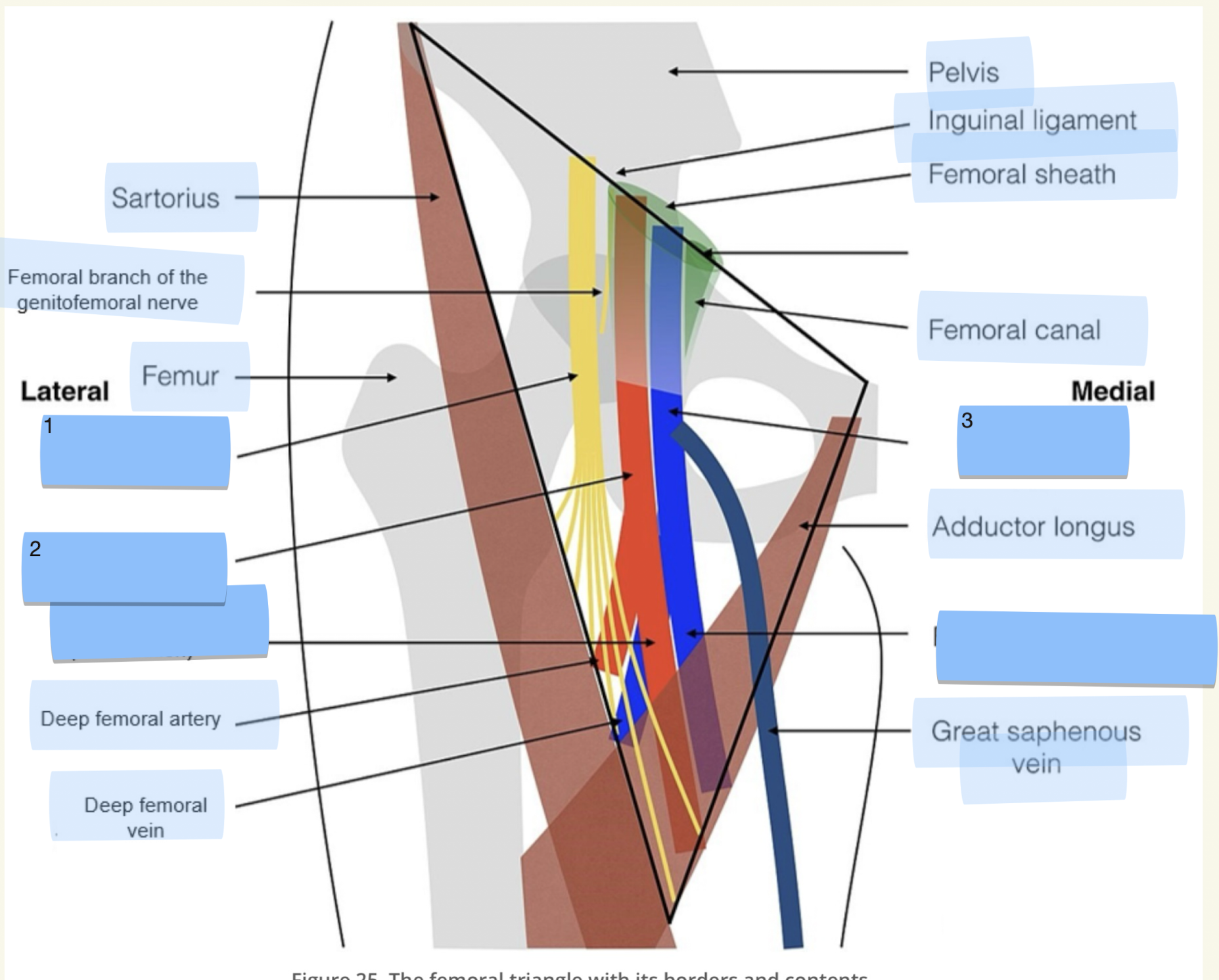
Label structures 1-5
1) great saphenous vein 2) femoral canal 3) Deep Femoral Vein 4) Deep femoral artery 5) Femoral branch of the genitofemoral nerve
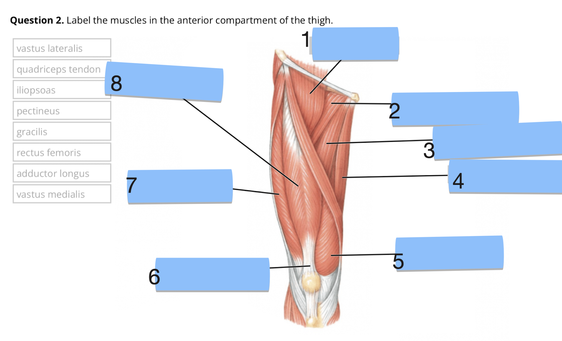
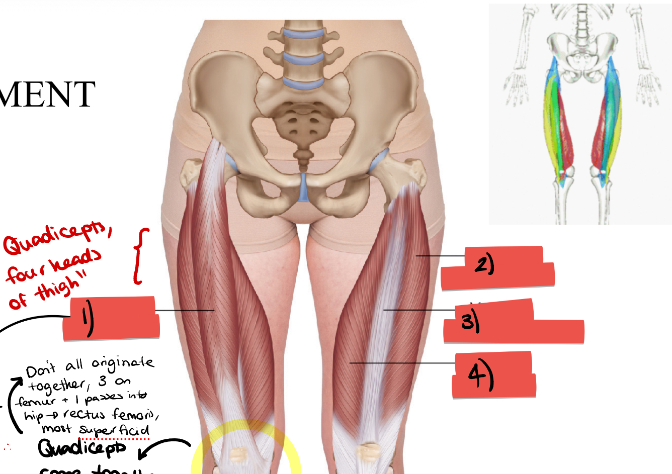
Label structures 1-8
1) iliopsoas 2) pectinous 3) adductor longus 4) gracilis 5) vastus medius 6) quadriceps tendon 7) vastus laterallis 8) rectus femoris
List the muscles in the anterior compartment of the thigh
iliopsoas, pectinous, adductor longus, gracilis, vastus medius, quadriceps tendon, vastus lateralis, rectus femoris, vastus intermedius
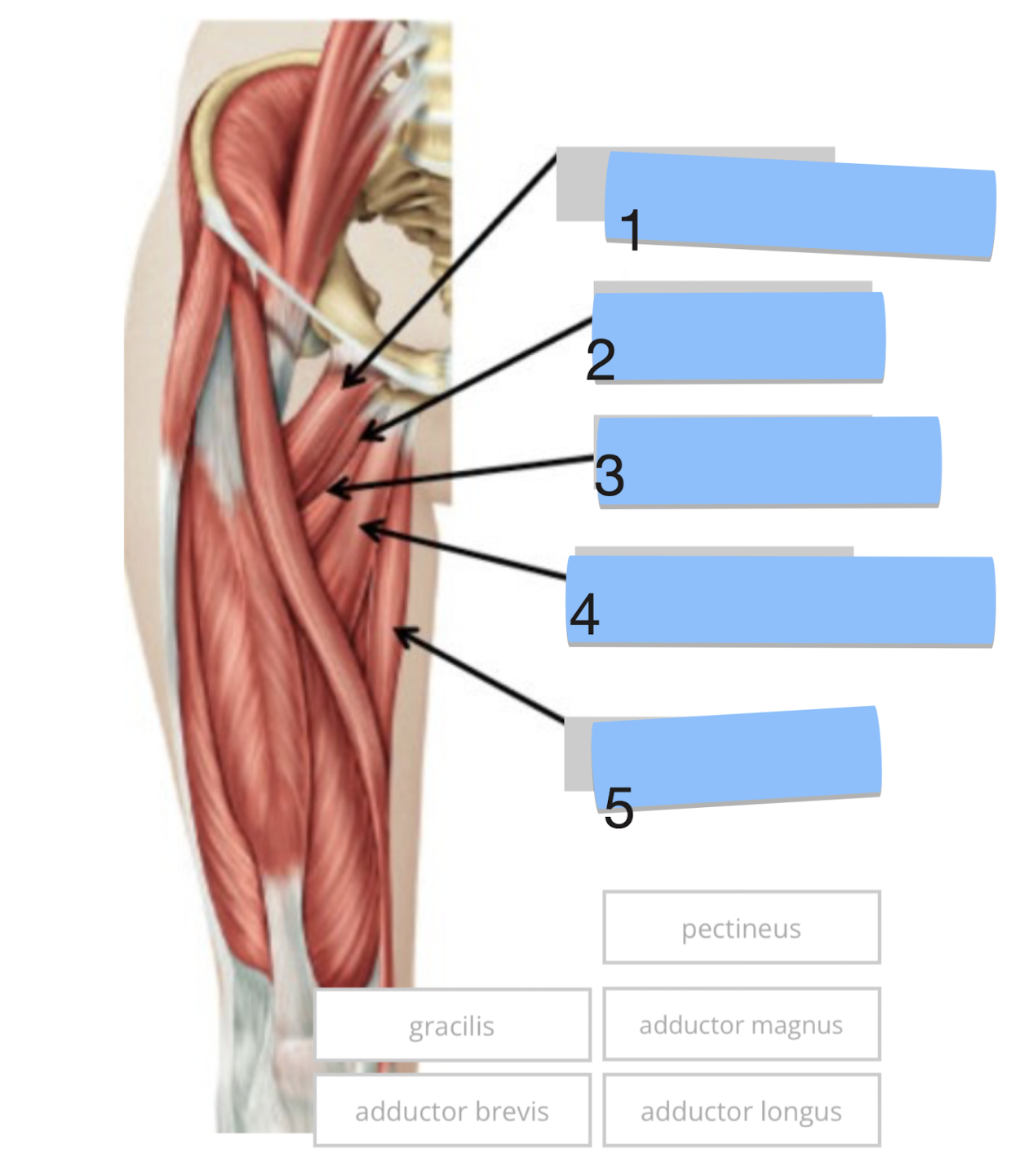
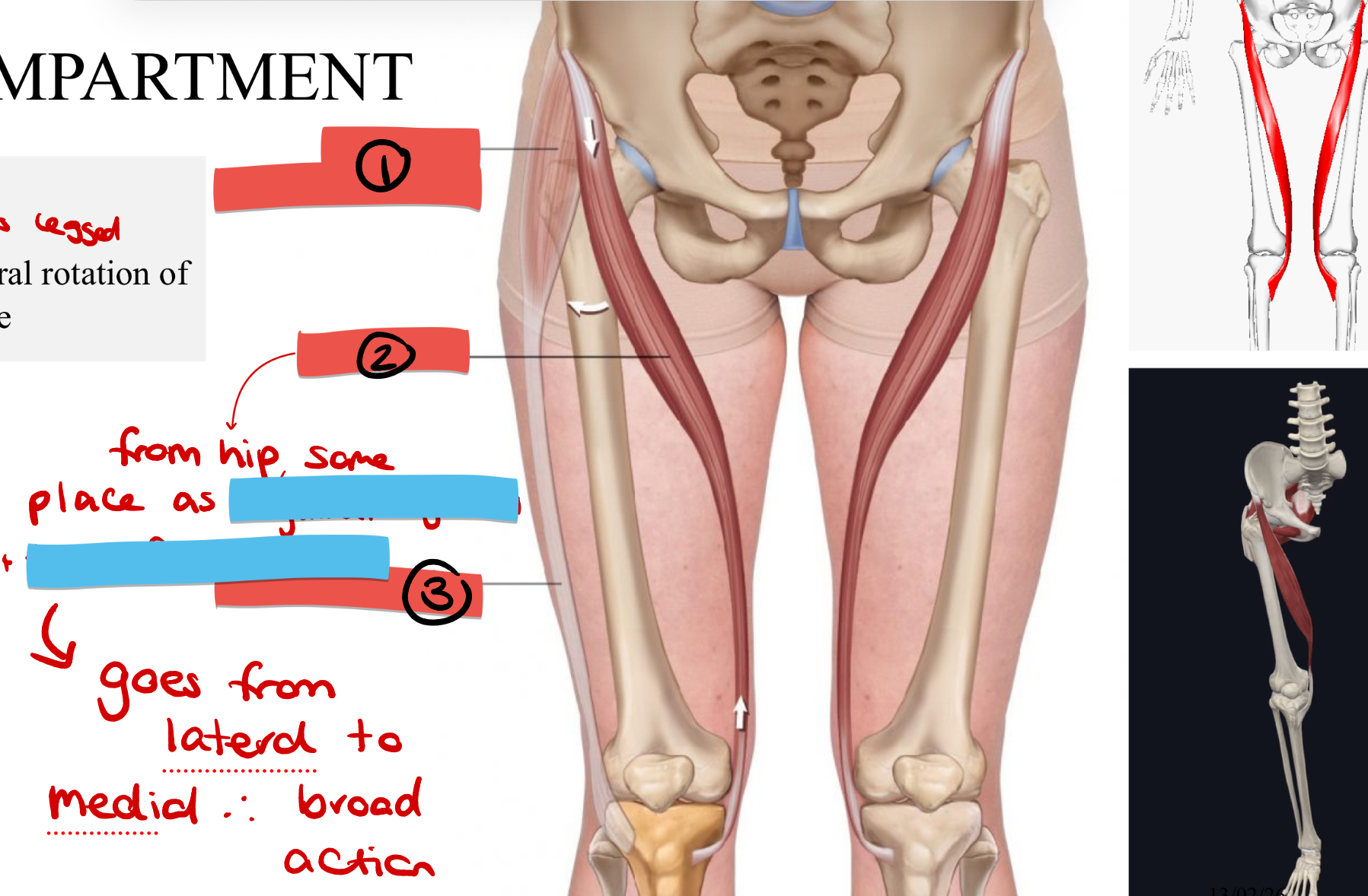
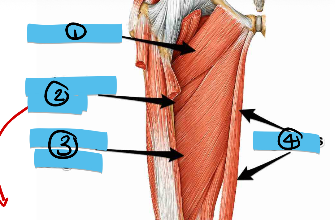
Label the muscles
Pectinius, Adductor Magnus, Adductor brevis, Adductor Longus, Gracillis
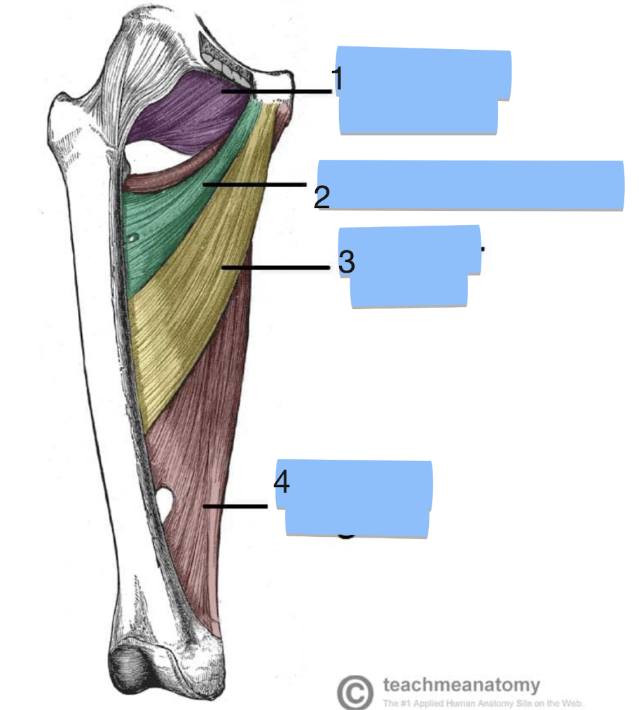
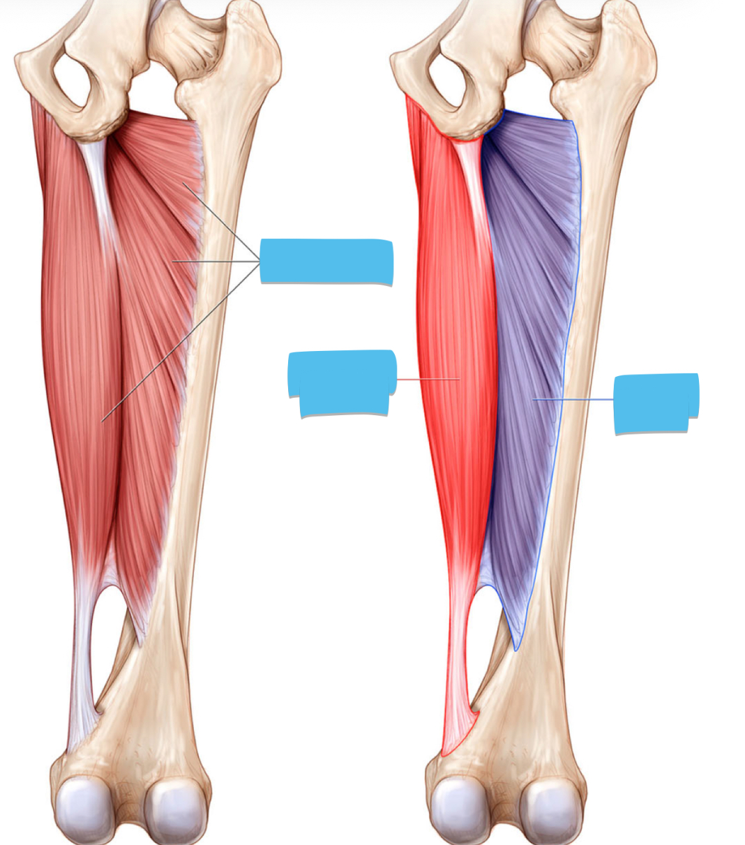
Label the muscles
1) obturator externus 2) adductor brevis 3) adductor longus 4) adductor Magnus
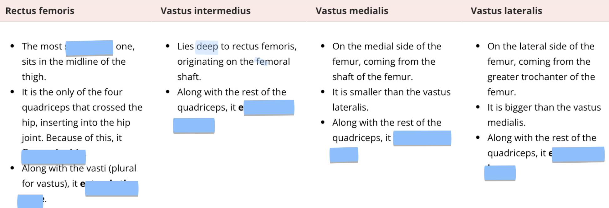
Fill in the blanks
1) superficial 2) flexes the hip 3) extends the knee 4) extends the knee 5) extends the knee 6) extends the knee
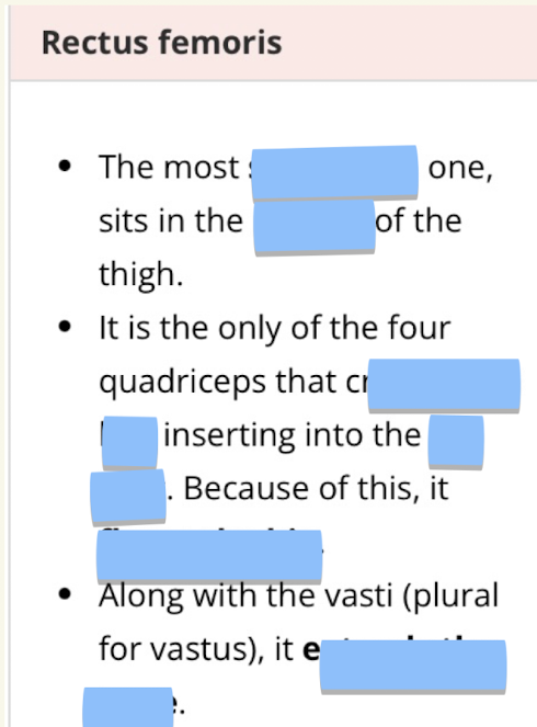
Fill in the blanks for the Rectus Femoris
1) superficial 2) midline 3) Crosses the hip 4) hip joint 5) flexes the hip 6) extends the knee
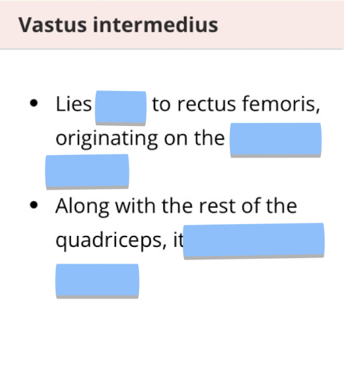
Fill in the blanks
1) deep 2) femoral shaft 3) extends the knee
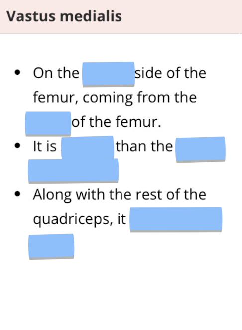
Fill in the blanks
1) medial 2) shaft 3) smaller 4) vastus lateralis 5) extends the knee
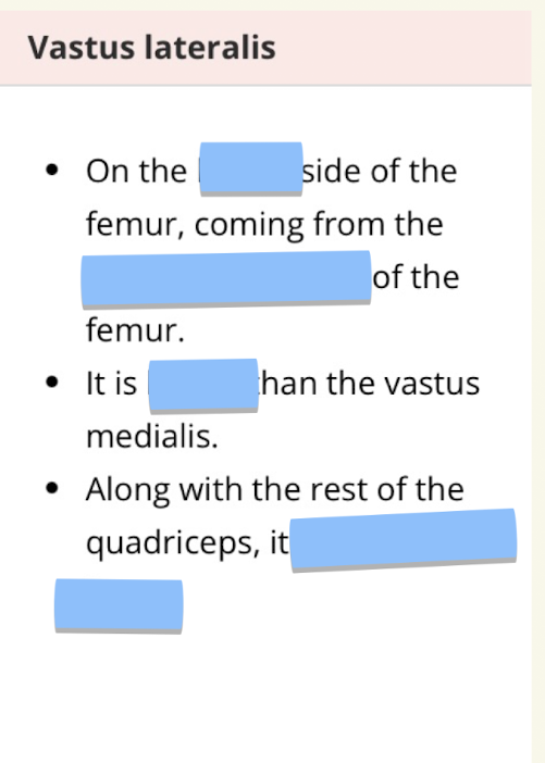
Fill in the blanks
1) lateral 2) greater trochanter 3) bigger 4) extends the knee

label terms 1-5
1) head 2) neck 3) greater trochanter 4) lesser trochanter 5) shaft
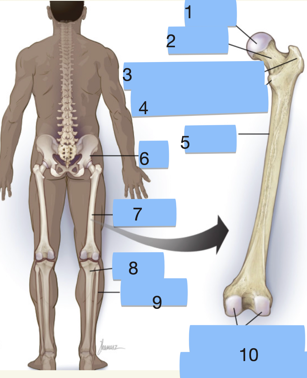
label terms 6-10
6) hip 7) femur 8) tibia 9) fibula 10) medial and lateral condyles
Where does the ligament of the head of the femur insert the femoral head into the acetabelum?
the fovea capsis
where’s the linea aspera and what does is do?
line down the shaft of the femur, for muscle attachment
where’s the patellar surface and what does it do?
anterior surface on distal femur, articulates with posterior surface of patella
what will muscles inserting on the greater trochanter do and why?
abduct the hip, due to location on lateral aspect
what is the lesser trochanter the primary insertion site for?
the illiopsoas muscle
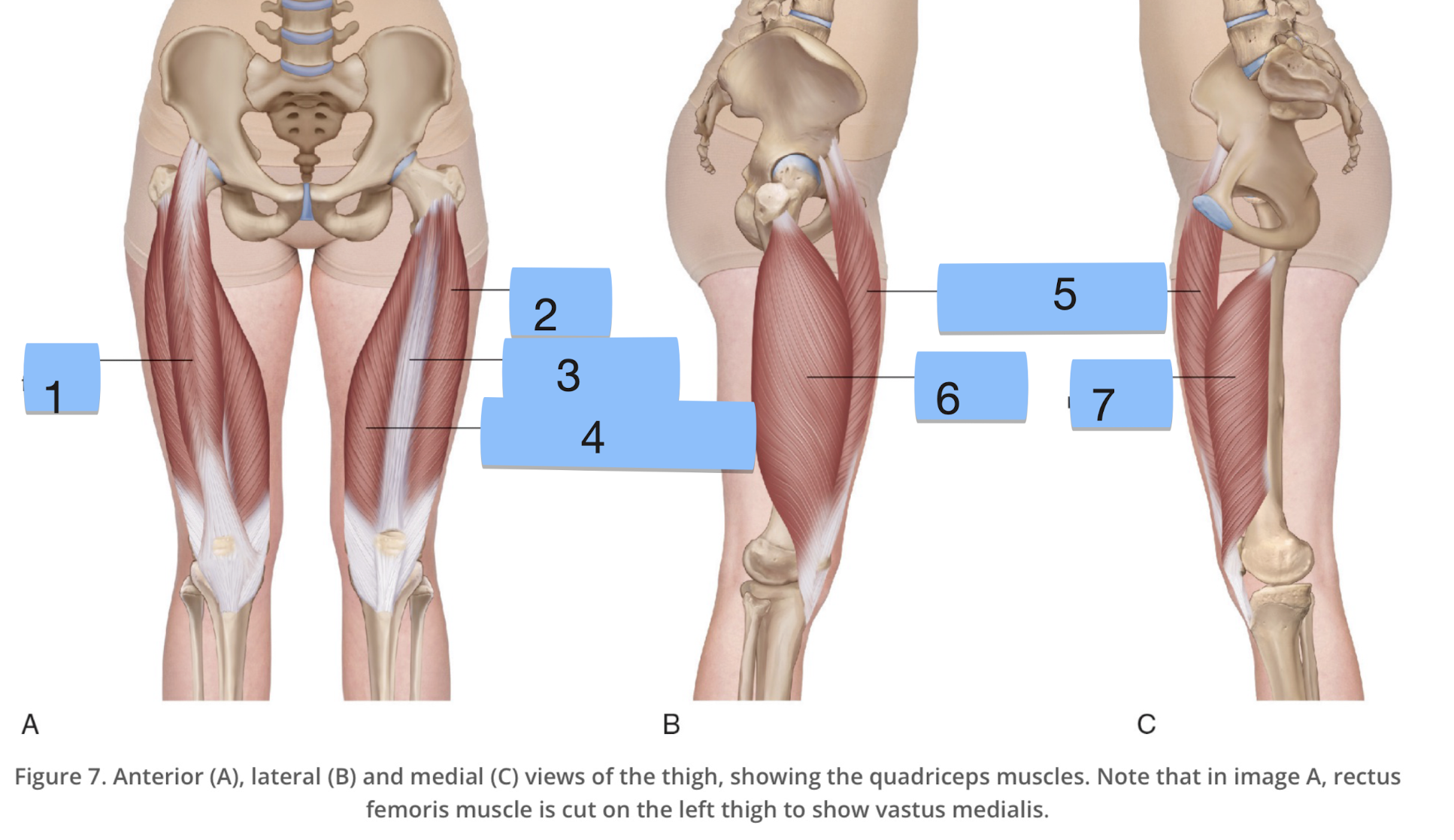
label structures 1-7
1) rectus femoris 2) vastus lateralis 3) vastus intermedius 4) vastus medialis 5) rectus femoris 6) vastus lateralis 7) vastus medialis
name the quadriceps (from medial to lateral)
vastus medialis, vastus intermedius, rectus femoris, vastus lateralis.
what nerve and what artery supplies the quadriceps
the femoral nerve and the femoral artery

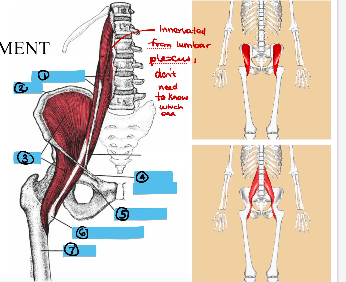
fill in blanks 1-7
1) iliacus 2) psoas major 3) internal 4) hip bone 5) inguinal ligament 6) lesser trochanter
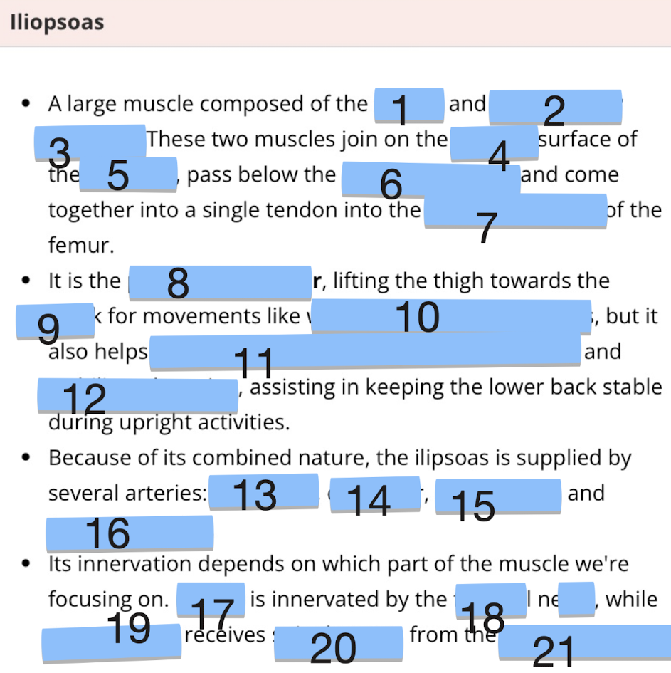
fill in blanks 8-12
8) primary hip flexor 9) trunk 10) walking, running and sit ups 11) rotates hip outwards 12) stabilises the spine
What is the angle of inclination at birth and in adulthood?
birth- 150, adulthood- 120-130
Whats it called when the angle of inclination is too inverted?
coxa vara
whats it called when the angle of inclination is too high
coxa valga
What femoral position causes the foot to be tilted inwards?
Femoral Anteversion
What femoral position causes the foot to be tilted outwards
femoral retroversion
Is the femur medially or laterally rotated in femoral ante version?
medially
Is the femur medially or laterally rotated in femoral retro version?
laterally
Label structures 1-4 in the anterior compartment of the thigh
iliotibial band, vastus lateralis, vastus intermedius, rectus femoris

Label structures 5-9 in the anterior compartment of the thigh
kin, Cassius medialis, sartorius, fasia lata, femoral vessles and nerve
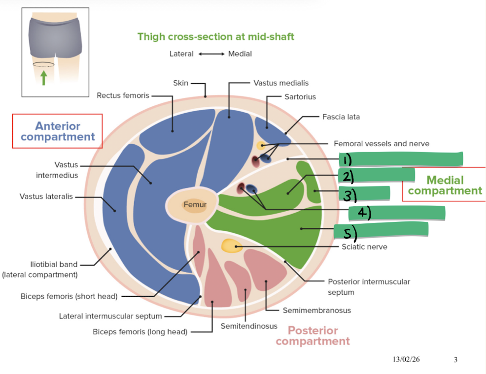
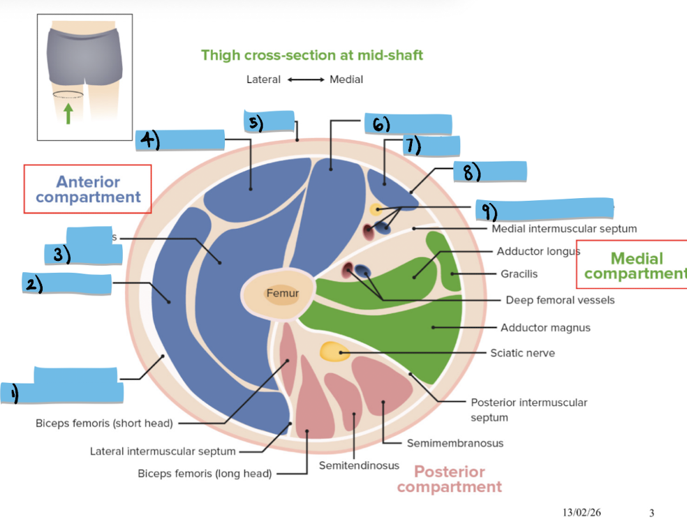
Label structures 1-5 of the medial thigh
medial intermuscular septum, adductor longus, gracilis, deep femoral vessels, adductor Magnus
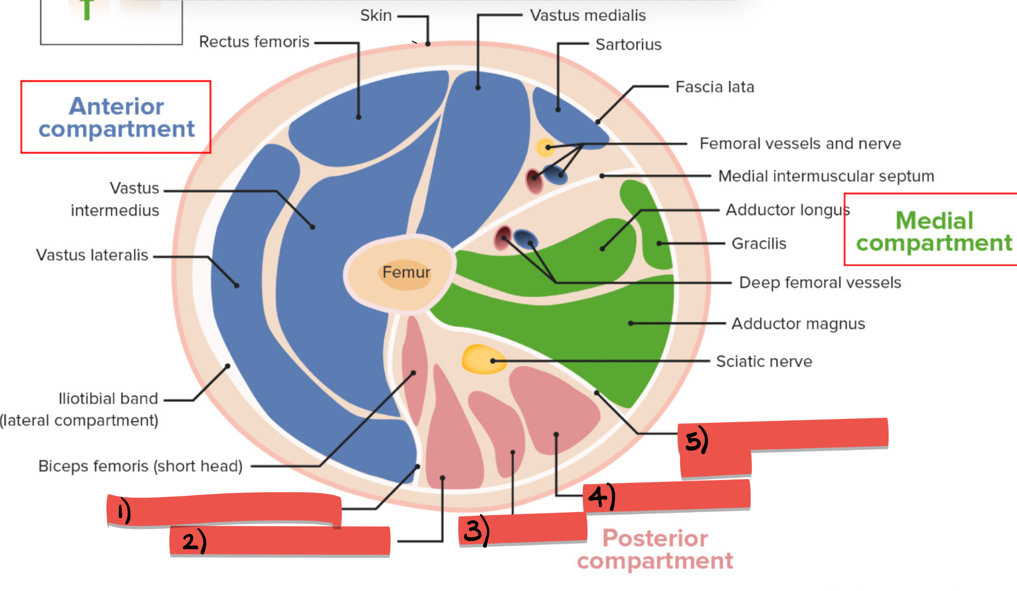
Label structures 1-5 of the posterior thigh
lateral intermuscular septum, biceps femoris, semitendinosus, smeimembrinosus, posterior intermuscular septum

Label structures 1-4
pelvis, acetabulum, femoral head, articular cartilage
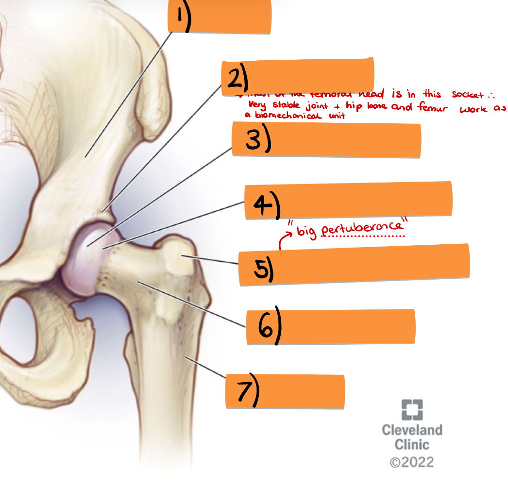
label structures 5-7
greater trochanter, femoral neck, femur
Which nerve supplies the posterior thigh?
sciatic
Which nerve supplies the medial thigh?
obturator
Which nerve supplies the anterior thigh?
femoral
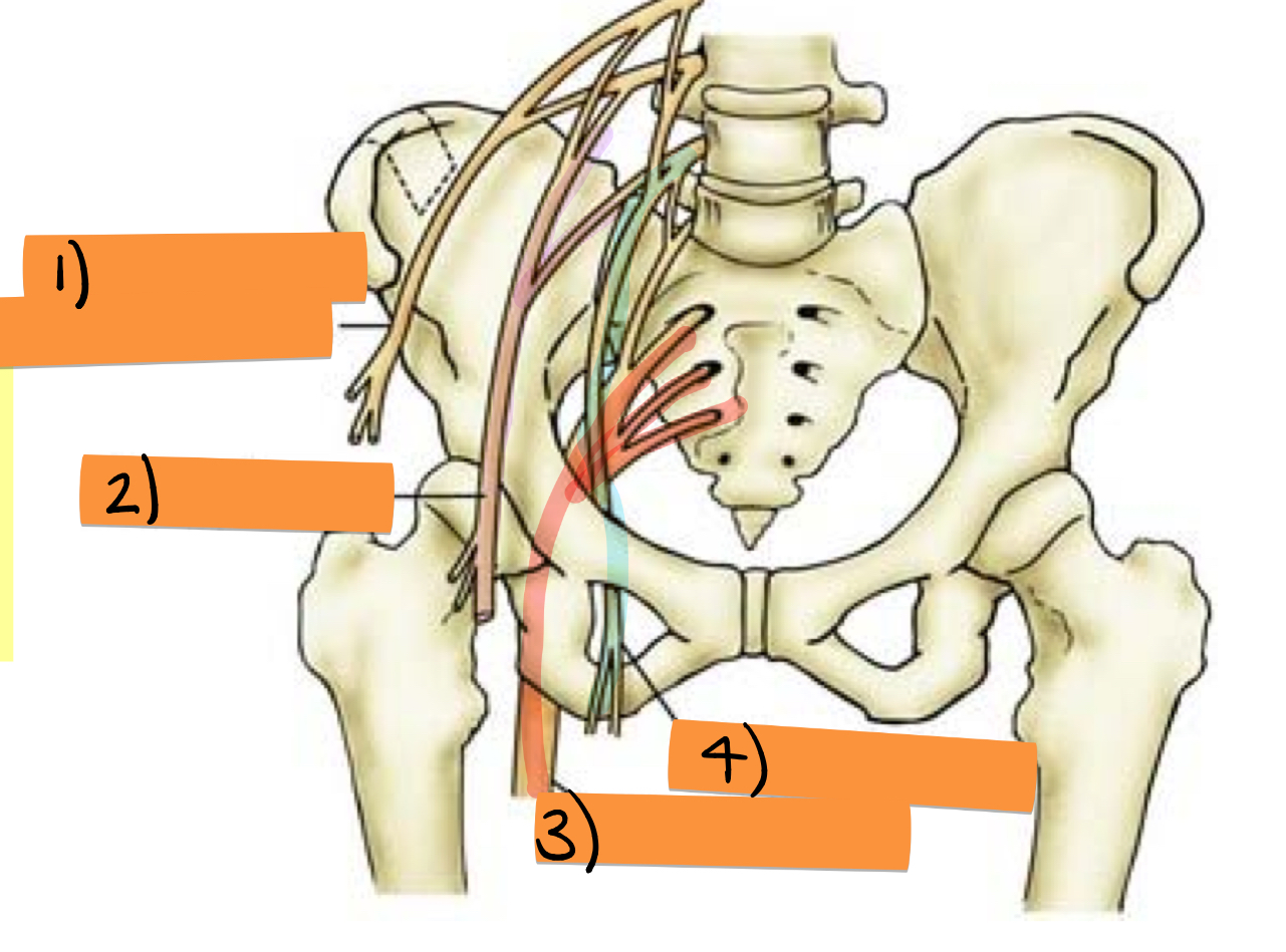
Label nerves 1-4
lateral femoral cutaneous, femoral, sciatic, obturator

label structures 1-5
gluteus maximus, gluteus medius, piriformis, greater trochanter, quadratus
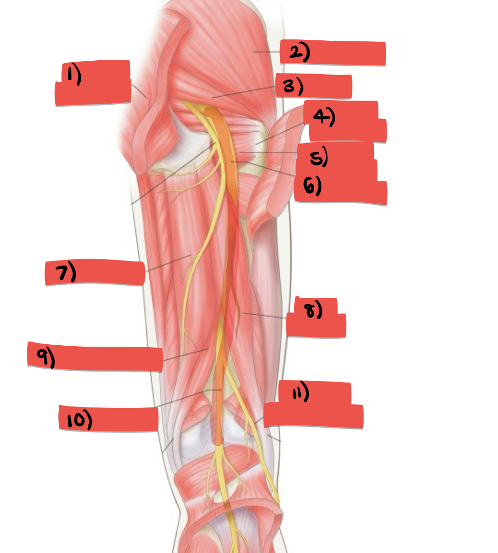
label structures 6-8
sciatic nerve, semitendinosus, biceps femoris

label structures 9-11
semimembranousus, tibial nerve, fibular nerve
Which compartment is this and what are structures 1-4
rectus femoris, vastus lateralis, vastus intermedius, vastus medialis
What does rectus femoris do to the knee
extension
what does rectus femoris do to hip
flexion
What does flexion do?
decreases the angle between two bones
what does extension do?
increases the angle between two bones
what affect does vastus intermedius have on the knee?
extension
what affect does vastus intermedius have on the knee
extension
what does vastus lateralis do to the knee
extends and stabilises
Label structures 1-3
vastus lateralis, rectus femoris, vastus medialis
Label structures 1-3
tensor fasciae latae, sartorius, iliotibial band
what does the sartorius do to the hip
flexion, abduction and lateral rotation
What does the sartorius do to the knee
flexion
Whats the role of the iliopsoas?
main flexor of the hip
What is the blood supply to the anterior thigh
femoral artery
What is the innervation to the anterior thigh
femoral nerve
what is the blood supply to the thigh?
iliolumbar, obturator, external iliac, femoral artery
What is the innervation to the illiopsoas?
lumbar plexus and femoral nerve
What is the mnemonic to remember blood supply to the illiopsoas?
I Often Eat Fries
What compartment of the thigh is the iliopsoas part of?
anterior
Whats affect does the iliopsoas have on the hip?
main flexor
Label structures 1-4
psoas major, ilium, iliacus, inguinal ligament
Label structures 5-7
pubic bone, lesser trochanter, femur
Label structures 1 and 2
pectinous, adductor longus
What does pectinous do?
flexion and adduction of the hip, stabilises the pelvis
What is the limit of pectineus flexion?
45 degrees
What compartment is pectineus in?
anterior/ medial
What is the blood supply to pectineus?
obturator artery
What is the innervation of pectineus?
femoral nerve
What muscle is pectineus often confused with?
adductor brevis
Label structures 1-4
pectineus, adductor brevis, adductor longus, gracilis
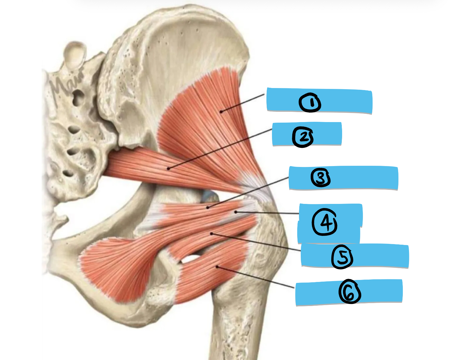
Label structures 1-6
gluteus minimus, piriformis, gemellus superior, obturator interns, gemellus inferior, quadratus femoris
What does the obturator externus do to the thigh?
lateral rotation plus abduction of the thigh when hip flexed
What is hip abduction
movement of thigh away from bodies midline
What is the innervation and blood supply of the obturator externus
obturator nerve, obturator artery
What group is the obturator part of
lateral rotator muscles
What does the adductor brevis do?
adducts and flexes hip, stabilises pelvis
What does the adductor longus do?
adducts and flexes hip, stabilise pelvis
What are the two portions of the adductor Magnus?
ischiocondular portion, pubofemoral portion
What do the portions of the adductor Magnus do? (ishiocondular and pubofemoral)
both adduct hip, ishiocondular extends hip, pubofemoral flexes the hip
How do you remember the difference between extension and flexion?
extension extends angle between two body parts by straightening joint, flexion reduces angle by bending joint
What supplies the adductors?
deep femoral artery, obturator nerve
What thigh compartment are the adductors part of?
The medial
The pubofemoral region of the adductor Magnus is activated and innervated as a adductor what is the ishioondular portion activated and innervated as?
a hamstring
Label these structures from left to right
adductor magnus, ischiocondular portion, pubofemoral portion
What memorable action does gracilis do (different from standard flexion, adduction etc…)?
Helps you not pee yourself by providing stable anchor for pelvic floor to brace against
What does gracilis do?
flexion and adduction of hip, flexion and medial rotation of knee
What innervates and supplies the medial compartment?
obturator nerve, deep femoral artery
Which compartment is gracilis part of
medial
What is the pes anserinus?
conjoined tendons of semitendinous, sartorius and gracilis
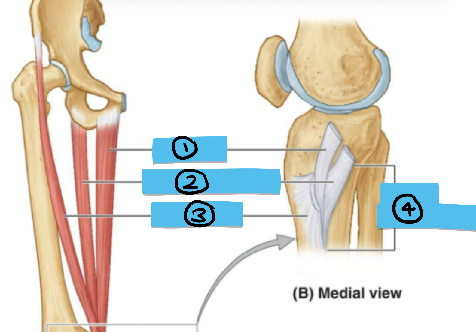
Label structures 1-4
gracilis, semitendinosos, sartorius, pes anserinus
What does pes anserinus mean in English (helps remember job)?
goose foot
How many structures are in the femoral triangle and what types?
4, 1 nerve, 2 vessels, 1 space
What is the memory device for remembering the boundaries of the femoral triangle?
SAIL
What are the borders of the femoral triangle
sartorius muscle (lateral), adductor longus muscle (medial), inguinal ligament (superior), Fascia latte and skin (roof)