Part 2
 Call Kai
 Learn
Learn Practice Test
 Spaced Repetition
 Match
Match Flashcards
 Knowt Play
1/61

There's no tags or description
Looks like no tags are added yet.
Name | Mastery | Learn | Test | Matching | Spaced | Call with Kai |
|---|
No analytics yet
Send a link to your students to track their progress
62 Terms
1,2
Cell Count
Any cell count should be performed immediately
WBCs & RBCs begin to lyse within an ____ hour
40% of WBCs disintegrate within ___ hours
WBC count
Cell count
Routinely performed on CSF
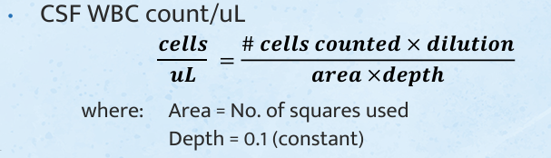
0-5, 0-30
Cell count
Normal values for WBC count for
Adults:______ WBCs/uL
Neonates:______ WBCs/uL
3% acetic acid with methylene blue
Cell count
Diluting fluid for WBC count
RBC count
Cell count
Done only in cases of traumatic tap and correction for WBCs or proteins is needed

total cell count
Cell count
Cells are counted in the 4 corner squares and the center square of the hemocytometer
NSS
Cell count
Diluent of Total cell count
WBC correction
Cell count
A calculation is used to correct CSF WBC counts which are falsely increased due to a traumatic tap
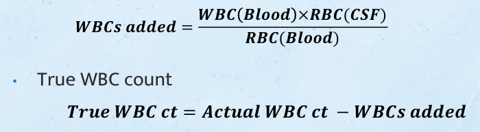
1, 750
Cell count : WBC Correction
If the patient’s peripheral WBC and RBC counts are within normal limits, some laboratories use the following formula:
Subtract ____white cell from the CSF WBC count for each_____ RBC counted in the spinal fluid
sedimentation, filtration, cytocentrifugation, centrifugation
Differential Count
Performed on stained smear
Specimen should be concentrated before smearing by using what methods (4)
___________
___________
___________- 0.1 mL of CSF combined with 1 gtt of 30% albumin produces adequate cell yield
___________- 5- 10 min
lymphocytes, monocytes, neutrophils
Differential count
What normal cells that are found?
__________- predominant in ADULTS (L:M ratio - 70:30)
__________- predominant in CHILDREN
__________- rarely found (If present causes; bacterial meningitis)
pleocytosis
Differential count
Increased numbers of normal cells are considered as ABNORMAL finding
LYMPHOTIC PLEOCYTOSIS: 90% lymphocytes, 10% monocytes
MONOCYTIC PLEOCYTOSIS: 90% monocytes, 10% lymphocytes
lymphocytes and monocytes
Predominant Cells in CSF
normal, all stages of development may be found
monocytes mixed with lymphocytes
increased in viral, tubercular, & fungal meningitis
Multiple Sclerosis
neutrophils
Predominant Cells in CSF
increased in bacterial meningitis & in early cases of viral, tubercular, & fungal meningitis
also found in cerebral hemorrhage
cell disintegrate rapidly
eosinophils
Predominant Cells in CSF
associated with parasitic infections, fungal infection (C. immitis), introduction of foreign material
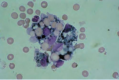
macrophages
Predominant Cells in CSF
found with RBCs in spinal fluid
may contain phagocytized RBCs (erythrophages) appearing as empty vacuoles, hemosiderin granule, etc.
increased number indicates previous hemorrhage
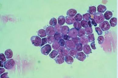
blasts
Predominant Cells in CSF
found in acute leukemia
either in lymphoblast, myeloblast, or monoblast form
lymphoma cells
Predominant Cells in CSF
found in disseminated lymphoma
resemble lymphocytes with cleft nuclei
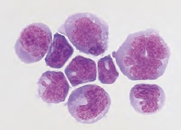
plasma cells
Predominant Cells in CSF
found in multiple sclerosis
due to lymphocyte reactions
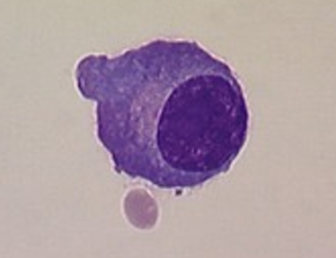
ependymal, choroidal and spindle shaped cells
Predominant Cells in CSF
for diagnostic procedures
seen in clusters with distinct nuclei & distinct cell walls
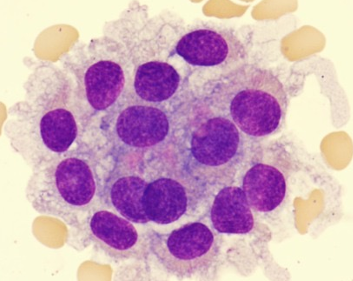
malignant cells
Predominant Cells in CSF
found in metastatic carcinomas & primary CNS carcinomas
seen in clusters with fusing of cell borders & nuclei
CSF protein
Most frequently performed chemical test on CSF
15-45 mg/dL
CSF protein
NV?
Values are higher in infants and older persons
CSF proteins are < 1% compared to serum
hemorrhage and meningitis
CSF protein
Is increased in;
______________ - most common, due to damage to the blood brain barrier
multiple sclerosis
CSF protein
Is increased in;
____________- due to production of immunoglobulins within the CNS
CSF leakage
CSF protein
Is decreased in; ?
albumin, pre albumin
CSF protein
Major CSF Protein?
Second most prevalent?
haptoglobin & ceruloplasmin
CSF protein
Alpha-globulins?
b2 transferrin
CSF protein
Beta-globulins?
Carbohydrate deficient transferrin, also known as “tau”
Note: This is the identifying protein for CSF, meaning this indicates that the specimen is really CSF
Not found in blood, only in CSF
IgG and some IgA
CSF protein
Gamma-globulins?
IgM, fibrinogen and lipids
CSF protein
Not found in normal CSF
3 % TCA
CSF protein : TURBIDIMETRIC
preferred method, precipitates both albumin & globulin
3 % SSA
CSF protein : TURBIDIMETRIC
precipitates albumin only
to precipitate globulin, add SODIUM SULPHATE
coomassie brilliant blue
CSF protein : DYE BINDING
protein binds to dye which turns RED to BLUE
csf serum albumin index
CSF protein : PROTEIN FRACTIONS
Evaluates the integrity of the BBB
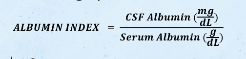
< 9
CSF protein : PROTEIN FRACTIONS
Normal CSF/serum albumin index?
Abnormal: ≥ 9
indicates destruction of the BBB
csf serum igG index
CSF protein : PROTEIN FRACTIONS
Measure IgG synthesis within the CNS

< 0.77
CSF protein : PROTEIN FRACTIONS
Normal CSF/serum IgG index?
Abnormal: ≥ 0.77
indicates increased IgG production within the CNS (ex. Multiple Sclerosis)
electrophoresis
CSF protein : PROTEIN FRACTIONS
Agarose gel electrophoresis followed by CBB (Commasie) staining – most frequently performed
Methods of choice when it is necessary to determine if the fluid is CSF (“tau” identification)
Done in conjunction with serum electrophoresis
For detection of OLIGOCLONAL BAND
Indicates immunoglobulin production
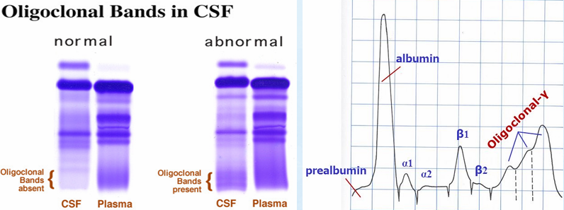
multiple sclerosis
CSF protein : PROTEIN FRACTIONS
In Electrophoresis, the presence of 2 or more oligoclonal bands in CSF but NOT in serum is valuable for the diagnosis of?
Other conditions:
neurosyphilis,
encephalitis,
neoplastic disorders,
Guillain-Barre syndrome
csf glucose
Done in conjunction with blood glucose
Specimen for blood glucose should be drawn 2 hours PRIOR to spinal tap
2 hours prior
CSF Glucose
Specimen for blood glucose should be drawn _________to spinal tap
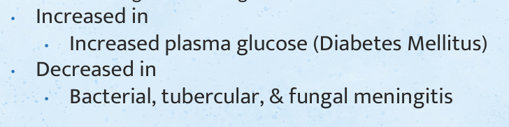
60 – 70%
CSF Glucose
Normal Value?
csf lactate
Aids in diagnosis and management of meningitis case
Inversely proportional
CSF Lactate
___________to glucose
Glucose is utilized by bacterial or fungi converting it to energy and producing lactate as a by-product
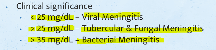
10-22mg/dL
CSF Lactate
Normal Value:
CSF glutamaine
Chemical test frequently performed in CSF but not in blood
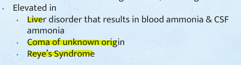
INDIRECT measure of CSF Ammonia
decreases
CSF glutamaine
INDIRECT measure of CSF Ammonia
As CSF ammonia increases, alpha-ketoglutarate ________ due to conversion into glutamine, resulting in coma
Brain tissue destruction
CSF Enzymes
Increased LD1 & LD2–
Viral Meningitis
CSF Enzymes
Increased LD2 & LD3
Bacterial Meningitis
CSF Enzymes
Increased LD4 & LD5
creatine kinase
CSF Enzymes
Elevated in stroke, MS, brain tumors, viral & bacterial meningitis, & epileptic seizure
AST
CSF Enzymes
Elevated in intracerebral & subarachnoid hemorrhage
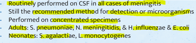
gram stain
CSF Microbiological Exam
blood culture
CSF Microbiological Exam
Done because the causative agent will be in both blood and CSF
acid fast / fluorescent antibody stains
CSF Microbiological Exam
For suspected cases of tubercular meningitis
india ink
CSF Microbiological Exam
Detects the presence of Cryptococcus neoformans
produce a star-burst pattern in gram stain
limulus lysate test
CSF Microbiological Exam
Detects gram negative bacterial endotoxins in body fluids
Principle:
In the presence of endotoxins, the amoebocytes (WBCs) will release lysate (protein)
(+) clumping/clot formation
horseshoe cram blood
CSF Microbiological Exam
Limulus lysate test reagent? It contains hemocyanin
VDRL
CSF Serologic exam
Recommended by CDC due to its ability to detect active cases of syphilis
FTA-ABS
CSF Serologic exam
Used with care to avoid contamination with blood because it remains positive in the serum of treated cases of syphilis
RPR
CSF Serologic exam
Not recommended because it is less sensitive than VDRL