Bone Physiology (MODS- Exam 1)
1/18
There's no tags or description
Looks like no tags are added yet.
Name | Mastery | Learn | Test | Matching | Spaced | Call with Kai |
|---|
No analytics yet
Send a link to your students to track their progress
19 Terms
List and describe the functions of bones
Protection for vital structures.
Support for the body and its vital cavities.
Mechanical basis for movement.
Storage for salts, specifically calcium and phosphate.
Continuous supply of new blood cells.
Describe the classes of bones
Long bones
Short bones
Flat bones
Irregular bones
Sesamoid bones
What are long bones?
Tubular structures, such as the humerus and femur.
What are short bones?
Cuboidal in shape and only found in the ankle and wrist.
What are flat bones?
Serve protective functions, such as the cranium
What are irregular bones?
Have various shapes other than long, short, or flat, such as facial bones and vertebrae.
What are sesamoid bones?
Develop in certain tendons to provide protection from excessive wear and change the angle as they pass through attachments, such as the patella.
Describe the process of bone formation and development
All bones are derived from mesenchyme (embryonic connective tissue) and form through either intramembranous ossification or endochondral ossification.
Osteogenesis= the process of bone formation
Describe the process of intramembranous ossification
directly converts the mesenchymal tissue to bone and forms the flat bones of the skull, clavicle, and most of the cranial bones
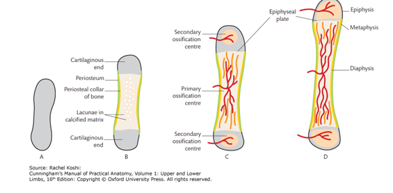
Describe the process of endochondral ossification (chronological order)
(cartilaginous bone formation), bone is preformed in cartilage. Cartilage cells are buried in a matrix and grow by the proliferation of cells and production of matrix. Over time, the cartilage is replaced by bone.
Supporting shell of bone laid down → cells die, leaving empty space in the calcified cartilage (periosteum) → invaded by blood vessels → bone laid down by osteoblasts → primary ossification center, which is in middle of the long bones → spreads toward the end in both directions → secondary ossification center develop at the end of each cartilaginous model→ ossification proceeds in all directions.
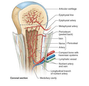
Discuss the vasculature and innervation of bones and joints
Veins: accompany arteries through the nutrient foramina (passes through periosteum). Large veins leave through foramina near the articular ends of bones
Nerves: accompany blood supply
Periosteum richly supplied with periosteal nerves (carry pain fibers) and Vasomotor nerves (cause constriction and dilation of blood vessels → regulates blood flow)
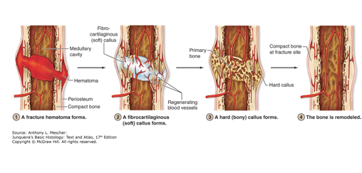
Describe how bone repairs itself after a fracture
Bone repair and growth involve the continuous resorption of previously formed bone tissue and the simultaneous laying down of new bone at a rate that exceeds removal.
Osteoclasts (large, multinucleated, motile cells) handle matrix resorption and remodeling.
This fracture repair process relies on osteogenesis= sum of osteoblasts and osteoclast activities.
A fracture hematoma forms
Fibrocartilaginous (soft callus forms)
Hard (bony) callus forms
Bone is remodeled
Describe fibrous joints
Articulation of bones by fibrous tissue where movement depends on the length of the fibers.
Types include the syndesmosis type (unites bones with a sheet of fibrous tissues, like the radius/ulna and coronal suture) and gomphosis/ dentoalveolar syndesmosis (a fibrous joint in teeth where a peg-like process fits into a socket)
Describe cartilaginous joints
Articulation of bones by hyaline cartilage or fibrocartilage.
Synchondroses (primary cartilaginous joints) permit growth of the length of the bone and allow slight bending until the epiphyseal plate closes (e.g., the hip joint). Symphyses (fibrocartilage/secondary cartilaginous joints) are strong, slightly mobile joints (e.g., the intervertebral disc).
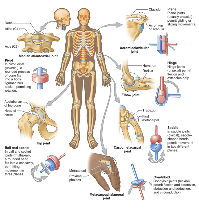
Describe synovial joints
The most common type of joint, characterized by an articular cavity containing synovial fluid. This fluid nourishes the articular cartilage and lubricates the joint surface. They are classified by the shape of the articulating surface and/or the type of movement they permit
List and describe the types of synovial joint movements
•Pivot: rounded process of bone that fits into ligamentous sockets; allows rotation
•Plane: uniaxial; permit gliding or sliding movements
•Hinge: uniaxial; permits flexion and extension ONLY
•Saddle: biaxial: shaped heads permit movement in two different planes
•Condyloid: biaxial; permits flexion, extension, Abduction, Adduction, and circumduction
•Ball and sock: multiaxial; rounded headed that fits into a concavity permitting movement in 3 planes
(Proud PAs Help Sick Children Breathe)
Describe the axial skeleton and major parts
Consists of:
•Head: cranium or skull
•Neck: cervical vertebrae
•Trunk: ribs, sternum, vertebrae, sacrum
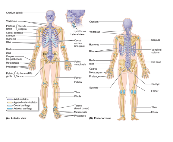
Describe the appendicular skeleton and major parts
Consists of:
•Bones of limbs
•Pectoral girdle
Pelvic girdle

Describe how an imbalance of calcium can affect bone tissue.
Could affect the formation/ remolding of bone tissue
If blood calcium drops, parathyroid hormone (PTH) triggers osteoclasts to break down the bone matrix to release calcium into the bloodstream, leaving the bones porous and brittle. Conversely, if blood calcium is high, calcitonin signals osteoblasts to halt bone resorption and deposit the excess calcium back into the bone tissue.