lab practical I
1/102
There's no tags or description
Looks like no tags are added yet.
Name | Mastery | Learn | Test | Matching | Spaced | Call with Kai |
|---|
No analytics yet
Send a link to your students to track their progress
103 Terms
hormones
chemical messengers that affect specific organs and tissues (targets)
endocrine glands?
anterior pituitary gland, thyroid, adrenals, parathyroids
endocrine organs?
pancreas, hypothalamus, gonads, thymus
follicle stimulating hormone (FSH)
development of sperm and ovarian follicles
luteinizing hormone (LH)
secretion of sex hormones
adrenocorticotropic hormone (ACTH)
stimulates the adrenal glands to produce and release cortisol (stress hormone)
thyroid-stimulating hormone/thyrotropic (TSH)
influences activity of thyroid hormone
melanocyte-stimulating hormone
stimulate melanocyte production
growth hormone (GH)
general metabolic hormone that plays a role in body size
prolactin (PRL)
stimulates breast development and promotes lactation after childbirth
oxytocin
stimulates uterine contractions during birth and milk ejection in lactating mothers
antidiuretic hormone (ADH)
cause the kidneys to reabsorb water, reducing urine output and conserving water
thyroxine (T4) and triiodothyronine (T3)
control over rate of metabolism and cellulr oxidation
calcitonin
decreases blood Ca2+ by stimulating calcium salt deposit into bones
parathyroid hormone
when blood Ca2+ levels are too low, causes calcium salts to be broken down from bone deposits
gonadocorticoids (androgens)
consist mainly of weak androgens and minor amounts of estrogens, which play key roles in libido, pubic hair development, and secondary sex characteristics
glucocorticoids
stress resistors, primarily increase blood glucose levels
mineralocorticoids
regulate water balance via sodium regulation in kidneys
insulin
when blood glucose is too high, this is released and helps transport glucose out of blood into the cells
glucagon
when blood glucose is too low, this is released and stimulates the liver
estrogen
responsible for growth of secondary sex characteristics; also acts in tandem with progesterone to bring cyclic changes of the uterine lining
progesterone
works with estrogen and helps prepare breast tissue for lactation
testosterone
responsible for growth of secondary sex characteristics
thymus
active and large during childhood but atrophies during maturation
pineal gland
produces melatonin which plays a role in biological rhythms
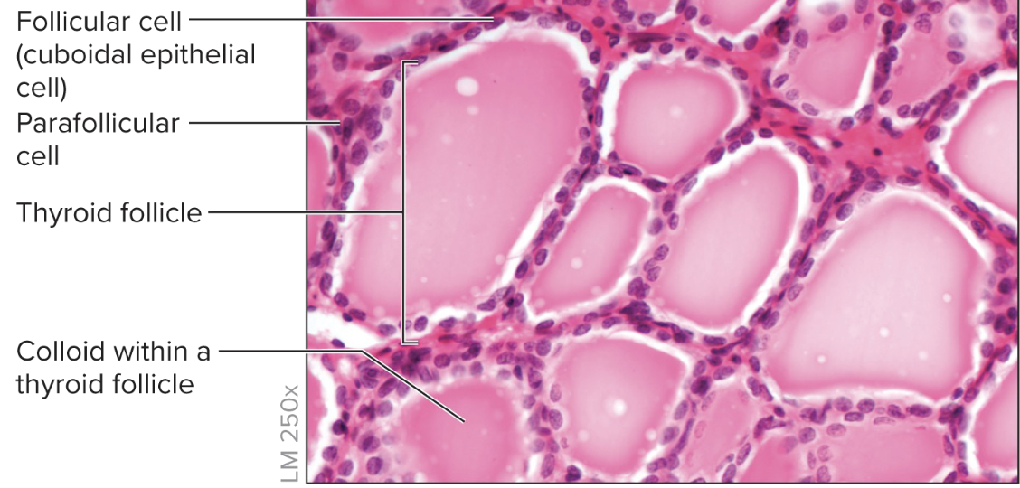
thyroid gland
what is this?
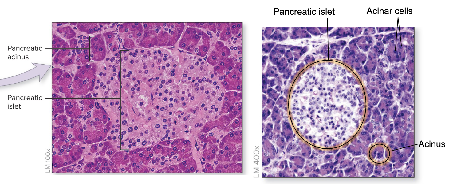
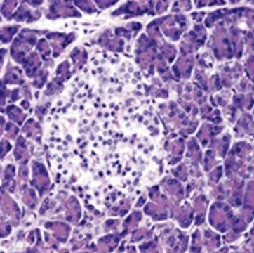
pancreas
what is this?
adrenal gland
consists of a cortex and medulla, with each part having different embryological origin
thyroid gland
consists of follicles lined with a simple cuboidal epithelium
parathyroid glands
four small endocrine glands that secrete a hormone that regulates blood calcium level
hypothalamus
secretes hormones to regulate hormone release by the anterior pituitary gland
pineal gland
secretes the hormone melatonin
thyroid gland
where are parafollicular cells are located?
luteinizing hormone
hormone released by the anterior pituitary gland that induces ovulation in females
epinephrine and norepinephrine
increased heart rate, contractility, and vasoconstriction
cortisol
increased glucose synthesis through protein breakdown, gluconeogenesis, and lipolysis
aldosterone
increased sodium and water retention by the kidneys
androgens
similar in structure and function to testosterone
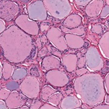
thyroid gland
The following micrograph is a specimen from what gland?
pancreas
The following micrograph is a specimen from what gland?
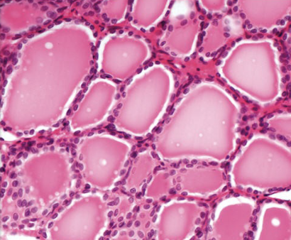
thyroid follicles
what part of thyroid histology is shown?
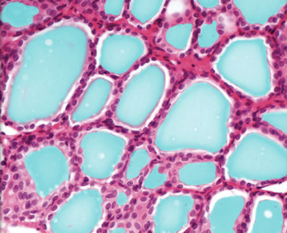
colloid
what part of thyroid histology is shown?
follicular cells
what part of thyroid histology is shown?
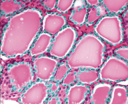
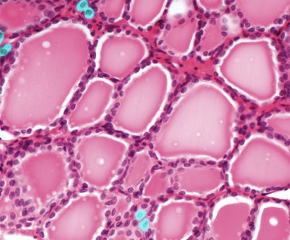
parafollicular (C) cells
what part of thyroid histology is shown?
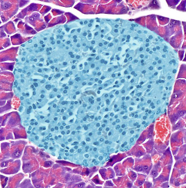
pancreatic islet of langerhans
what part of pancreas histology is shown?
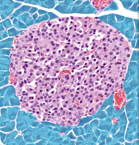
pancreas acini (exocrine pancreas)
what part of thyroid histology is shown?
plasma
55% of blood; 90% water and acts as a solvent, important for osmotic balance
albuminin
major plasma protein used for osmotic balance
fibrinogen
precursor protein for clotting
globulins
plays a role in liver function, blood clotting, and fighting infection
erythrocytes (RBCs)
transport oxygen and carbon dioxide; aid in blood pH regulation
platelets
plays a role in hemostasis (blood clotting)
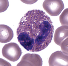
monocytes
circulating cells that migrate out of the bloodstream to become large, phagocytic cells called macrophages
lymphocytes
responsible for the adaptive immune response to infection
eosinophils
fight parasitic infections and mediate (neutralize) the effects of histamines; phagocytic cells
basophils
release histamine and heparin; involved in the inflammatory response
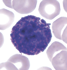
eosinophil
which part of blood cells are shown?
basophil
which part of blood cells are shown?
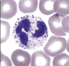
neutrophil
which part of blood cells are shown?
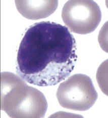
monocyte
which part of blood cells are shown?
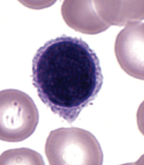
lymphocyte
which part of blood cells are shown?
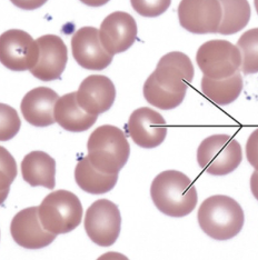
erythrocytes
what is being pointed at?
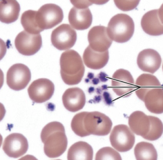
platelets
which part of blood cells are shown?
pericardial sac
connective tissue sac external to epicardium
epicardium
connective tissue layer that contains blood vessels, lymphatic vessels, and nerves
myocardium
cardiac muscle, contractile tissue of the heart
endocardium
epithelial layer that lines the chambers and valves
tricuspid valve
prevents backflow of blood into right atrium
pulmonary semilunar valve
prevents backflow of blood into right ventricle
bicuspid valve
prevents backflow of blood into left atrium
aortic semilunar valve
prevents backflow of blood into left ventricle
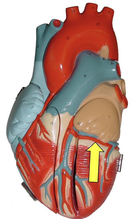
coronary arteries
branch from aorta, supply oxygenated blood to heart when heart is relaxed
cardiac veins
return deoxygenated blood to right atrium for recirculation
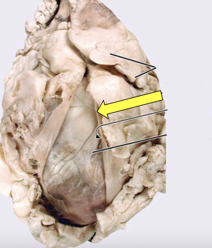
fibrous pericardium
what part of the sheep heart is shown?
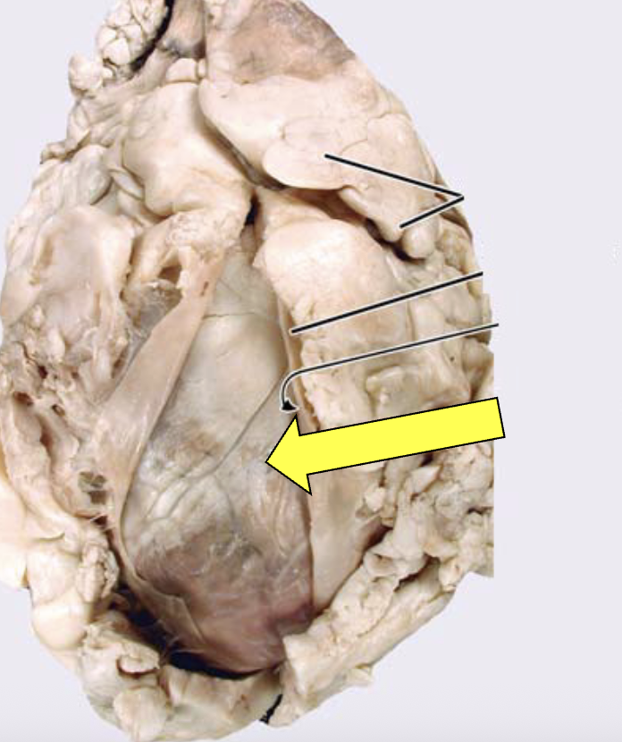
epicardium (visceral pericardium)
what part of the sheep heart is shown?
myocardium (cardiac muscle)
what part of the sheep heart is shown?
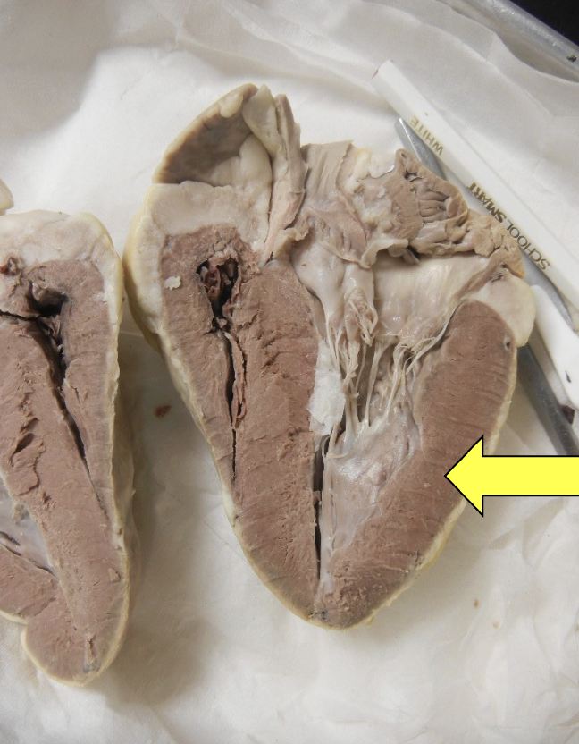
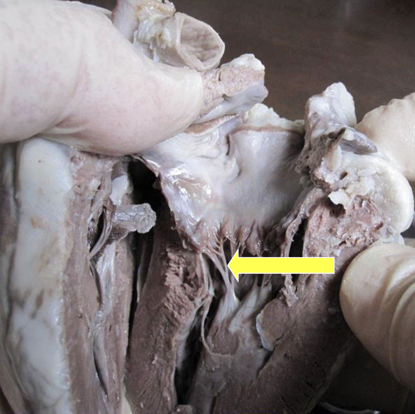
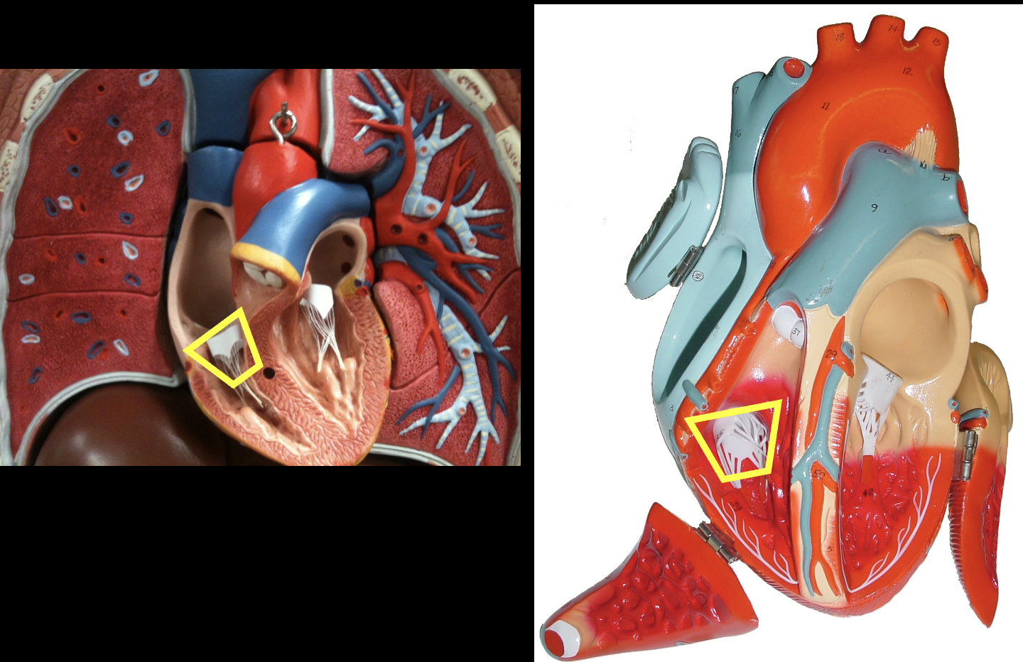
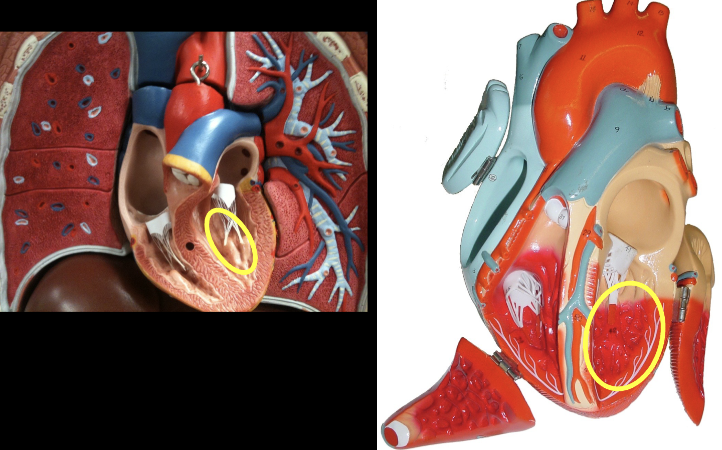
papillary muscle
what part of the sheep heart is shown?
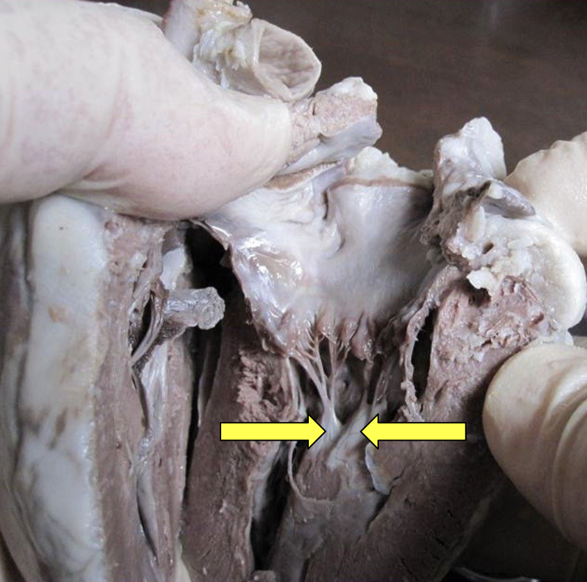
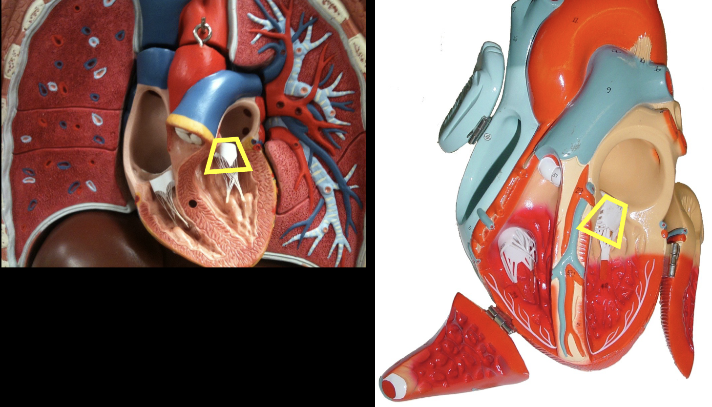
chordae tendineae
what part of the sheep heart is shown?
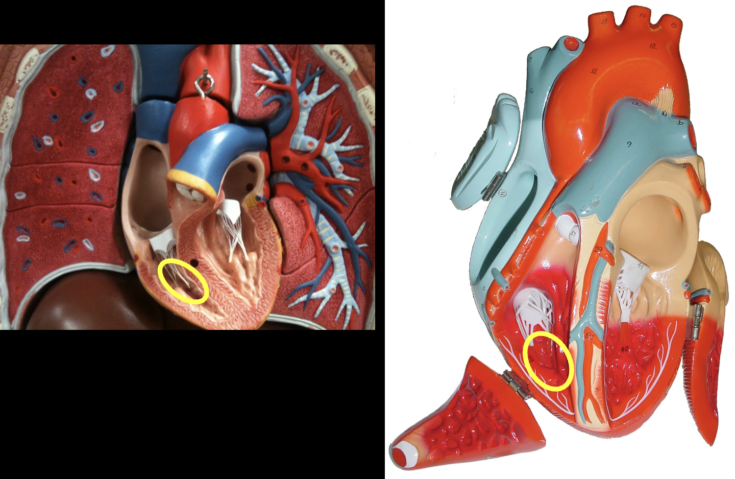
trabeculae carneae
what part of the sheep heart is shown?
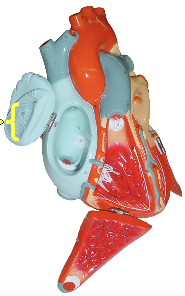
musculi pectinate (pectinate muscle)
what part of the heart muscle is shown?
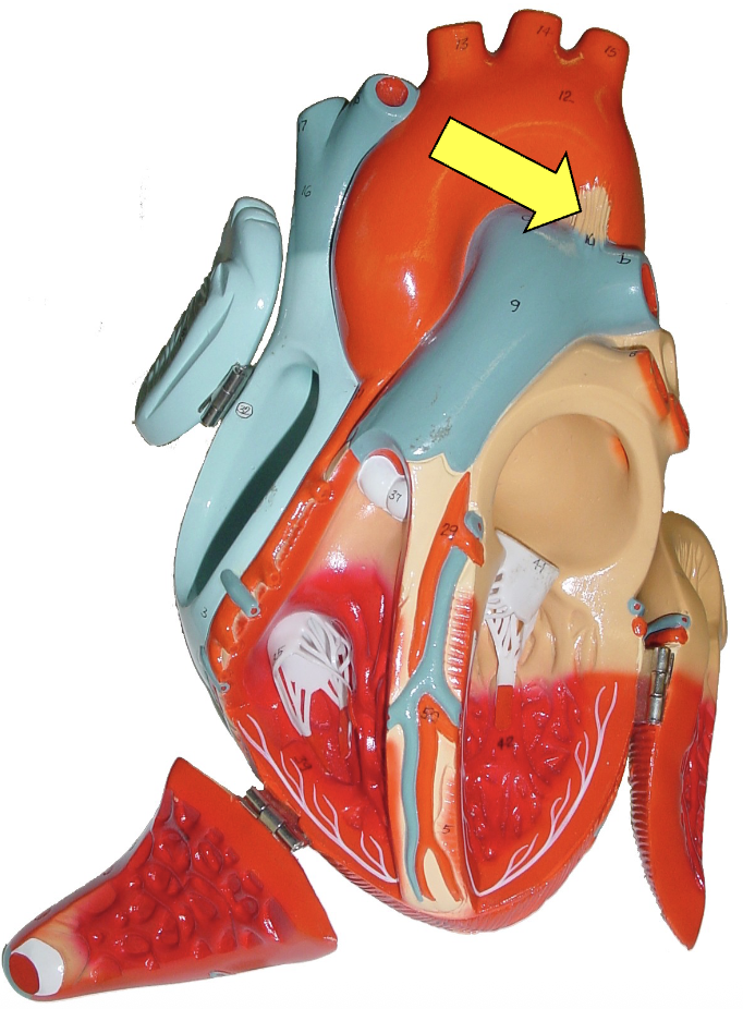
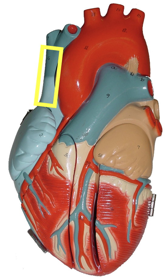
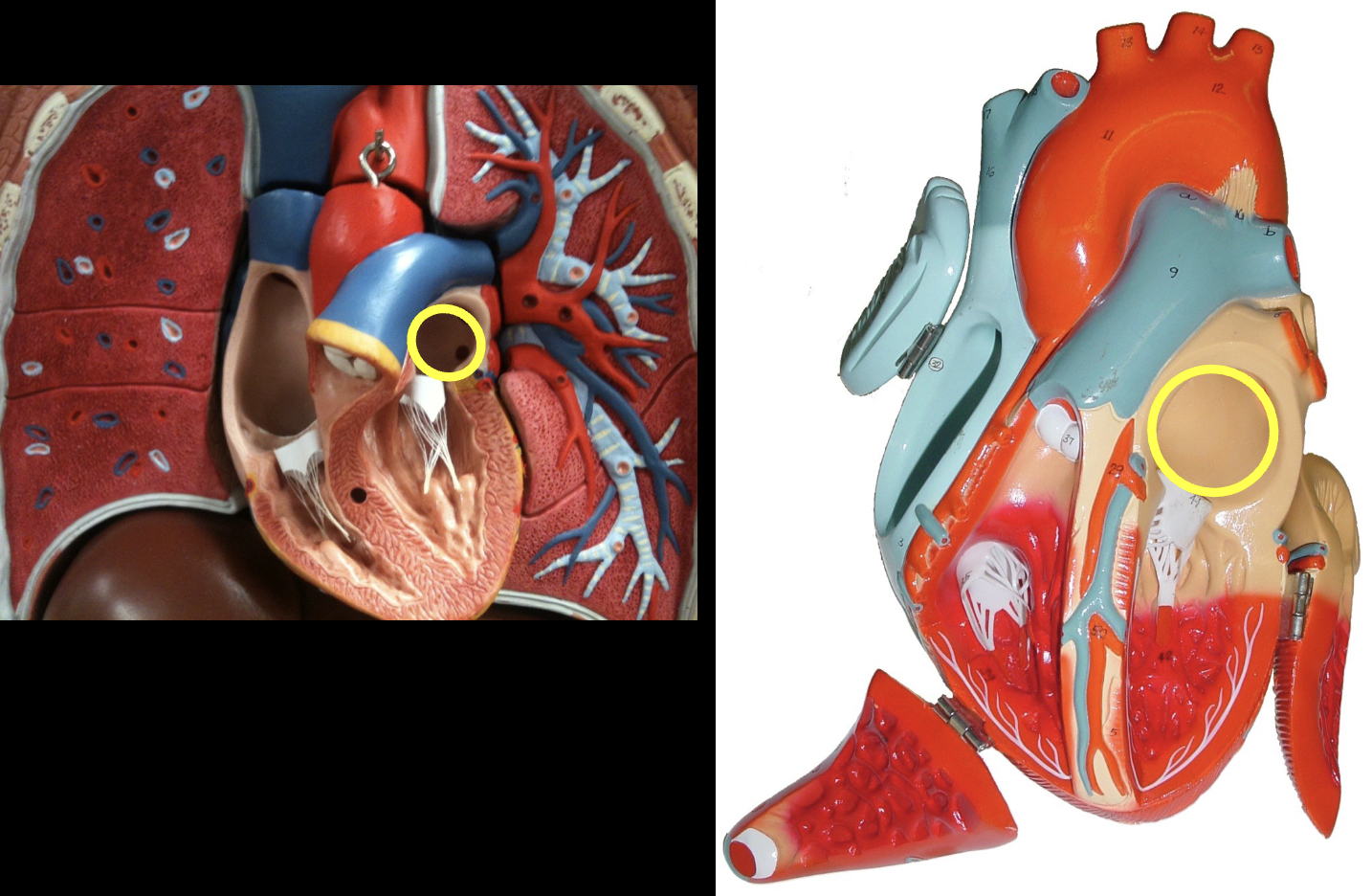
ligamentum arteriosum
what part of the heart muscle is shown?
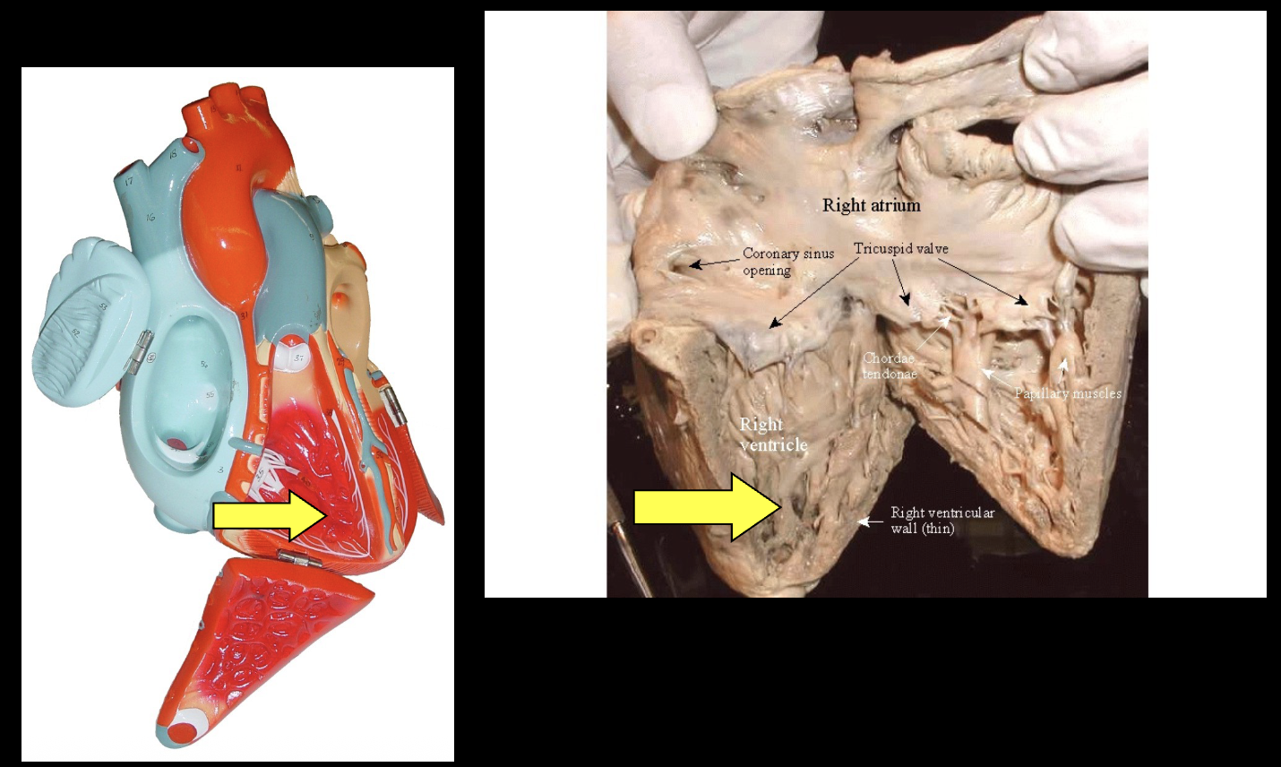
right atrium
what part of the heart muscle is shown?
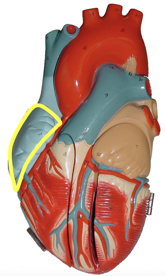
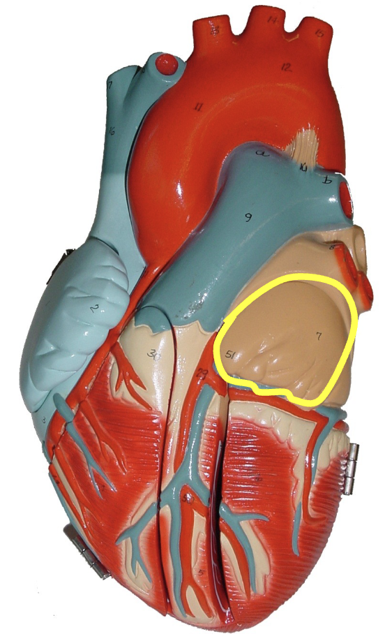
right auricle
what part of the heart muscle is shown?
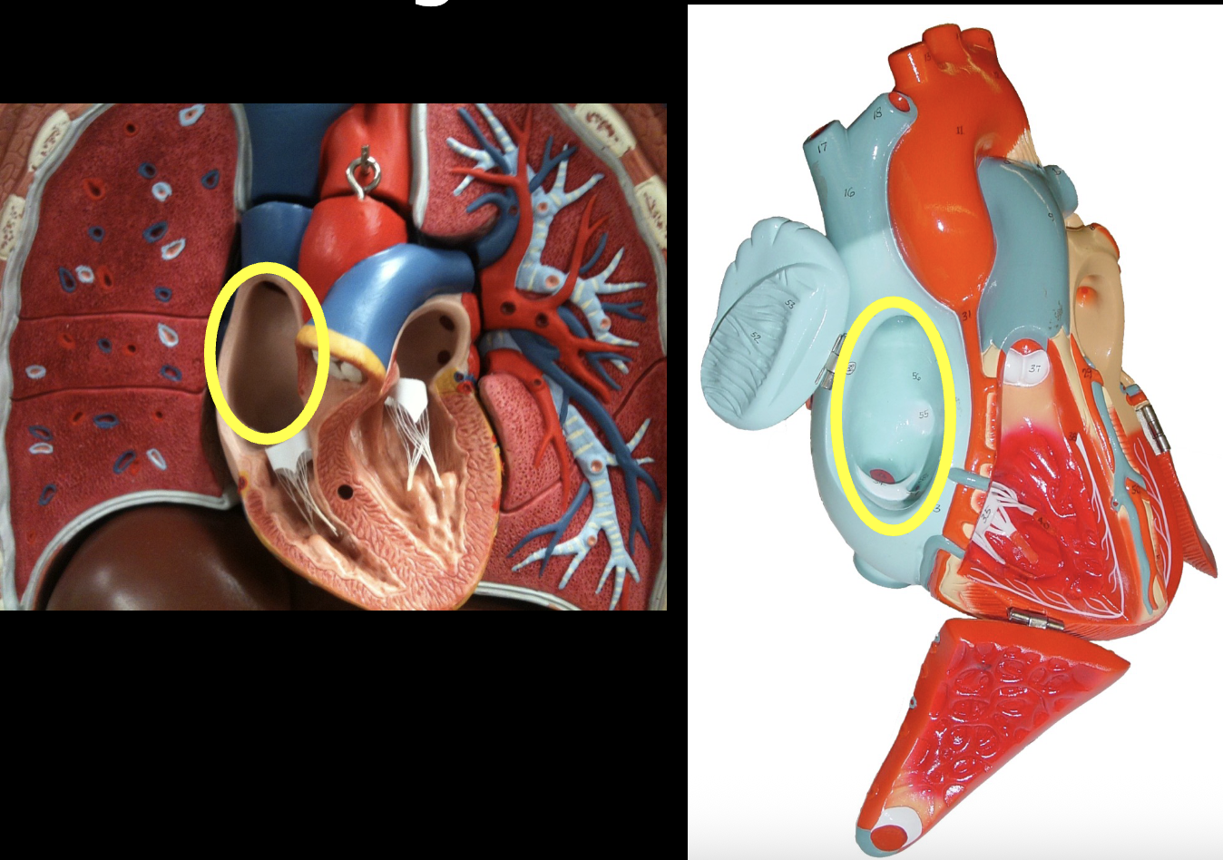
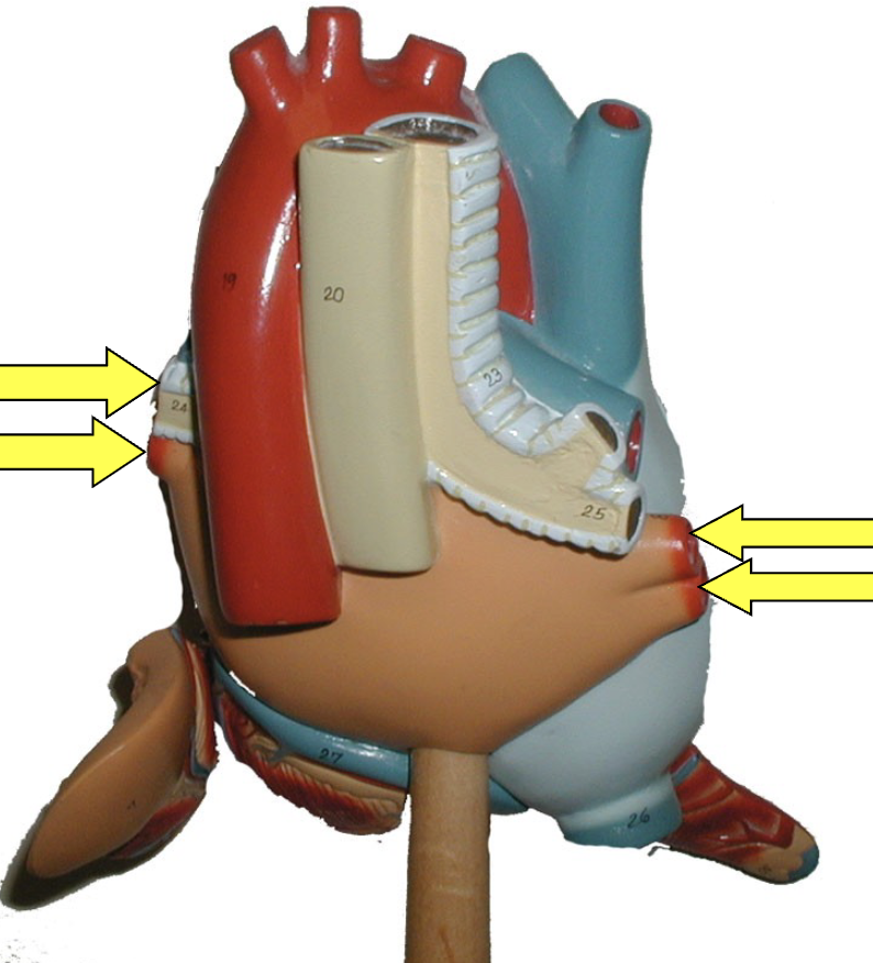
superior vena cava
what part of the heart muscle is shown?
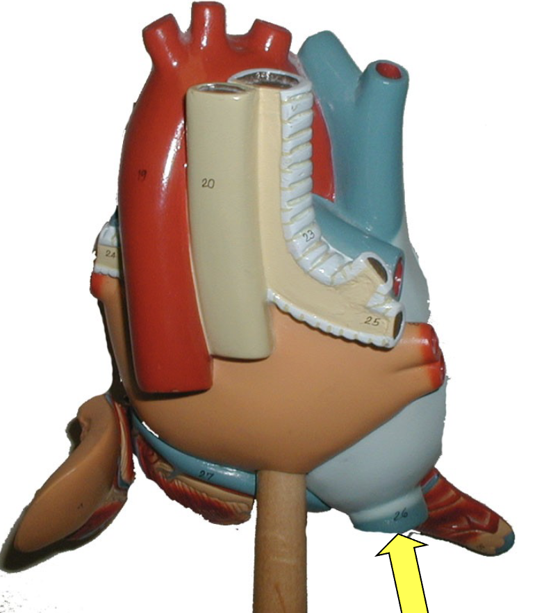
inferior vena cava
what part of the heart muscle is shown?
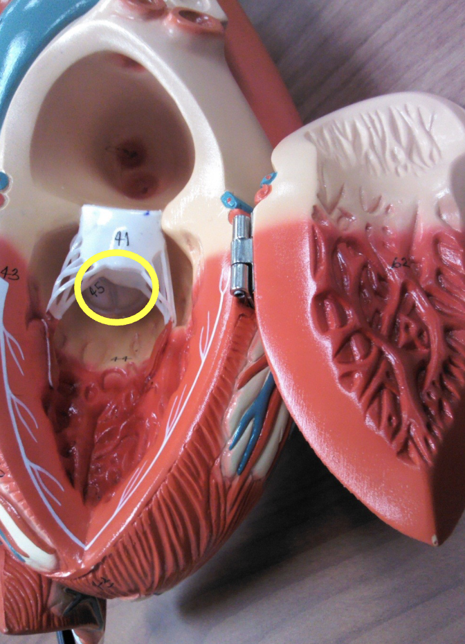
tricuspid valve
what part of the heart muscle is shown?
right ventricle
what part of the heart muscle is shown?
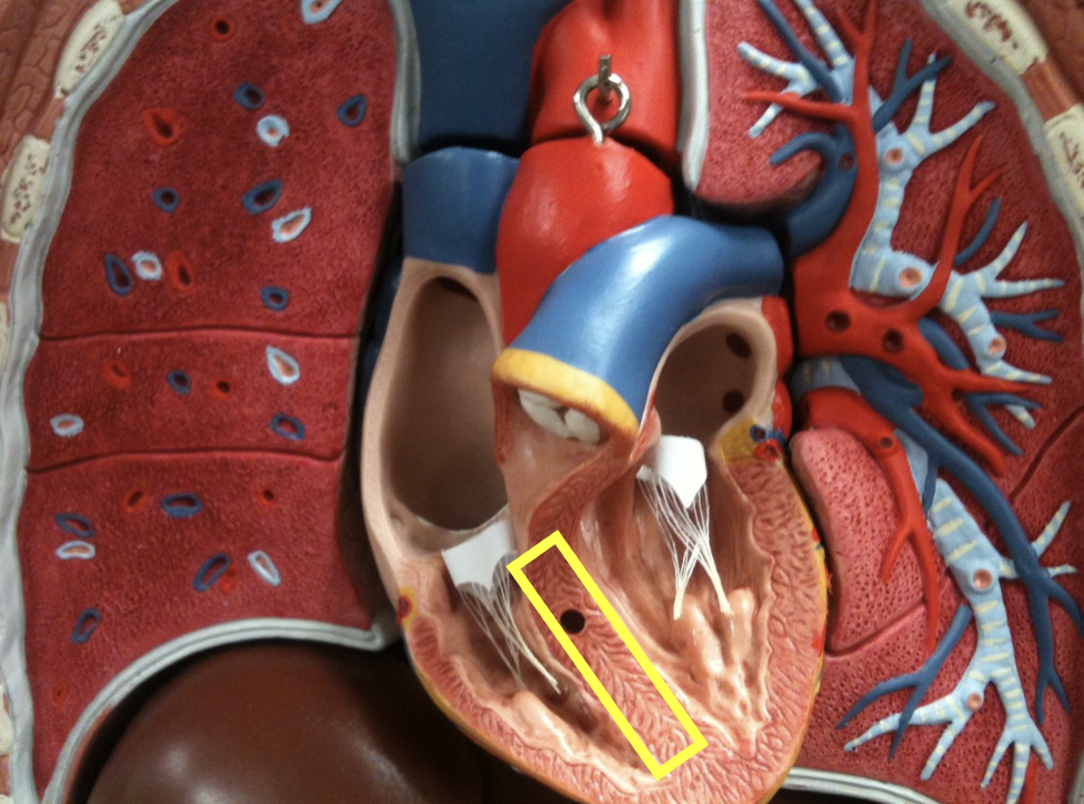
interventricular septum
what part of the heart muscle is shown?
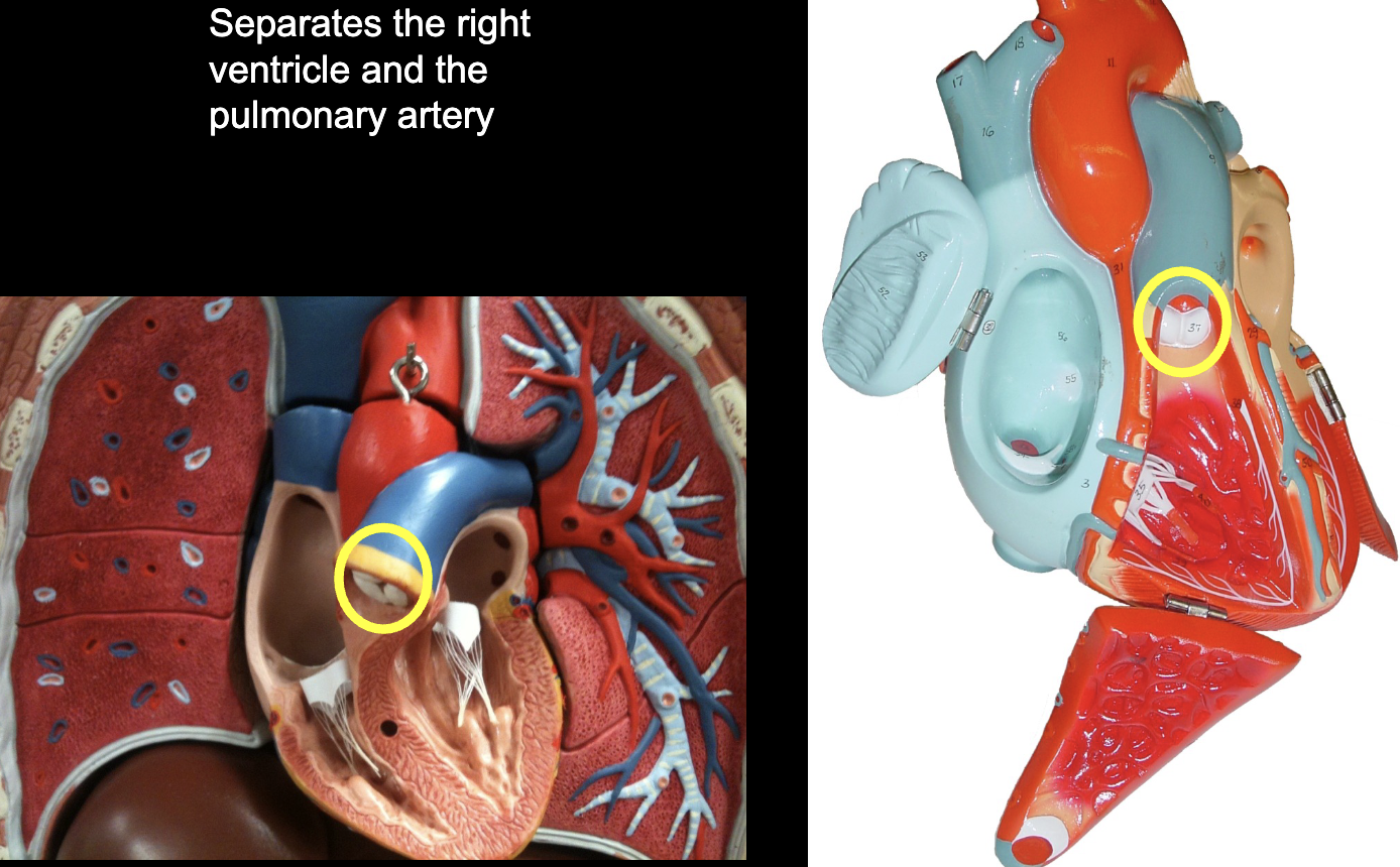
pulmonary (semilunar) valve
what part of the heart muscle is shown?
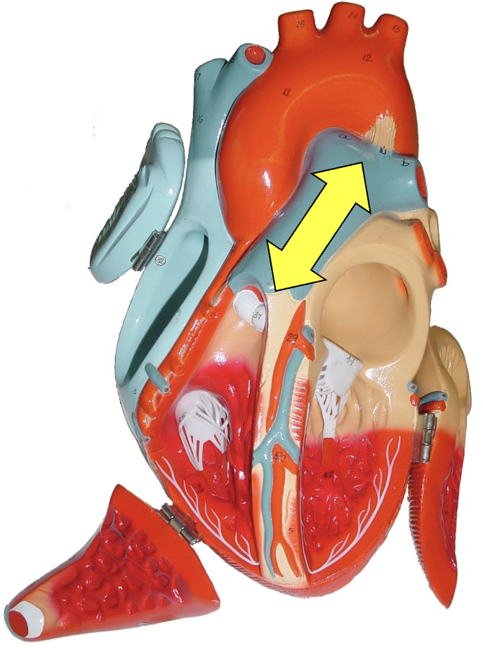
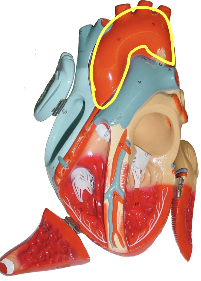
pulmonary trunk
what part of the heart muscle is shown?
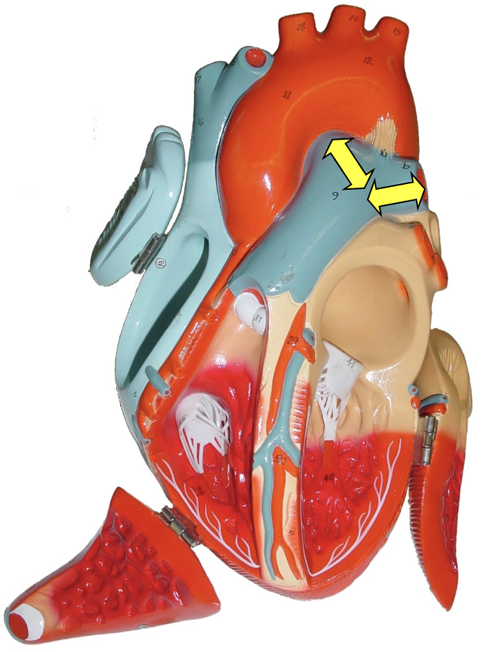
pulmonary arteries (l & r)
what part of the heart muscle is shown?
left atrium
what part of the heart muscle is shown?
left auricle
what part of the heart muscle is shown?
pulmonary veins
what part of the heart muscle is shown?
mitral (bicuspid) valve
what part of the heart muscle is shown?
left ventricle
what part of the heart muscle is shown?
aortic (semilunar) valve
what part of the heart muscle is shown?
aorta
what part of the heart muscle is shown?
circumflex artery
what part of the heart muscle is shown?