A&P252 UNC lab 4
1/95
Name | Mastery | Learn | Test | Matching | Spaced | Call with Kai |
|---|
No analytics yet
Send a link to your students to track their progress
96 Terms
Acromioclavicular joint
a fixed joint, where the acromial end of the clavicle articulates with the acromion process.

Coracoid process
muscle (coracobrachialis) attachment

Acromion
process where scapula articulates with acromial end of clavicle
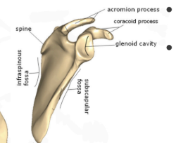
Spine of Scapula
separates the infraspinous and supraspinous fossa
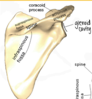
Supraspinous Fossa
posterior fossa that house rotator cuff muscles and stabilize glenohumeral joint in the shallow glenoid cavity - above spine of scapula

Infraspinous Fossa
posterior fossa that house rotator cuff muscles and stabilize glenohumeral joint in the shallow glenoid cavity- below spine of scapula

Subscapular Fossa
anterior fossa that house rotator cuff muscles and stabilize glenohumeral joint in the shallow glenoid cavity

Glenoid cavity
site of artculation with humerus

Head of Humerus

Greater Tubercle
GT-hint
found near the glenohumeral joint

Lesser Tubercle
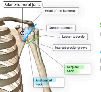
intertubercular Groove

Anatomical Neck
at epiphyseal plate that separates the head of humerus from diaphysis

Surgical neck
narrower region much more commonly fractured

Glenohumeral joint
ball and socket joint, due to the shallowness of the glenoid cavity, this joint is unstable and has a wide range of motion

Trochlea
where the ulna lines up with
-Tro

Capitulum
Where the radius lines up with
-Cap

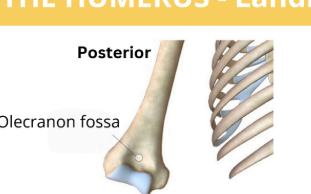
Olecranon Fossa
hold the olecranon process of the ulna and allows for full extension of the elbow joint
Medial Epicondyle
attachment point for most anterior forearm muscles(flexors)
-Me
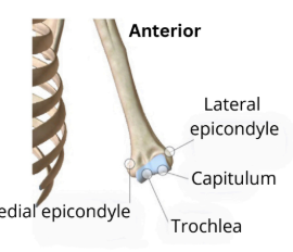
Lateral Epicondyle
attachment point for most posterior forearm muscles(extensors)
-la

Humerus

Ulna
uh
lines up with the medical epicondyle and trochlea
Uh oh, Me-Tro
pinky side

Radius
Rad
lines up with the capitulum and lateral epicondyle
Rad Cap, Lad

interosseous membrane
facilitates pronation & supination at the proximal radioulnar joint

styloid process

coronoid process
also found in the mandible
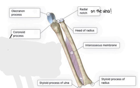
Olecranon process

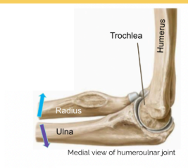
Humeroulnar joint
is a hinge joint capable of extension and flexion
found near humerus and ulnar + radius
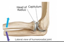
Humeroradial joint
is a ball and socket joint but has limited rotation due to surroundings bones and interosseous membrane.
Can extend, flex, pronate, and supinate.
head of radius

Radioulnar joint
the proximal - is a pivot joint that allows the rotation of the radius, which result in pronation and supination
distal is a fixed joint
radial notch
smooth surface on the ulna where it articulates with the radius

radiocarpal joint
aka the wrist
is a condyloid joint, which allows abduction, adduction, flexion, and extension

scaphoid
carpal found in the proximal row from lateral to medial
Some

lunate
carpal found in the proximal row from lateral to medial
lovers

triquetrum
carpal found in the proximal row from lateral to medial
try
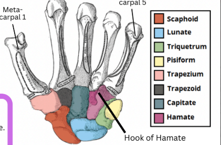
pisiform
carpal found in the proximal row from lateral to medial
positions

trapezium
carpal found in distal row from lateral to medial
that

trapezoid
carpal found in distal row from lateral to medial
they

capitate
carpal found in distal row from lateral to medial
cant

hamate
carpal found in distal row from lateral to medial
handle
hook or hamate - near pinky (metacarpal 5)

metacarpals
clap

metacarpophalangeal joints
between metacarpals and proximal phalanges
condyloid joint, which allows for abduction, adduction, extension, and flexion
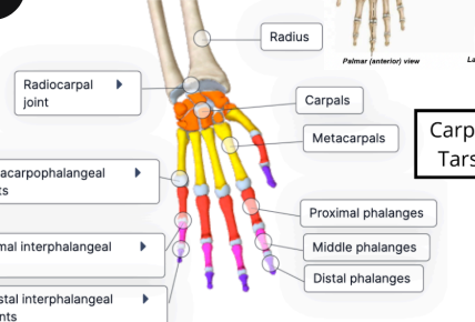
distal interphalangeal joints
between distal and middle phalanges
hinge joint, which allows for the extension and flexion

Proximal/Intermediate/Distal Phalanges

pollex
anatomical name for the first digit of the hand, thumb
ilium
helps forms the coxa/hip bone, which contributes to the acetabulum

ischium
helps forms the coxa/hip bone, which contributes to the acetabulum
isch
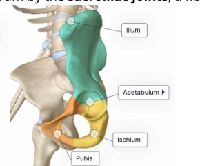
pubis
helps forms the coxa/hip bone, which contributes to the acetabulum
p

acetabulum
the socket for the femur

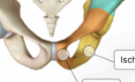
pubic symphysis
fibrocartilaginous joint, where 2 hips bones connected anteriorly
iliac crest
superior crest of ilium

anterior superior iliac spine
noticeable bump on the lateral end of the ilium that serves as the attachment for the inguinal ligament
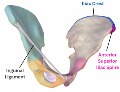
inguinal ligament
tough band stretching from the anterior superior iliac spine too the pubi
creates a protected passageway for the neurovascular structures, have one on each coxa

superior ramus
joints the ilium
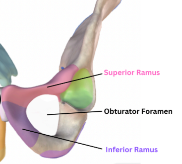
inferior ramus
joins the ischium

obturator foramen
a large opening that is covered by connective tissue from which muscles attach, but allows for a small nerve and blood vessels to pass through

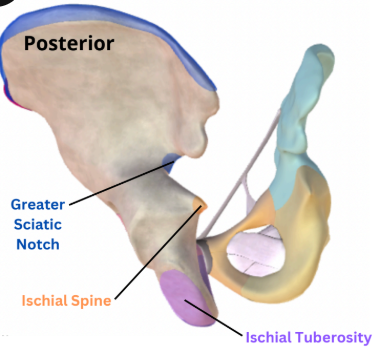
ischial tuberosity
rough patch on most inferior region of the pelvis and site of muscle attachment for hamstrings on the posterior thigh

ischial spine
protects posteriorly from the acetabulum

greater sciatic notch
large notch superior to the ischial spine and the passageway for the body’s largest nerve, the sciatic nerve
pelvic inlet
aperture formed by the superior ramus of pubis, ilium, and sacrum
larger and rounder in biological female - childbirth
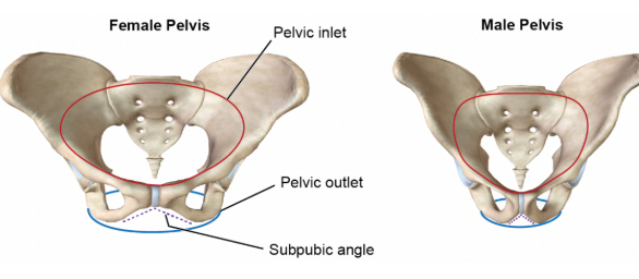
pelvic outlet
broader outlet in biological females

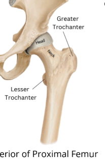
head of femur

neck of femur

greater trochanter

lesser trochanter
LT - hint
medial condyle
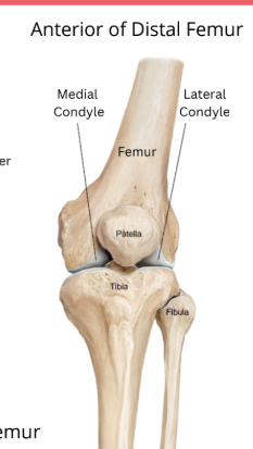
lateral condyle

patellar surface

medial/lateral meniscus
articulates with femoral medial and lateral condyles
there is additional articular cartilage and menisci at the knee joint

tibial tuberosity
attachment site for patellar tendon from the thighs quadriceps muscles

head of fibula

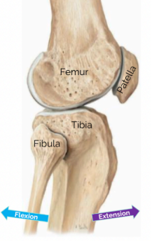
tibia
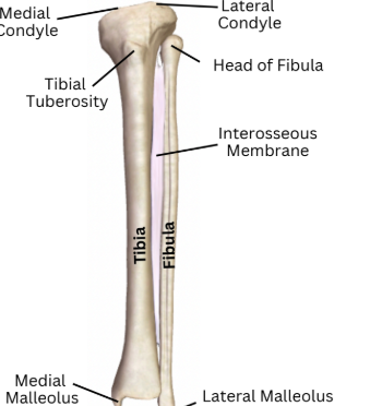
fibula

femur
medial/ateral malleolus
surrounding the talus bone of the ankle to support the talocrural joint

talocrural joint
saddle joint between the tibia/fibula and talus bone
movements includes eversion, inversion, dorsiflexion, and planar flexion
proximal/intermediate/distal phalanx

metatarsophalangeal joint
between metatarsals and proximal phalanx
condyloid joint that allows these movements abduction, adduction, extension, flexion
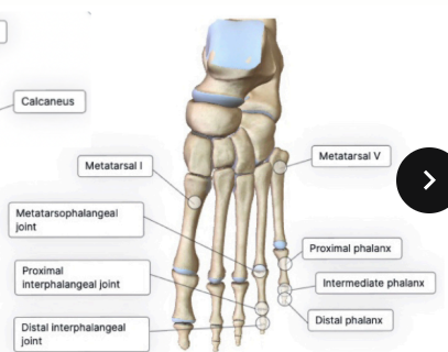
proximal interphalangeal joint
between proximal and intermediate phalanx
hinge joints that allow extension and flexion

distal interphalangeal joint
between the distal and intermediate phalanx
hinge joints that allow extension and flexion

talus

calcaneus
attachment site of the calcaneal tendon

calcaneal tendon

navicular

cuboid

medial/intermediate lateral cuneiform
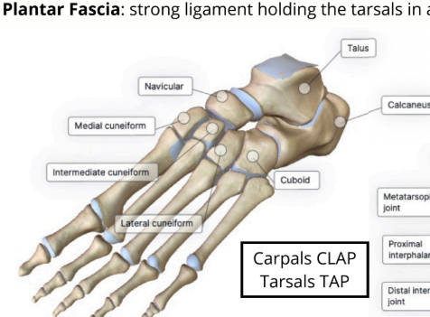
plantar fascia
strong ligament holding the tarsals in an arch (navicular, medial cuneiform, intermediate cuneiform, and lateral cuneiform, cuboid)

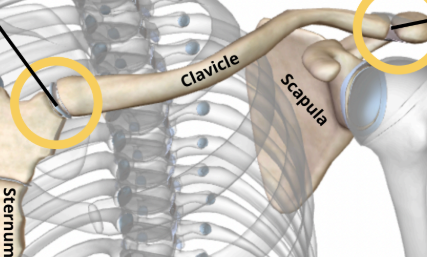
sternoclavicular joint
a saddle joint, where the sternal end of clavicle articulates with the sternum
proximal interphalangeal joints
between proximal and middle phalanges

sacroiliac joints
fibrous joint, 2 hip bones are connected posteriorly to the sacrum
knee joint
is a hinge joint between the femur, tibia, and patella
allow extension of knee, which move leg forward
allow extension of knee, which move leg backwards
hallux
anatomical name for the first digit of the foot
hallucis
big toe region
pollicis
thumb region