muscular microanatomy
1/8
There's no tags or description
Looks like no tags are added yet.
Name | Mastery | Learn | Test | Matching | Spaced | Call with Kai |
|---|
No analytics yet
Send a link to your students to track their progress
9 Terms
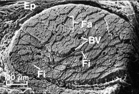
Skeletal muscle produces voluntary movement and is made of multinucleated, striated fibers (Fi) grouped into fascicles (Fa). Epimysium (Ep) surrounds the whole muscle, perimysium surrounds each fascicle, and endomysium surrounds each fiber. Blood vessels (BV) and nerves run through these layers to supply each fiber.
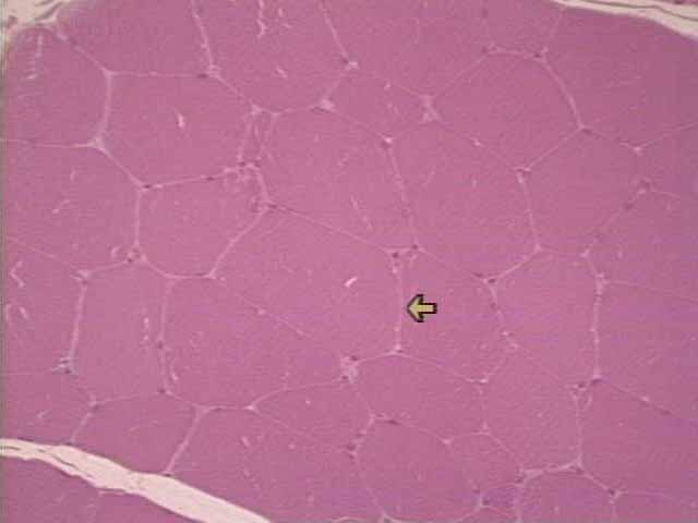
Slide 3 shows a magnified view of muscle fibers in a fascicle. The arrow points to a thin, light endomysium, and small purple nuclei are seen just under the muscle fiber membrane.
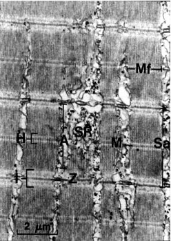
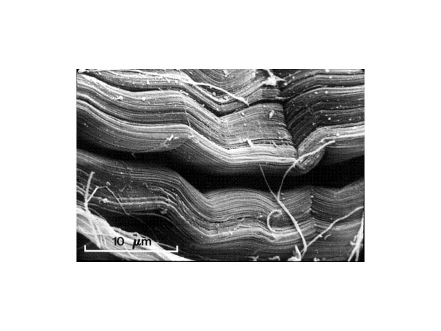
Slide 1 is a high-resolution transmission electron micrograph of four fibrils, illustrating the detailed structure of the sarcomere (between the arrows). Note that the arrows point to the Z-lines which are the demarcations of each sarcomeric unit. The structure "A" is the sarcoplasmic reticulum, a network of flattened membrane tubules which release calcium to the sarcomeres. It is this calcium that acts as the immediate trigger for muscle contraction.
Slide 1 is a high-resolution transmission electron micrograph of four fibrils showing sarcomeres between the arrows. The arrows point to the Z-lines, which mark the boundaries of each sarcomere. Structure “A” is the sarcoplasmic reticulum, which releases calcium that triggers muscle contraction.
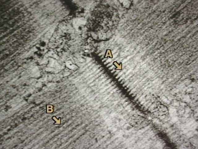
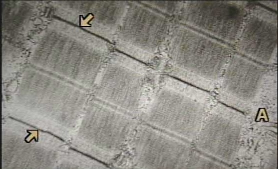
Slide 2 shows the orderly arrangement of actin (A) and myosin (B) filaments within sarcomeres. Actin filaments attach to the dark Z-line, and the myosin filaments are noticeably thicker than the actin filaments.
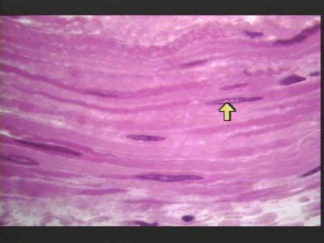
Slide 2 shows smooth muscle with flattened purple nuclei in the center of spindle-shaped cells. The cell borders are hard to see, but the cells are tightly packed, allowing excitatory signals to pass directly from one cell to another.
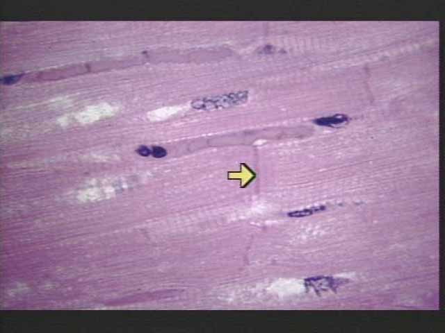
Cardiac muscle is found in the heart wall and has branching, striated cells with a central nucleus and intercalated discs. These discs connect cells and transmit both force and electrical signals. Slide 1 shows faint horizontal striations and dark blue nuclei, and the arrow points to an intercalated disc that links cardiocytes and allows electrical conduction between them.
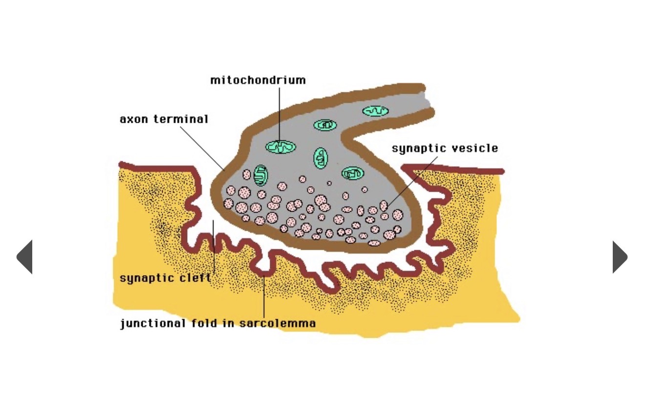
Myoneural (neuromuscular) junction is where a motor neuron axon terminal contacts the muscle sarcolemma, separated by a synaptic cleft. The axon terminal contains synaptic vesicles with acetylcholine and mitochondria. Acetylcholine is released into the cleft, binds receptors on junctional folds, and depolarizes the sarcolemma to trigger a muscle action potential and contraction.
Slide 1 shows dense regular connective tissue in a tendon made of tightly packed, parallel collagen fibers that resist pulling force in one direction. Dense irregular connective tissue has fibers arranged in many directions and resists stress from all directions, found in the dermis and dura mater. Tendons are adapted for strong unidirectional force due to their parallel collagen bundles.