2. Week 1: Cell Cycle (copy)
 Call Kai
 Learn
Learn Practice Test
 Spaced Repetition
 Match
Match Flashcards
 Knowt Play
1/23

Earn XP
Description and Tags
Lecture 2 - 643 - Faridi
Name | Mastery | Learn | Test | Matching | Spaced | Call with Kai |
|---|
No analytics yet
Send a link to your students to track their progress
24 Terms
What are the categories of cells that are in vivo (3)
Cells that highly specialized and lack the ability to divide, once differentiated
Neurons, muscle cells, RBCs
Cells that do not divide normally but can cycle with an appropriate stimulus
Liver cells (surgery)
Lymphocytes (antigen interaction)
Highly mitotic cells - these are constantly dividing
Male gametes
Hematopoietic stem cells
Epithelial cells
Recall the overall pathway from growth factor binding to cell response
Growth factors and RTKs
Growth factors bind to receptor tyrosine kinases (RTKs)
RTKs are typically monomers before binding
Ligand binding causes:
Dimerization
Trans-autophosphylation
Key concept: Ligand binding
growth factor = ligand
ligand binds outside the cell (in this image ligand does not enter cell - signal transduction)
triggers signal transduction inside the cell according to image
Signal transduction
activates mitogenic signals - mitogenic is growth-promoting signals
these signals involve intracellular biochemical processes
leads to cell cycle activation
cell cycle = process of cell growth and division
What is cancer
arises through a series of somatic alterations in DNA that result in unrestrained cellular proliferation
Recall the eukaryotic cell cycle - what are the phases involved?
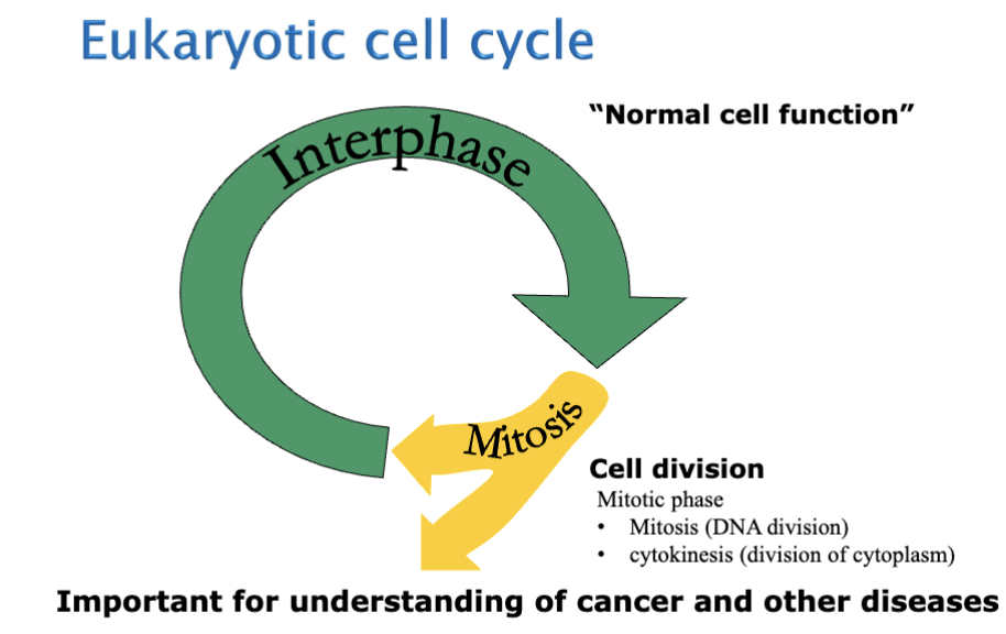
Interphase - normal cell function, and then the cell divides into the mitotic phase
In the Mitotic phase includes
Mitosis (DNA division) and Cytokinesis (division of cytoplasm)
Recall the processes of Interphase
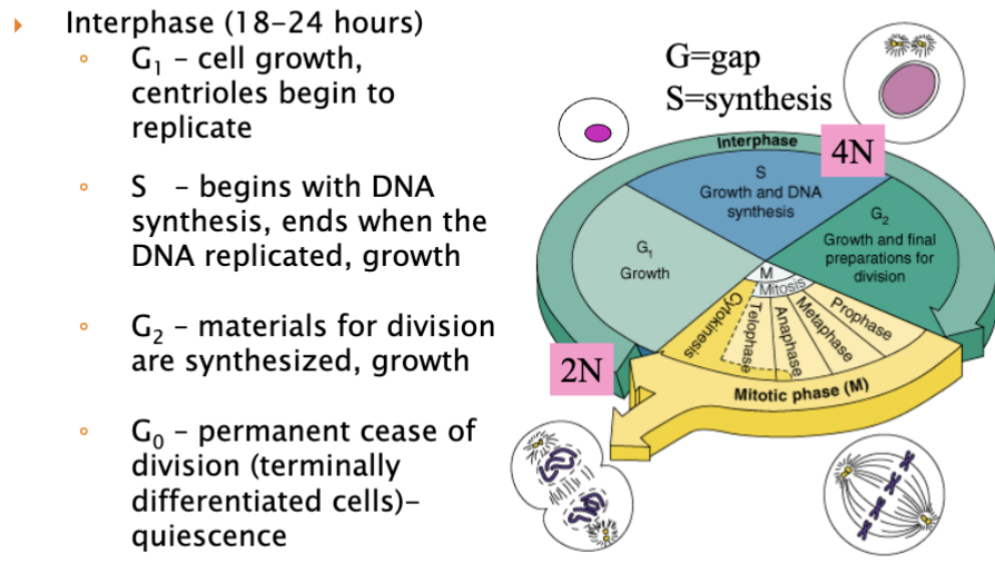
Gap/growth phase “nothing happening”
G1 - a lot of cell growth
2N (N = # of genes per chromosome) - cell starts as 2N and begins to grow
S phase (synthesis)
begins with DNA synthesis, and it ends when the DNA is fully replicated; more growth is occuring within this cell
at the end of S-phase, 2N has doubled to 4N - doubled the amount of DNA, getting ready for the cell to dividde
G2
gets materials ready for cell division
G0 - typically ignored, not seen in image
this is what neurons are undergoing - have left the cell cycle; these are NOT cycling cells
What are the 4 phases of the M phase? What is the 5th phase? When does the 5th phase begin and what does it form?
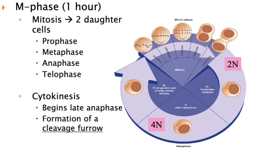
The cleavage furrow is important in cytokinesis because it is the structure that helps divide one cell into two daughter cells.
Completes the final step of cell division after mitosis
List the DNA damaging agents and its pharmacological class (3)
Cyclophosphamide
type of alkylating agent
damages cells DNA and may kill cancer cells
may also lower body’s immune response
Doxorubicin
type of anthracycline antibiotic
type of topoisomerase inhibitor
Cisplatin
type of platinum analogue
Identify DNA inhibiting/division agents and its pharm class
Fluorouracil
type of antimetabolite
stops cells from making DNA and may kill cancer cells
Methotrexate (Trexall, Rheumatrex)
antimetabolite and antifolate
stops cells from using folic acid to make DNA and may kill cancer cells
Gemcitabine
antimetabolite
stops cells from making DNA and may kill cancer cells
Identify DNA inhibiting/division agents
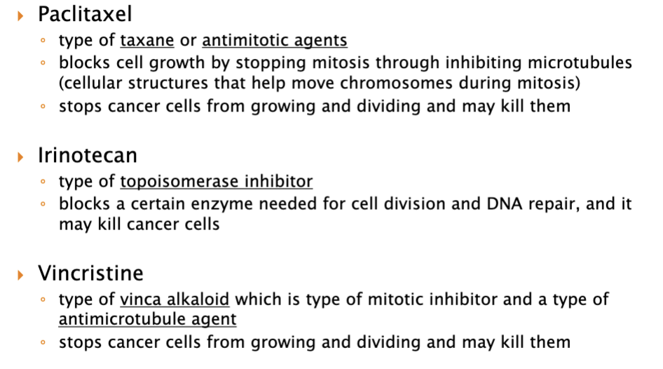
What are the key regulators of the cell cycle (2)
main way is through this protein complex
cyclin-cdk complex
which is a heterodimeric protein kinases
cyclin
cyclin-dependent protein kinases (cdk)
it’s function is that it controls the timing of the cell cycle
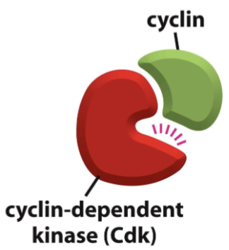
Explain what each member of the heterodimer complex does (the Cyclin-cdk complex)
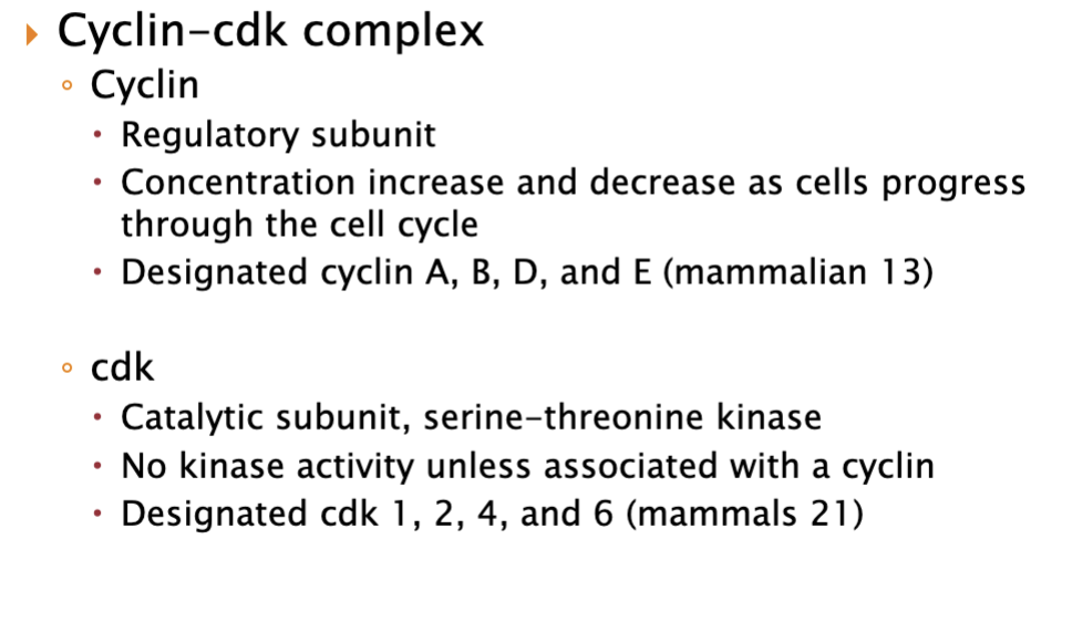
Cyclin = the regulatory subunit
Its levels go up and down during the cell cycle.
It controls when the complex becomes active.
Different cyclins appear at different stages of the cell cycle.
CDK (cyclin-dependent kinase) = the catalytic subunit
It is an enzyme (a kinase) that adds phosphate groups to proteins.
CDKs are usually present in the cell all the time, but they are inactive alone.
They become active only when bound to the correct cyclin.
Together:
Cyclin binds CDK → forms the cyclin-CDK complex
The activated complex phosphorylates target proteins
This pushes the cell into the next stage of the cell cycle
So in simple terms:
Cyclin = the “on/off timing controller”
CDK = the “worker enzyme” that carries out the action
What individuals cyclins and cdks pair up and what is each of their functions?
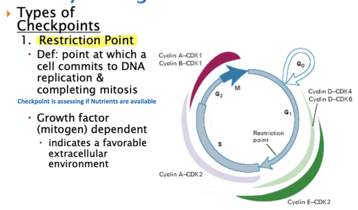
Where can the restriction point be found? - what cyclins and kinases pair up to move past this?
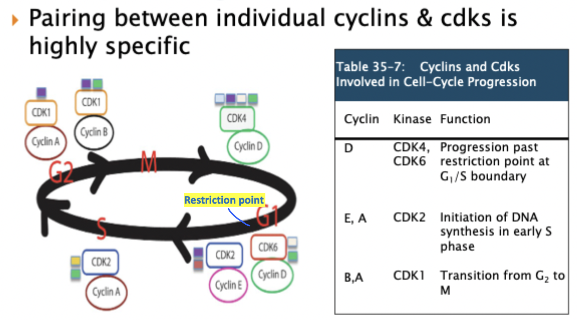
paring between cyclin and cdk is very specific
remember if cells are not cycling, they are referred to as G0
G1
Cyclin D binds to CDK6 and drives/allows for it to go past the restriction point and move into the next phase
Cyclin E and Cyclin A will bind to CDK2 - initiates DNA synthesis in early S phase
Cyclin B and A bind to CDK1 to transition from G2 into M
restrict point is somewhere in late G1 - example of a cell cycle “check point”
What factors regulate CDKs in order to activate the cyclin-cdk complex?
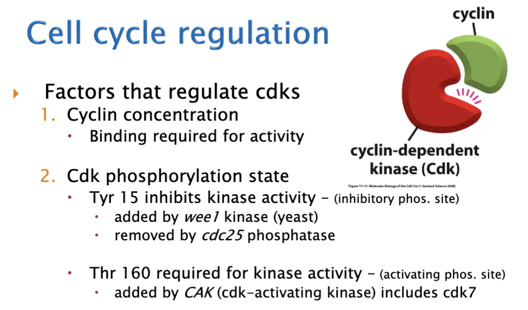
CDK concentrations do not change, Cyclin Concentrations is what changes
cyclin concentration is the first factor that comes into play
CDK has two phosphorylation sites
1st site - tyrosine (tyr) 15 - if there is a Phosphate (P) here, it inhibits the activity of CDK
the Phosphate gets here by a kinase → wee1 (yeast)
a phosphatase is an enzyme that is used to remove the tyr 15, more specifically cdc25 phosphatase allowing for cdk activation
2nd site - threonine (thr) 160 - Phosphate here allows for cdk activity that is added by CAK
cdk7 is a specific CAK
Cell cycle regulation in total
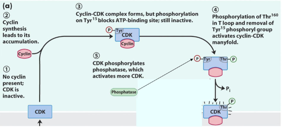
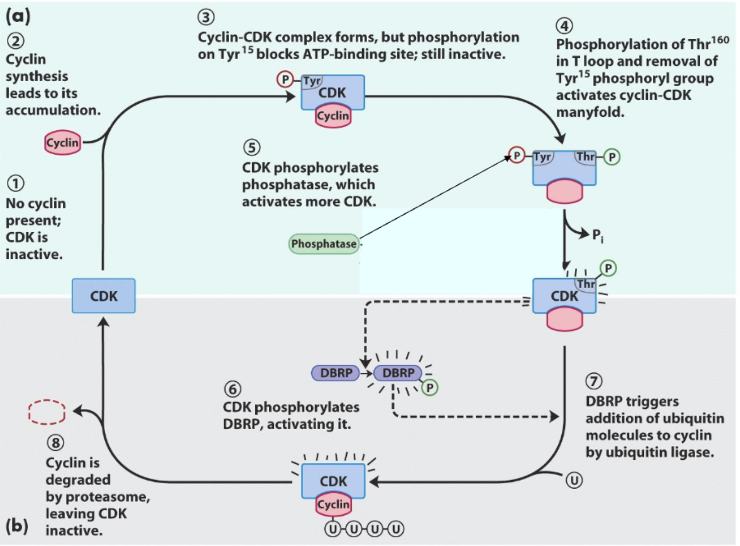
no cyclin present, so cdk is inactive
stimulation
still inactive tyr-15 inactives the complex, must be removed by a phosphatase (cdc25) to remove the P on tyr-15
still inactive because the P in tyr-15 is still not removed
active since P on thy-160 and there is no P on tyr-15
What factors regulate CDKs after the cyclin-cdk complex has been activated?
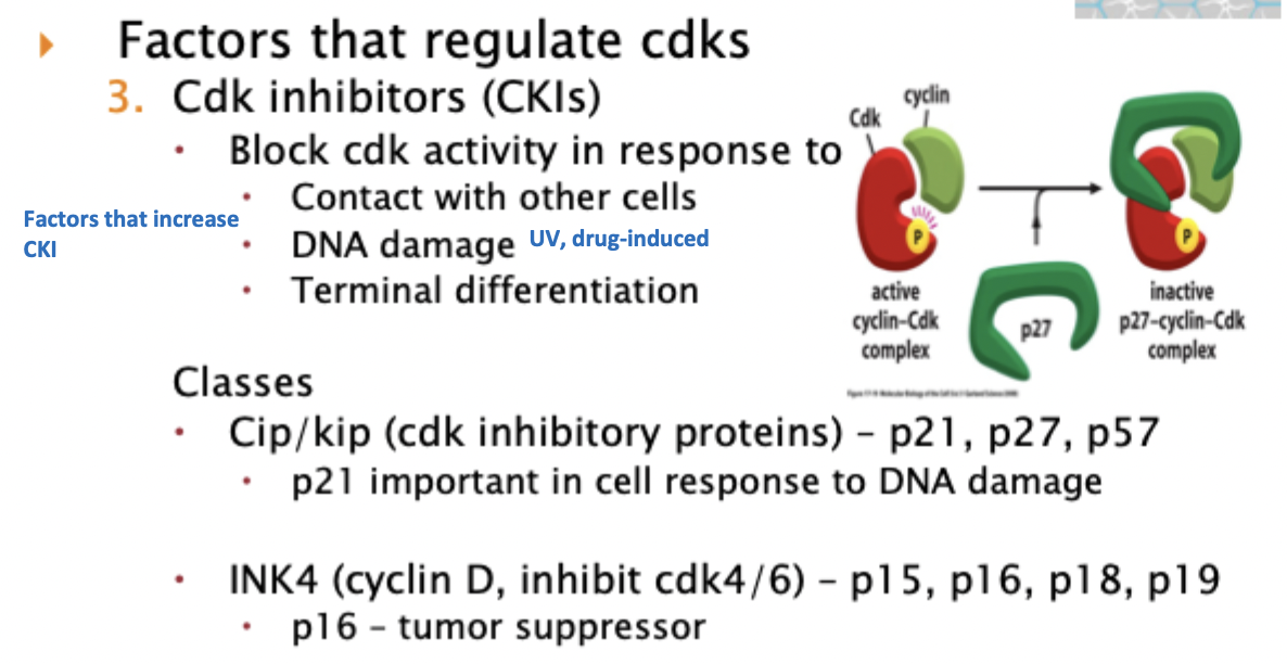
CKIs - these are endogenous proteins within a cell
a CKI blocks cdk activity in response to certain things listed in the image
why would we want the cell cycle to be off?
because not all cells should be dividing
overgrowth/contact example: in hepatocytes when cells are touching their neighbors and there’s no space left, they stop dividing = CKIs increase and cell cycle stops
DNA damage example: in DNA damage such as sun exposure, do not want damaged cells copying themselves, so CKIs increase and cell cycle stops
CKI levels very high in neuronal cells, not dividing cells
Understand what CDK inhibitory proteins (cip or kip) and INK4 proteins do
p16
p21
Mechanism of action of CDK4/6 inhibitors in breast cancer
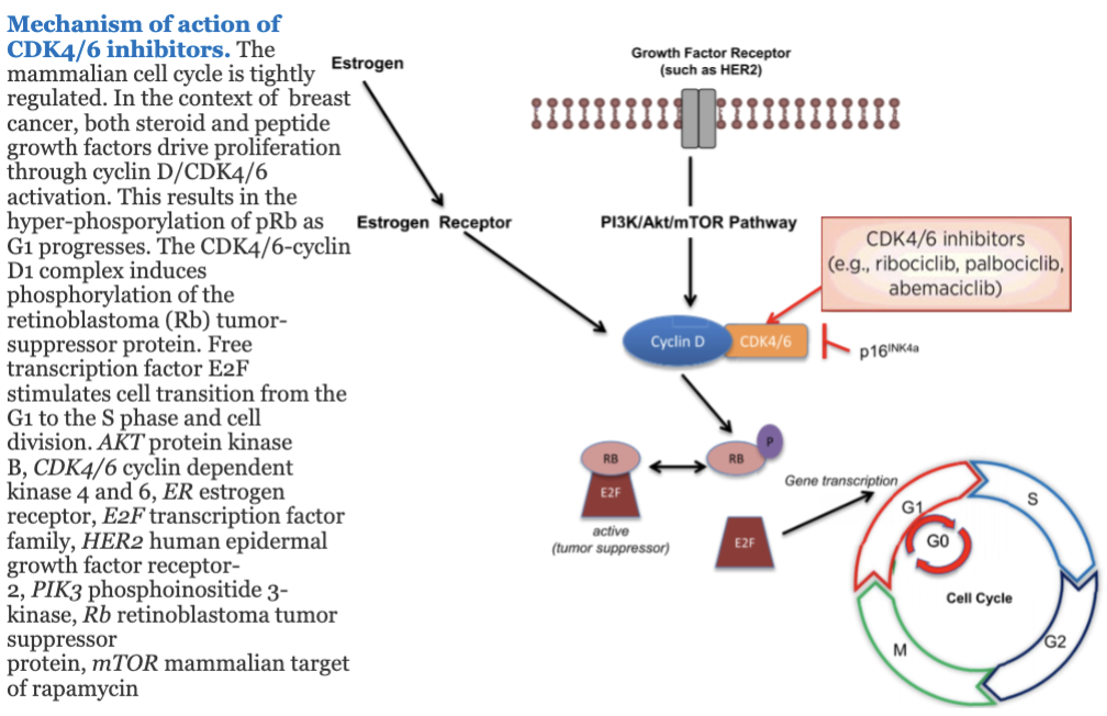
used in certain types of breast cancer like HR-positive, HER2-negative breast cancer
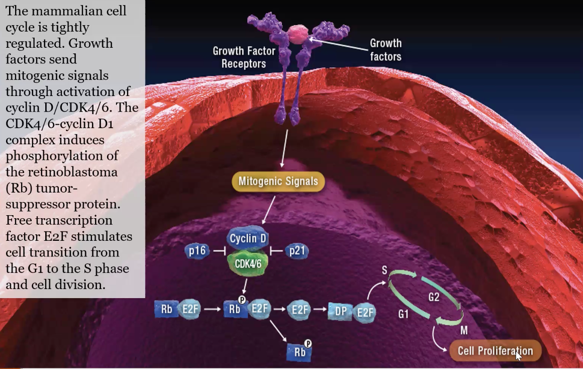
when CDK4/6 is inhibited, this allows for Rb to stay active/”on” so that cancer cells cannot go past the restriction checkpoint from G1 to S phase
Rb is a tumor suppressor protein
Cyclin-dependent kinase (CDK) inhibitor agents
Palbociclib (Ibrance)
type of CKD inhibitor
blocks certain proteins, which may help keep cancer cells from growing
Ribociclib (Kisqali)
Abemaciclib (Verzenio)
Examples of antiestrogens and estrogen modulators
Tamoxifen
antiestrogen (selective estrogen receptor modulator, SERM)
Fulvestrant (pure antiestrogen)
binds to proteins and blocks estrogen receptors
Anastrazole (Arimidex)
aromatase inhibitor
lowers amt of estrogen made by the body
Examples of inhibitors of mitogenic receptors or pathways
Trastuzumab
Monoclonal antibody and type of HER2 receptor antagonist
binds to a protein called HER2, which is found on some cancer cells
Erlotinib
type of EGFR tyrosine kinase inhibitor (TKI)
blocks protein called EGFR
Idelalisib
type of PI3K tyrosine kinase inhibitor
blockers certain proteins
What factors regulate CDKs - what happens when cyclin (and CKI) concentrations decrease?
recall the first 3 cell cycle regulation factors
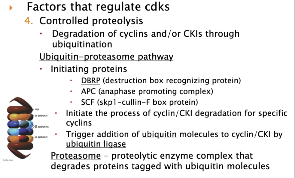
it is targeted for degradation when cyclin concentrations go down by Ubiquitin-proteasome pathway
know the three initiating proteins and that they initiate the process of cyclin or CKI degradation for specific cyclins
Ubiquitin ligases include E1, E2, E3 - come and puts ubiquitin on
once ubiquitin molecules made, the proteasome recognizes this and degrades cyclin and/or CKI
Structure of the proteasome
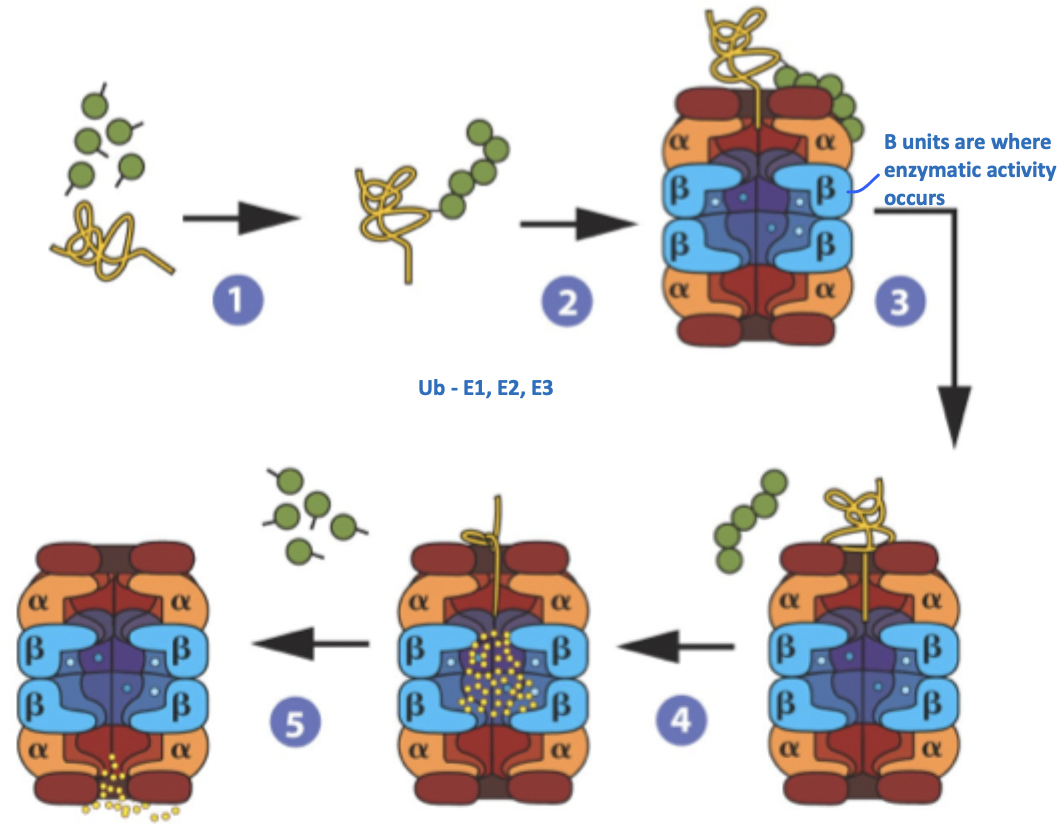
green circular structure - Ub
yellow structure - cyclin
illustrates how cyclins are degraded
knowing this, can we therapeutically target cancers where too much degradation is occurring?
yes, in myeloma cancer - too much degradation
use agents that target this
Bortezomib (Velcade, PS-341)
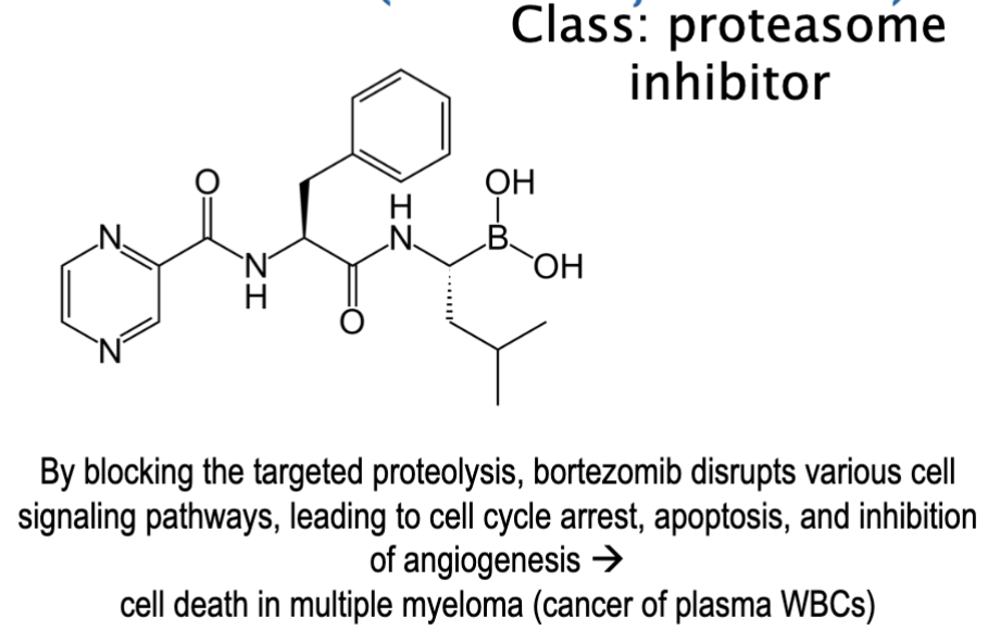
What happens when the proteasome is blocked?
Proteins accumulate inside the cancer cell:
damaged/misfolded proteins
pro-apoptotic proteins
cell-cycle inhibitors
This creates severe cellular stress.
Result: cell cycle arrest, unfolded protein response, apoptosis (cell death)
define the significance of checkpoints
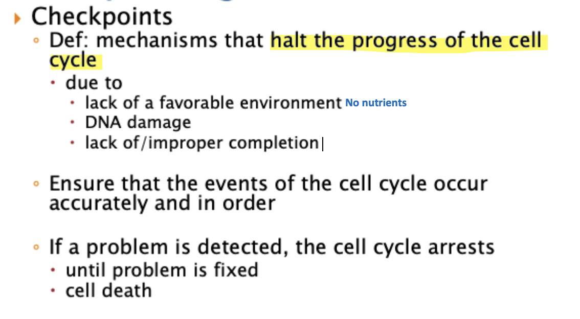
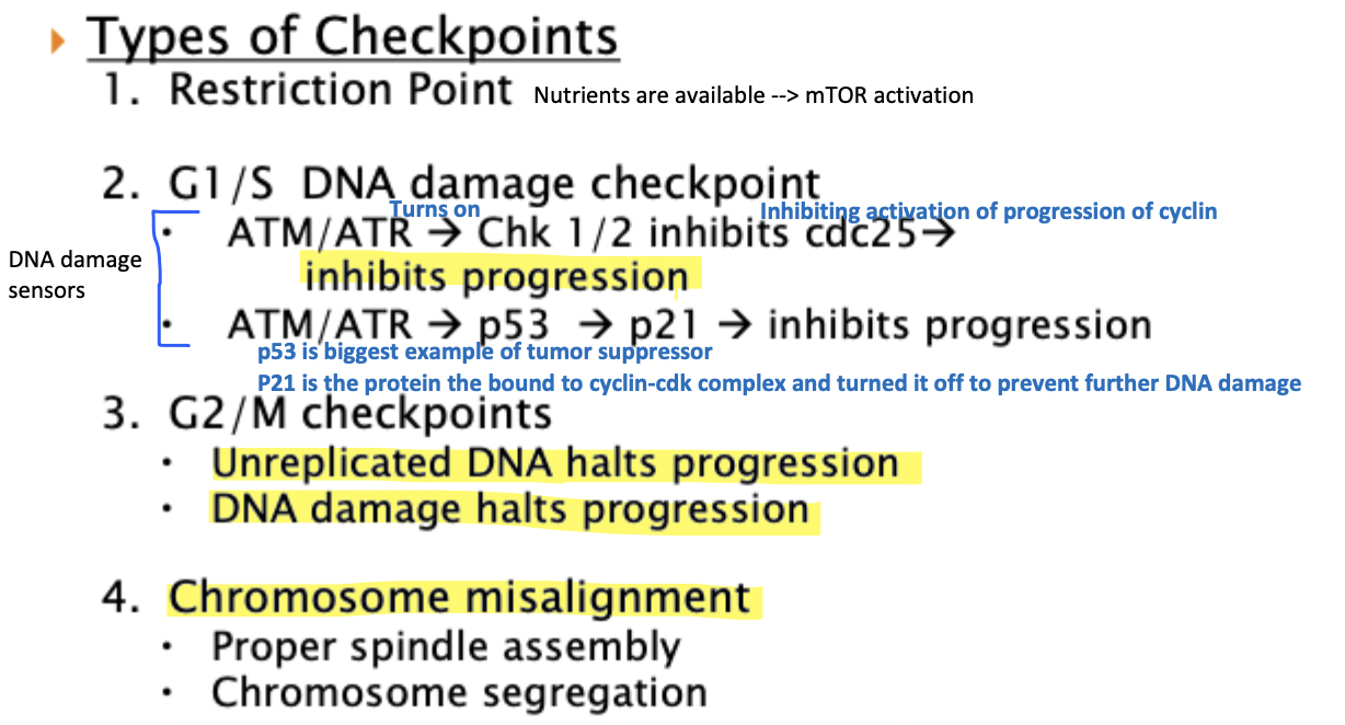
What are the types of checkpoints