BME 301 - Bioelectricity (FINAL EXAM)
1/199
Earn XP
Description and Tags
Contains contents from lectures 8-12 of BME 301. Preparation/flashcards for the final exam on Mon, May 18, 5:30 PM
Name | Mastery | Learn | Test | Matching | Spaced | Call with Kai |
|---|
No analytics yet
Send a link to your students to track their progress
200 Terms
This is the beginning of Lecture 8: The Heart
Sensory, motor, and interneurons
What are the 3 kinds of neurons?
These cells are charged up and ready to fire a electric surge/action potential
What does it mean when neurons and muscle tissue are excitable?
In muscle cells, it leads to a movement/contraction of the cell
What is the main difference between the membrane potential in nerves and in muscle cells?
Smooth muscle, skeletal (striated) muscle, and cardiac muscle
What are the 3 kinds of muscles?
It is able to spread to surround muscle, and communicate in the process.
The electrical activity of cardiac muscle is able to what? (hint: communication)
The orientation of the tilt of the heart
What about the heart causes its conductivity to flow in a certain direction?
The 4 chambers are the left and right atria, and the left and right ventricles. The atria are on the top, and the ventricles are on the bottom.
Name the 4 chambers of the heart. Which are on the top and which are on the bottom?
The left ventricle, because it pumps all the blood throughout the body and it needs a lot of energy to push all the blood throughout. ECG/EKG signals are dominated by the work of the left ventricle.
The _____ chamber of the heart is the most important. Explain why.
The septum
What separates the electrical activity of the left and right ventricles?
Because it does not have to do as much work
Why is the right ventricular wall smaller than the left ventricular wall?
Because when blood needs to be pushed out, it can be squeezed out.
Why are the walls of the heart (myocardium) in a spiral?
Pulmonary valve
What part of the heart makes sure the blood goes from the right ventricle and goes out of the pulmonary artery?
It is at the same time, but it very particularly coordinated.
Describe the movement of the heart (beating)
A heart muscle cell
What is a myocyte?
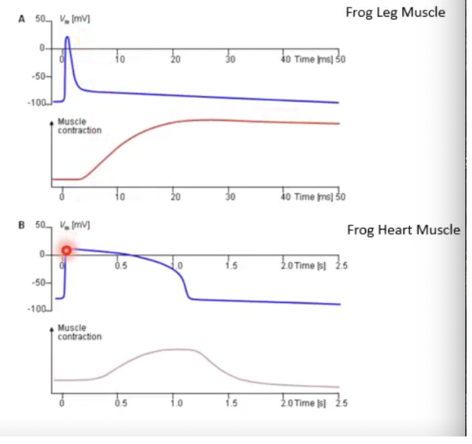
The action potential of heart cells is MUCH longer than that of a neuron. This is because the length of an action potential is related to the function that the body needs to perform.
What is the main difference between the action potentials of heart cells and neurons, and why?
Leg muscle cells have shorter action potentials than heart muscle cells, but heart muscle cells have shorter muscle contractions than leg muscle cells
Describe the difference between the action potential of a leg muscle cell and a heart muscle cell.
After the peak of muscle contraction
After an action potential occurs in a heart cell, when can it fire another one?
Consistently being at the peak of a muscle contraction
What is tetanus?
It is the relationship between the possibility of an action firing vs. the contraction of a muscle
Explain the concept of excitation-contraction coupling.
True
T/F Activation of a muscle cell can propagate from cell to another cell IN ANY DIRECTION (it can depolarize any muscle around it).
The atria and ventricles, because this would make the heart work out of sync
What is the only exception to the rule that an activated muscle cell can propagate from cell to cell in any direction?
Sinus node
What node of the heart controls action potentials? (hint: it is self excitatory)
Pacemaker cells (set the pace of the heart independent of anyone’ control) ; 70
_____ cells can generate around ____ action potentials per minute
The AV node
What is the node of the heart that is responsible for propagating the signal down?
The sinus node is what is responsible for generating action potentials in the heart, and the AV node is between the atria and ventricles and is responsible for propagating those signals down (but can also fire action potentials on its own)
Explain the sinus and AV nodes, and their relationship with action potentials
It is dependent on the length of the action potential; it is NOT cell to cell dependent
What is repolarization in the heart dependent on?
It is a conduction pathway that transmits signals from the AV node to the ventricles (making sure they contract efficiently to pump blood)
What is the bundle of hiss?
It tells heart muscles to contract, and allows for synchronized contraction of the ventricles
What is a Purkinje fiber?
When the left ventricle contracts
What does the largest signals in an ECG reading represent?
The AV node pathway
What is the pathway in a healthy heart that allows for signals in the atria to reach the ventricle?
It is when sodium rushes into the cell, making the inside of the cell positive and a net negative outside of the cell (with a negative voltage outside the cell as well)
Describe depolarization in these heart cells
The local current, which is caused by the rushing in of sodium into the cell
What exactly about depolarization causes the propagation of one cell to its neighboring cell?
After depolarization, you need to repolarize to get back to the membrane potential
What is needed to get back to membrane potential?
FALSE - the origin is different from depolarization and repolarization
T/F The origin is the same for depolarization and repolarization
It is when potassium comes out of the cell slowly, the outside becomes a little bit more positive, and this happens a LOT slower than depolarization, with a wider activation interval
Explain what repolarization is
Because of the slow speed of repolarization
Why is the activation interval of repolarization much wider than that of depolarization?
It is a low resistance connection between cells that allow communication after depolarization
What is a gap junction/nexus?
It reads the electrical signals from the heart to ensure proper function, and can be used to diagnose heart conditions
What is an electrocardiogram?
Sinus/AV node cell action potentials are rounded (slow component), while muscle cell action potentials are pointed at their peak (fast component)
What is the difference in action potential shape between sinus/AV node cells and muscle cells?
The QRS complex, which shows ventricular depolarization (electrical activation of the ventricles, right before they depolarize)
What is the biggest signal of an ECG, and explain it.
Calcium
What ion is responsible for the constant long length of an action potential?
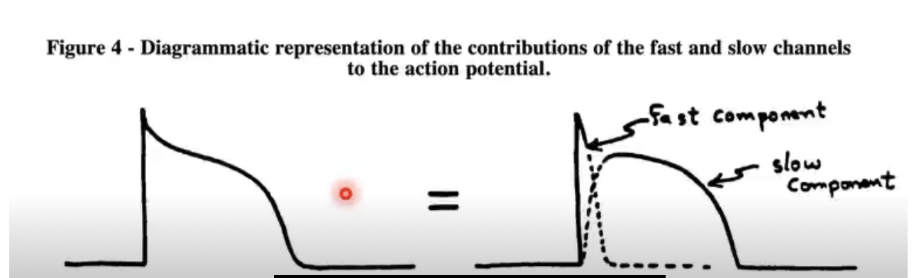
Tetrodotoxin can block the sodium channel, and this allows for the action potential to solely look like the slow component
What can block a sodium channel, and what affect does it have?
FALSE - you ideally would want to see each signal and its parts to identify each cell’s action potential, but it is impossible
T/F You can easily see every signal part of an ECG and its signals separately
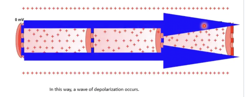
A wave of depolarization occurs through diffusion
What happens to a cell that is next to a cell that has depolarized?
RA (right arm), LA (right arm) and LL (left leg)
What are the 3 electrodes of Einthoven’s triangle?
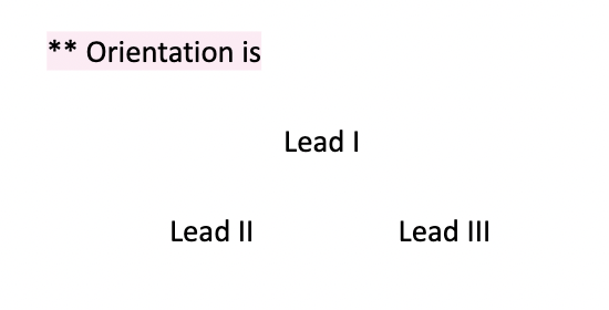
I + III = II (they are dependent on each other)
What is the relation between Leads I, II, and III? Also illustrate their orientiation.
They are 3 additional leads that bisect Leads I, II and III (for 6 leads in total)
What are the Goldberger Augmented Leads?
Unipolar chest leads, and their reference in an average of the original 3 Enthoven leads
For the 6 additional leads in a 12-lead ECG, where are they placed?
Because you can see the QRS the best (and it is Lead II)
Why is the bottom strip of an ECG the best?
The x axis is TIME (s) and the y axis is VOLTAGE (mV)
What are the x and y axes on an ECG reading?
Both show a blood blockage, but infarction is where tissue has died, and ischemia is where that tissue has not died yet
What is the difference between infarction and ischemia?
It is on the surface of the chest just above the left ventricle
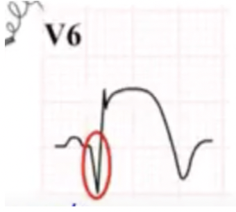
Why does V6 show the best insight to what is happening biologically?
The red circle is showing a large negative spike, showing the depolarization is moving away from the electrode
In this picture of an ECG of a myocardial infarction, what does the red circle represent?
An ST elevation
What is the hallmark of a myocardial infarction?
It is when cells depolarize when they are not supposed to (nothing went through the sinus node)
What is an ectopic heartbeat?
Atrial starts in the atria with two quick heartbeats, and ventricular starts in the ventricle and is abnormal repolarization. These are not dangerous, it just feels like a flutter in the heart
What is the difference between an atrial and ventricular ectopic heartbeat?
The resting heart rate is equal to or more than 100 beats per minute
What is tachycardia?
It is an irregular heart rhythm, where the atria are beating fast and chaotically, where blood may pool and clots can form.
What is Atrial fibrillation (Afib)?
They are devices that apply electric shocks to maintain heart rhythm, and if necessary, restart the heart
How do pacemakers work?
Implantable defribrillator
What does ICD stand for?
Cardiac resynchronization therapy implantable defibrillator (uses a third lead into the left ventricle)
What does CRT-ICD stand for?
The next 5 flashcards are the Lecture 8: The Heart Quiz!
The QRS complex is the depolarization and repolarization of the left ventricle, and it is the largest signal because of how large the left ventricle’s work is for the heart.
What does the QRS complex represent, and why is it the largest signal in the ECG?
Lead II has the best view of the QRS complex because of its angling towards the heart
What lead has the best view of the QRS complex, and why?
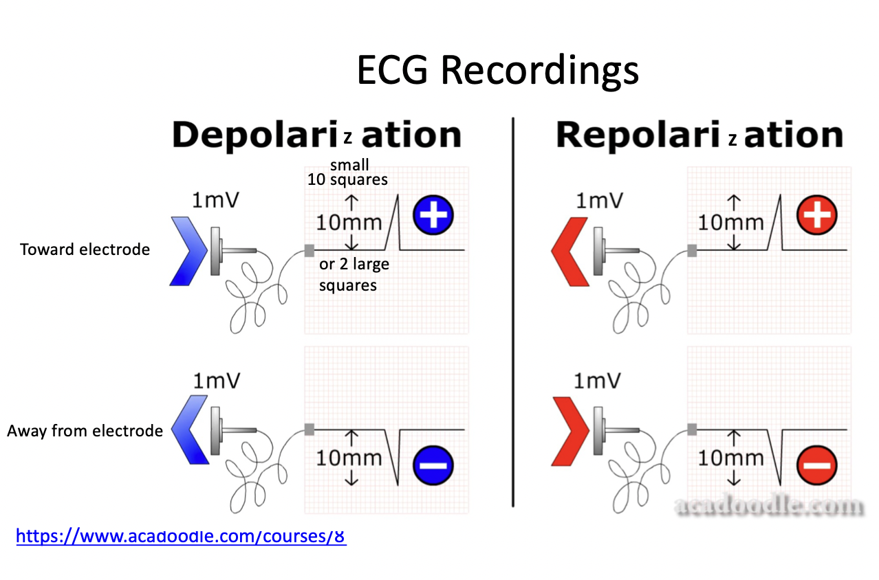
Depolarization toward an electrode - positive spike
Repolarization away from an electrode - positive spike
What is the difference in ECG signal when: a depolarization wave moves TOWARD an electrode vs. when a repolarization moves AWAY from an electrode?
Action potentials are shorter in outer cells than inner cells, so repolarization propagates in the opposite direction because the outer cells polarize first
Why is the direction of the depolarization wave opposite of repolarization?
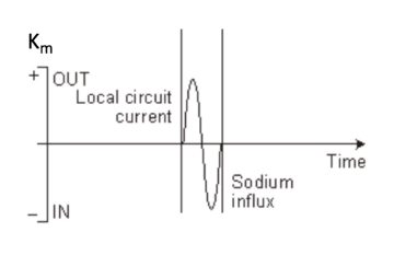
Km has that shape because it is proportional to the second derivative of voltage
In this image, why does Km have the shape shown on the right?
This is the beginning of Lecture 9: Muscles and EMG
An electromyography (electrical muscle graph recording)
What is an EMG?
A squeezing of the median nerve
What causes carpal tunnel syndrome?
The central nervous system is the brain and spinal cord, and the peripheral nervous system takes signals from the brain and spinal cord to the rest of the body
Describe the difference between the central and peripheral nervous system
Somatic neurons can be controlled, and autonomic cannot be controlled and are involuntary
Describe the difference between somatic and autonomic neurons
It records electrical activity of the skeletal muscles, and it is complex because its signal comes from neurons and muscles
What does an EMG measure?
It is a motor neuron and the muscle fibers that it innervates (can innervate multiple), and the firing of motor units is all or nothing
What is a motor unit?
It means that when an action potential fires, the muscle FULLY contracts
What does it mean when the firing of a motor unit is all or nothing?
It explains how within a muscle, muscle fibers and intermingled, so there is not a pattern with motor units. The pros of this is that fibers being distributed evenly through a muscle causes smooth muscle contractions
Explain the concept of motor units being intermingled
During a very weak muscle contraction
In what situation could you only observe 1 motor unit?
They are the number of muscle fibers in one motor unit, and they vary greatly (1:6 to 1:2000), and depends on the area of the body
Explain what an innervation ratio is.
This states that motor units are recruited based on ascending size order (small to large). Weak stimulation recruits small motor units, and stronger stimulation recruits large motor units. However, this relationship is not considered linear.
Explain the size principle
The nonlinear relationship is between the number of motor units being recruited and the force generated. If all the motor units were the same size, as you recruited more and more, the force induced by those units would linearly increase. But because the motor units are getting larger and larger as you recruit them, the force grows exponentially.
Explain why the size principle is not a linear relationship.
Slow twitch is dark meat with high myoglobin and slow to fatigue (uses oxygen and glucose to product a lot of energy). Fast twitch is white meat, low myoglobin, and fatigues quickly (uses fast and high tension contractions)
Explain the difference between show and fast twitch muscle
Rate coding is a measurement of how fast action potentials go from the central nervous system to the motor neurons
What is rate coding?
The increase of muscle strength and muscle mass (e.g. getting stronger when you weight lift), which was actually not mean to work biologically
What is synchronization?
ISI, or interspike interval variability, shows the recruitment of a motor unit and how it changed over time
Explain ISI
Action potentials
What causes muscle fibers and motor units to twitch?
It is the number of action potentials that a single motor unit can have over time
What is an impulse train?
Electrode placement, electronic amplifiers, line interference, biological noise detectors, fat in the body, etc.
Name common sources of noise that may interfere with EMG results
It is a decrease in maximal force or power production in response to contractile activity (during muscle fatigue, less and less motor units are being utilized)
What is muscle fatigue?
Motor neuron EMG is through the muscles in the skin (CMAP, or compound muscle action potential), and sensory neuron EMG is through the fingers (SNAP, or sensory nerve action potential)
What is the primary difference between EMG for motor neurons and EMG for sensory neurons?
Their high blood sugar and fats in blood damage nerves, and cause loss of sensation in their muscles
How does having diabetes affect nerves?
It is a process where a nerve is cut or crushed, and this neuron damage goes toward a motor unit, where it can either be regenerated or lost completely. The origin of the damage on the arm is hard to detect.
What is Wallerian degeneration?
It is when your peripheral nerves are being attacked, resulting in numbness, weakness, and sometimes paralysis (where the paralysis is ascending, starting in the legs and traveling upwards)
What is Guillain-Barre syndrome?
It takes out any background noise that the reading may pick up
What is the purpose of ground in an EMG reading?
Amplitude (microV), Distal and/or Peak Latency, and Conduction Velocity
What are the measured parameters of sensory nerve conduction?
Amplitude (mV), Distal Latency, Proximal Latency, Conduction Velocity
What are the measured parameters of motor nerve conduction?
A signal that indicates a dying muscle cell (active ongoing destruction)
What is fibrillation?
The discharge of a motor unit (in ALS or eye twitches)
What is fasciculation?
It is a method of prosthetics where nerves that used to be in the arm are transferred to residual muscle, which can control the prosthetic arm. Its barriers are that there is a lack of sensory feedback, background noise, and lack of accuracy when recording signals
What is target muscle reinnervation?
The next 5 flashcards are the Lecture 9: Muscles and EMG Quiz