PALM 309: Quiz 6
1/102
There's no tags or description
Looks like no tags are added yet.
Name | Mastery | Learn | Test | Matching | Spaced | Call with Kai |
|---|
No analytics yet
Send a link to your students to track their progress
103 Terms
The periodontium
3 tissues that hold the tooth in position
Alveolar bone proper
bone nearest to the tooth, studded w little holes where PDL fibers insert
Supporting alveolar bone
around alveolar bone proper, no PDL fibers
Resorbtion
lost tooth with no force applied to bone that surrounds it
located in the tooth root outside the dentin
Cementum
Cementoenamel junction
cementum and enamel meet
cementoblasts
risen from mesenchyme surrounding developing tooth
cementum contains 50% of what kind of minerals?
hydroxyapatite crystals
Cementum contains about 50% of what organic materials?
collagen type 1 fibers
cementum is very similar to what?
bone
When is cementum laid down?
after the dentin in the root forms
Cementoblasts that are surrounded by cementum located in lacunae are called?
cementocytes
Contains cementocytes and is found at the root apex
cellular cementum
does not contain cementocytes and is located near the crown
acellular cementum
Layer of cementoblasts located between cementum and PDL
regenerative cementum
the PDL contains
collagen type 1 and CT cells (fibroblasts, plasma cells, macrophages, and mast cells)
insert in cementum and the alveolar bone
principal fiber group
located nearest the crown, insert into acellular cementum (top rim of alveolar bone proper)
Alveolar crest fibers
below the alveolar crest fibers, extend horizontally between the cementum and alveolar bone
horizontal fibers
largest group of PDL, run obliquely between cementum and alveolar bone
oblique fibers
located at the root apex, extend between cementum and alveolar bone
apical fibers
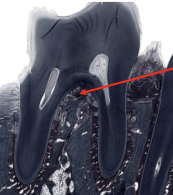
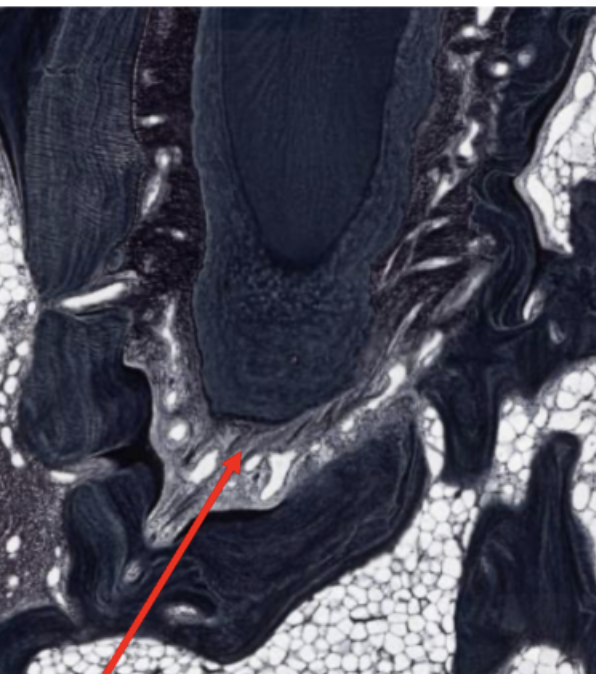
only found in multiroot teeth, extend between the alveolar bone and cementum at the bifurcation
interradicular fibers
insert into cementum OR alveolar bone OR don’t insert into either bone or cementum
gingival fiber group
located nearest the crown, extend between cementum of adjacent teeth
transseptal fibers
extend between the cementum near crown to gingiva
dentogingival fibers
Extend between the alveolar bone and the gingiva
alveologingival fibers
around the tooth and help keep attached gingiva attached to the tooth
circular fibers
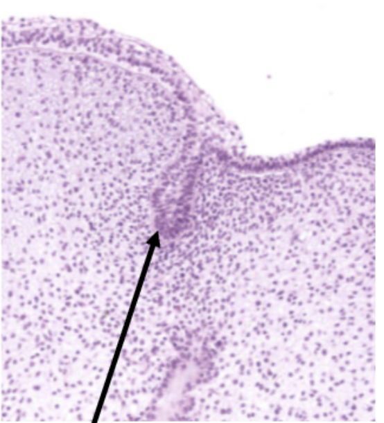
Acts as a barrier and prevents leakage of material into PDL
dentogingival junction
the free gingiva is lined by what?
sulcular epithelium
the epithelium that attaches the attached gingiva to the tooth enamel is called?
junctional epithelium
What can cause the junctional epithelium to detach from the enamel?
infection, poor oral hygiene, and aging
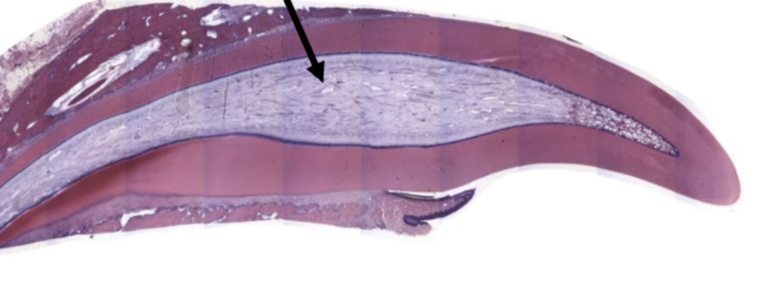
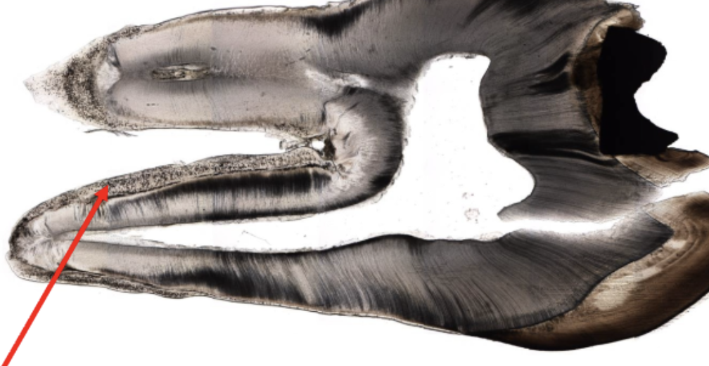
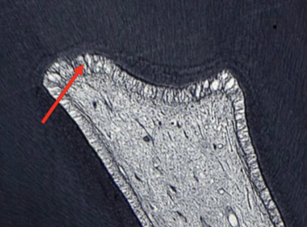
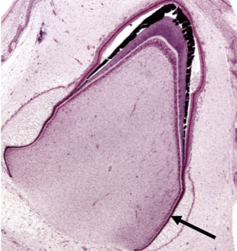
Where is the black arrow pointing to?
tooth pulp
CT core of the tooth with all typical CT cells, very vascular with many nerves
pulp cavity
located in the pulp, forming a layer adjacent to the dentin
odontoblast cell bodies
beneath the odontoblasts is an area called the ___ which has very few cells
cell free zone
beneath the cell-gree zone with many cells
cell rich zone
located in the cell rich zone and extends into the cell-free zone
subodontoblast nerve plexus (Raschkow’s plexus)
abnormal accumulations of dentin or minerals that form as we age
denticles (pulp stones)
denticles that are made of dentin
true denticles
denticles that are more common and made of mineral accumulations
false denticles
abnormal accumulations of cementum
cementicles
when a tooth is lost, there is no force applied to the opposite tooth so an excess of cementum will be laid down in the opposing tooth
hypercementosis
cementum attaches directly to alveolar bone with no PDL between
Ankylosis
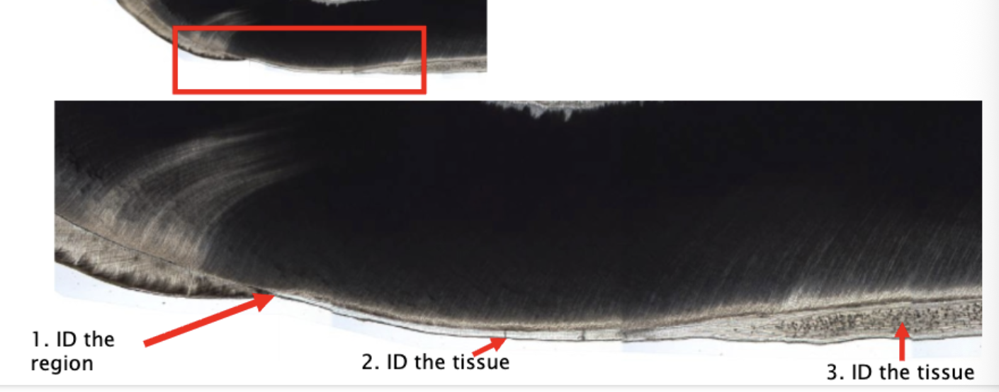
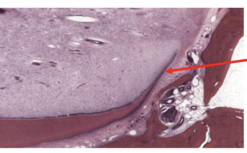
ID the region
cementoenamel junction

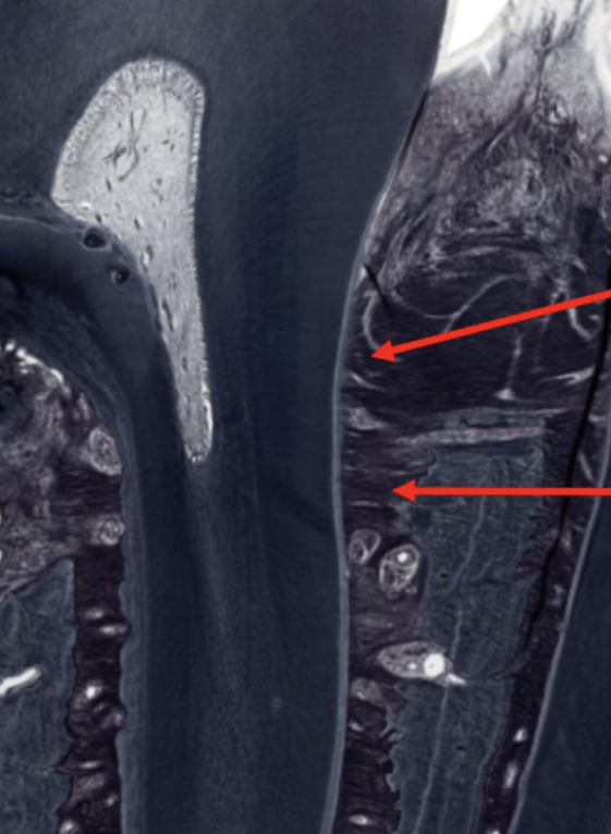
ID the tissue
acellular cementum

ID the tissue
Cellular cementum

ID the cells (top arrow)
odontoblasts
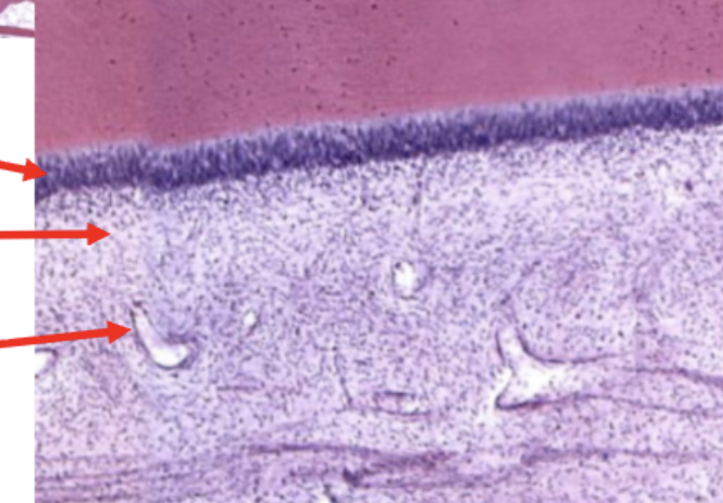
ID the region (middle arrow)
pulp cell-free zone

ID the region (bottom arrow)
pulp cell-rich zone
ID the black fibers at the point
sub-odontoblast nerve plexus

ID the fibers (bottom left arrow)
horizontal

ID the fibers (top Left arrow)
Transseptal fibers
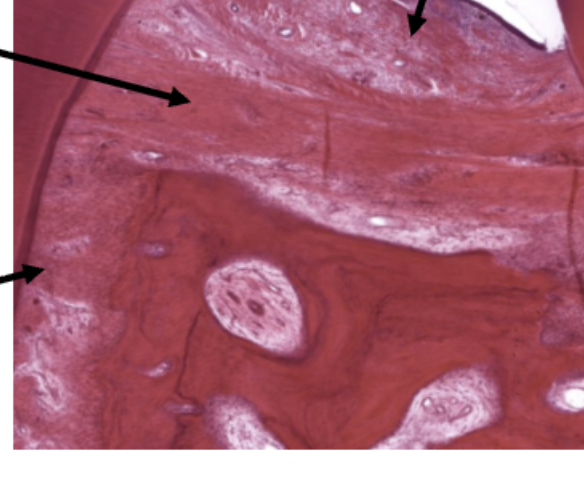
ID the fibers (right arrow)
dintogingival fibers
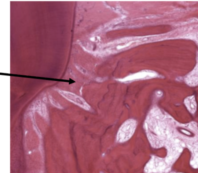
ID the fibers
alveolar crest
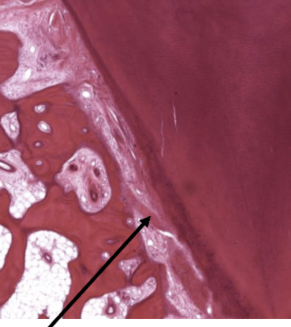
ID the fibers
Oblique fibers
ID the fibers
interradicular fibers
ID the fibers
apical fibers
ID the fibers (top right arrow)
alveolar crest fibers

ID the fibers (bottom right arrow)
horizontal fibers
ectoderm, endoderm, and mesoderm
3 primitive germ layers
Superficial layer that gives rise to the skin, oral cavity, lining, enamel and neuroectoderm
ectoderm
gives rise to components of CNS and PNS
Neuroectoderm
Gives rise to CT, muscle, circulatory system, lymphoid, organs, kidney, most of reproductive system and most of the tooth
mesoderm (mesenchyme)
gives rise to the lining of the GI tract and most digestive organs
Endoderm
loosely arranged star-shaped cells
stellate reticulum
ID the enamel organ stage
bud stage
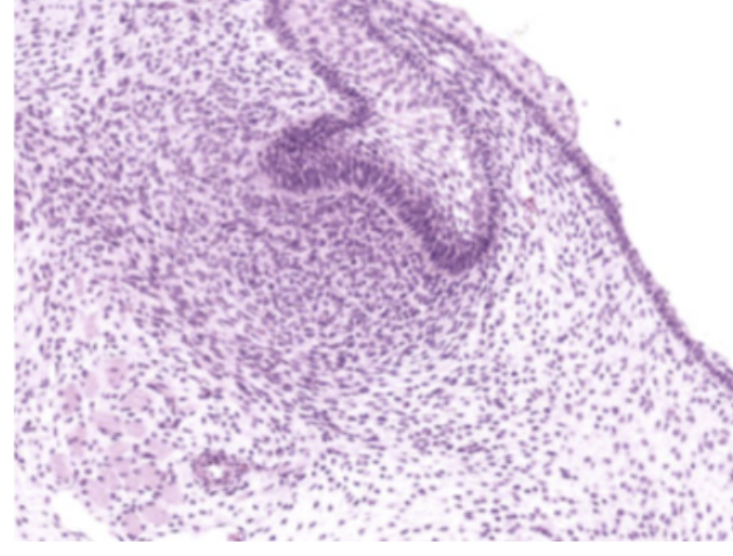
ID the stage
cap stage
ID the stage
bell stage

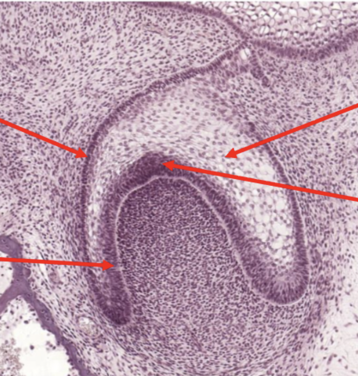
ID the cell layer (bottom left arrow)
inner enamel epithelium

ID the cell layer (top left arrow)
outer enamel epithelium

Id the cell layer (top right arrow)
stellate reticulum

Id the cell layer (bottom right arrow)
stratum intermedium
Id the cell region (top arrow)
Dental follicle

ID the cell region (bottom arrow)
Dental papilla
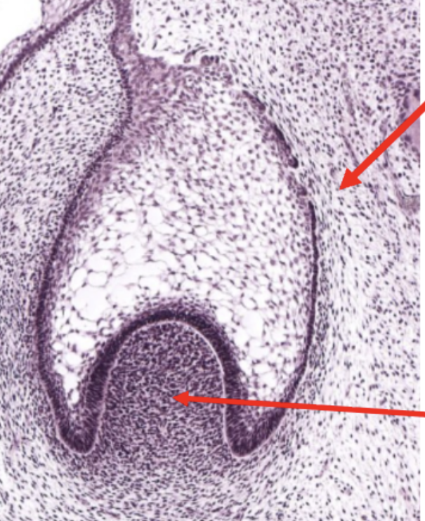
ID the structure
epithelial root sheath
ID the structure
epithelial diaphragm