Unit 3: Cell Biology
1/151
There's no tags or description
Looks like no tags are added yet.
Name | Mastery | Learn | Test | Matching | Spaced | Call with Kai |
|---|
No analytics yet
Send a link to your students to track their progress
152 Terms
Cell Theory
All living things are composed of cells and all cells come from other cells
Robert Hooke
First person to look at a plant through a microscope
‘Cellulae’: Tiny compartments
Light Microscopes (LM)
Earliest type of microscope
Visible light passes through a sample and glass lenses
Objective lens
Ocular lens
Light is bent to magnify the image of the specimen
Image projected into your eye or a camera
Magnification
The increase in an object’s image size compared with its actual size
Notation: LM 230X
Resolution
The measure of the clarity of an image. The ability to distinguish two nearby objects as being separate from each other
Electron Microscopes (EM)
Focuses beams of electrons (instead of light) through a specimen sample
Electromagnets bend the electron path and magnify the image
EM images are always black and white
Scanning EM (SEM)
Study the detailed architecture of a cell
The sample is coated with a thin film of heavy metal (ex, gold)
Electrons excite the gold atoms
Electrons are scattered and detected by a device that projects the image onto a video screen
3D Image
Transmission EM (TEM)
Electron beam passed through a very thin section of a specimen
Stains containing heavy metals coat certain types of cellular structures
Electrons scattered by the more densely stained parts
Scattered electrons are detected, and an image is produced
Prokaryotic cells
The first cells to evolve and lived for about 1.5 billion years before eukaryotic cells evolved
Bacteria and archaea
Small, simple cell structure. About 1/10 the size of eukaryotic cells
No internal membrane-bound structures
Ribosomes are smaller and have a slightly different structure than in eukaryotes
Eukaryotic cells
Evolved from prokaryotic cells about 1.8 billion years ago
All higher life forms such as plants, animals, and fungi
Larger, more complex cells
Structures common to all life
Plasma membrane, ribosomes, cytosol, DNA, cytoplasm
Plasma membrane
Membrane that surrounds the cell. Made mainly out of a phospholipid bilayer
Ribosomes
Machinery for protein synthesis
Cytosol
Aqueous solution that fills the cell
DNA
One or more chromosomes
Cytoplasm
The entire contents of the inside of the cells, excluding the interior of the nucleus (which only Eukaryotes have)
Prokaryotic Cell structure
Nucleoid
Cell wall
(Optional) Capsule
(Optional) Flagella
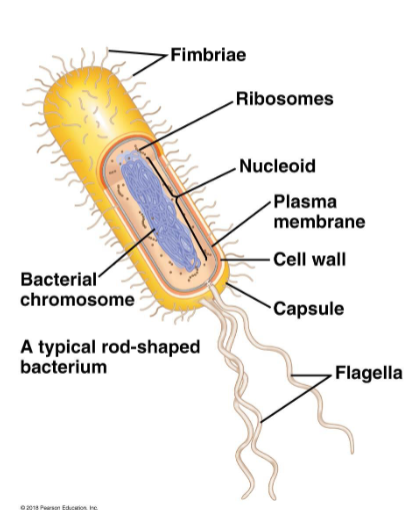
Nucleoid
Region of the cell where the chromosome is coiled
Cell Wall
Rigid, chemically complex shell surrounding the plasma membrane. Protects the cell and maintains the cell’s shape
Capsule
Sticky outer coat around the cell wall. Glues the cell to surfaces or to other cells. Not in all prokaryotes
Flagella
Long projection that propels a cell through its environment. Not in all prokaryotes.
Animal cell
Lysosome and centrosomes
Flagella and cilia (optional)
Very very rare in plant cells
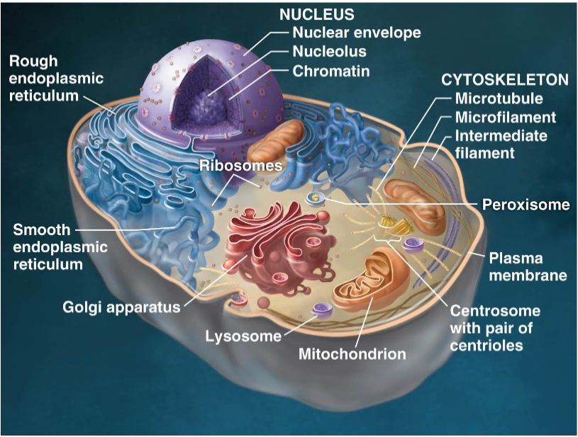
Organelles
Found in eukaryotic cells only. Membrane-bound structures that perform specific tasks. A cell may contain many copies of each organelle
Organelle Function
Genetic control of the cell - Nucleus and ribosomes
Manufacture, distribution, and breakdown of molecules - Endoplasmic reticulum, Golgi apparatus, lysosomes, vacuoles peroxisomes
Energy processing - Mitochondria, chloroplasts
Structural support, movement, and communication between cells - Cytoskeleton, plasma membrane, plant cell wall
Cellular metabolism
The chemical activities of the cell
Internal condition of an organelle
Each organelle maintains its own specific internal chemical conditions
Optimal for enzyme function
Organelles may have membrane-bound organelles
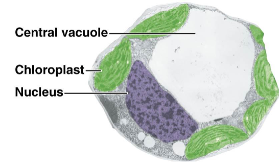
Plant Cell
Rigid cell wall (made of cellulose)
Plasmodesma
Chloroplasts
Large central vacuole
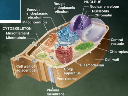
Plasmodesma
Cytoplasmic channels that link adjacent cells
Chloroplasts
Location of photosynthesis.
Large central vacuole
Stores water and chemicals
Function of Nucleus
Contains the cell’s genetic instructions (DNA)
Controls the cells activities by directing protein synthesis
DNA organization
Organized into chromosomes
Associates with many proteins
The proteins help coil the long strands of this to form a chromosome
A human cell has 46 separate chromosomes
Chromatin
Complex of proteins and DNA. Appears as a diffuse mass within the nucleus
Nucleus Structure
Nuclear envelope
Nucleolus
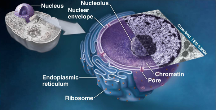
Nuclear envelope
Double membrane enclosing the nucleus. Has pore proteins
Pore proteins
Regulate the flow of large molecules and connect the nucleus to the endoplasmic reticulum
Nucleolus
The location where ribosomal RNA (rRNA) is synthesized. Proteins made in the cytoplasm are brought into the nucleus to assemble with the rRNA to make ribosomes
Ribosomes
The cellular components that use instructions form the nucleus to build proteins (non-membranous organelle). Interact with mRNA to build a protein.
Free Ribosomes
Suspended in the cytosol. Proteins made here generally function in the cytosol.
Bounded ribosomes
Attached to the outside of the endoplasmic reticulum and nucleus. Makes proteins that will be exported from the cell.
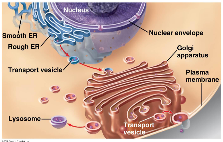
Endomembrane System Definition
Internal membrane involved in most cellular functions. Synthesis, storage, distribution, and export of molecules.
Membranes
Divide the cell into functional compartments. May either be physically connected or linked by transport vesicles.
Vesicle
Sac made of membrane
Organelles in the Endomembrane System
Nucleus, nuclear envelope, endoplasmic reticulum (rough and smooth), Golgi apparatus, lysosomes, vesicles, vacuoles, plasma membrane
Endoplasmic Reticulum (ER) - def and function
Network of flattened sacs and tubules.
Largest component of the endomembrane system.
Directly linked to the nuclear envelope (membranes are continuous).
Vesicles bud from the ER to travel to other organelles
Function: Major manufacturing site in the cell
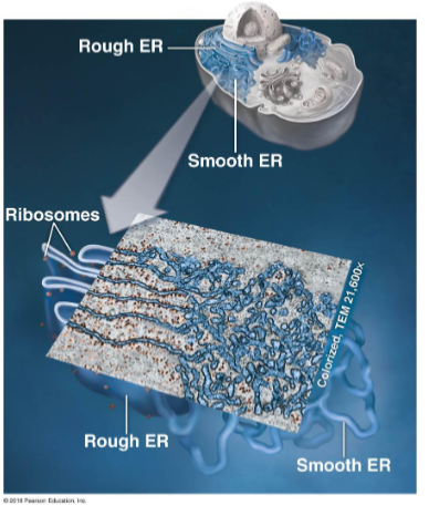
Smooth Endoplasmic Reticulum (ER)
Synthesis of lipids (oils, phospholipids, and steroids)
Storage of calcium ions
Detoxification
Rough Endoplasmic Reticulum (ER)
Proteins produced by ribosomes attached to the rough ER are often excreted from the cell (secretory proteins). Synthesizes new membrane fragments.
Grows its own membrane by adding phospholipids and membrane proteins
Completed membranes are transported as vesicles to the appropriate area of the cell
Secretory proteins
Proteins excreted from a cell. Produced by ribosomes attached to the rough ER.
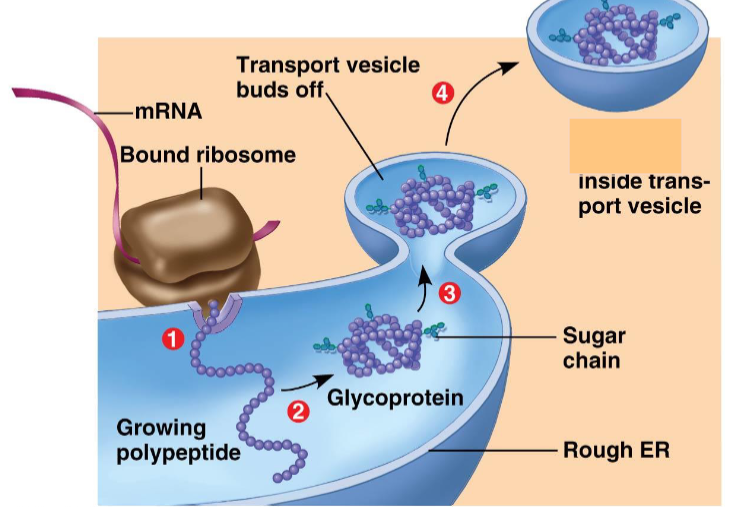
Secretory Protein Creation Steps
The bound ribosome (attached to the Rough ER) grows a polypeptide inside the Rough ER as specified by the mRNA.
Inside the Rough ER the polypeptide is folded and sugar chains are added, turning it into a glycoprotein
The membrane around the glycoprotein surrounds it and buds off, turning into a transport vesicle
Secretory protein inside the transport vesicle is sent to the Golgi Apparatus for further processing
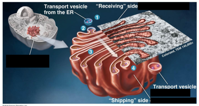
Golgi Apparatus
Stack of unconnected flattened sacs. A cell may contain hundreds of this organelle (cells active in protein excretion have more)
Function of Golgi Apparatus
Warehouse and processing station for molecules produced by the Endoplasmic Reticulum
→ Carbohydrate portion of a glycoprotein may be modified
→ Molecular identification tag may be added
Once processed, products are transported to their destination
Processing in the Golgi Apparatus
Docking station (received as a transport vesicle from the ER)
Vesicle adds its membrane and contents to the sac
Products are modified as they travel from one sac to the next
Shipping side (sent as a transport vesicle)
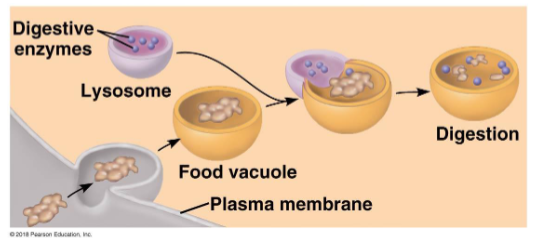
Lysosome
Membrane-enclosed sac of digestive enzymes. The membranes and enzymes are made in the endomembrane system. Provides an acidic envrioment for its enzymes to function. Protects the rest of the cell from the acidic conditions (compartmentalization)
Digestive functions
Food, pathogens, recycling of damaged cell components
Lysosomal Diseases
Lysosomal enzymes are missing. Lysosomes become engorged because they cannot break down their contents. Interferes with cellular function (Tay-Sachs disease)
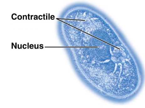
Vacuoles
Large vesicles with a variety of functions.
Food
Contractile vacuoles (collect water, main wheel expels the water, moving the cell)
Plant and fungal digestion
Plant seeds store reserves of proteins for starting growth
Flower petals hold pigments to attract pollinators
Plants store compounds that are poisonous or unpalatable to animals
Central vacuole
In plants, absorbs water allowing the cell to grow in size (more rigid). Stores vital chemicals and maybe toxic waste products.
Peroxisomes (def and func)
Metabolic, membrane-bound compartment that does not originate in the endomembrane system
Still unknown how they relate to other organelles
Function:
Break down fatty acids to use as cellular fuel
Detoxification of harmful compounds in your liver
Mitochondria
Carries out cellular respiration in all eukaryotic cells.
Mitochondria composition
Enclosed by two membranes
Each is a phospholipid bilayer with embedded proteins
Inner membrane is called cristae
The region between the inner and outer membranes is the intermembrane space
The region inside the inner membrane is the mitochondrial matrix.
Cristae
Inner membrane in mitochondria. Inner membrane is highly folded to increases the membrane surface area to maximize ATP production.
Intermembrane space
Region between the inner and outer mitochondria membranes.
Mitochondrial Matrix
Region inside the inner mitochondria membrane. Contains mitochondrial DNA, ribosomes and enzymes that catalyze reactions of cellular respiration
Chloroplast Structure
2 membranes separated by a thin intermembrane space
Inner membrane contains stroma and thylakoids
Photosynthesis
Coverts solar energy to chemical energy
Stroma
Thick fluid inside the inner membrane. Contains chloroplast DNA, ribosomes, and enzymes
Thylakoid
Network of interconnected membranous sacs. Chlorophyll is embedded in the membranes to trap solar energy.
Granum
Stack of thylakoids. Resembles stack of poker chips
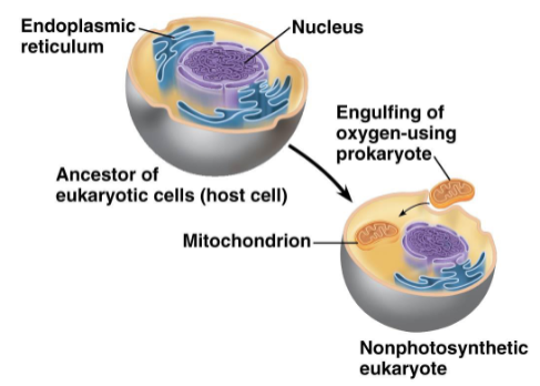
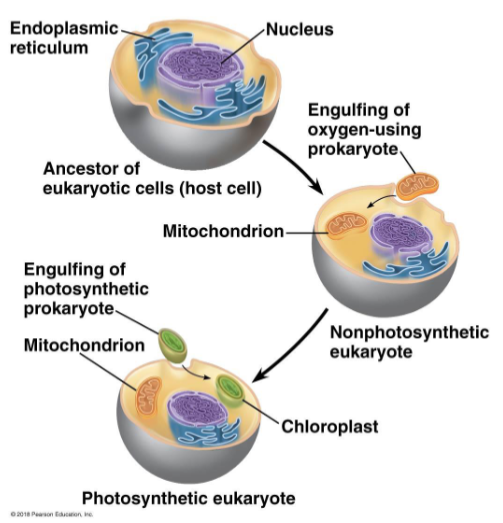
Endosymbiont theory - overall
Mitochondria and chloroplasts were once small prokaryotes that began living inside larger cells:
Grounds for Endosymbiont Theory
Mitochondria and Chloroplasts closely resemble prokaryotic cells
• Single circular DNA molecule
• Ribosomes similar to those of prokaryotes
• Reproduce inside the cells in a similar way to prokaryotes
Endosymbiont theory - mitochondria
Photosynthetic prokaryotes filled our atmosphere with oxygen
A large cell engulfed a small cell that could use oxygen to produce large amounts of energy
Benefit to host: high levels of ATP production
Benefit to small cell: protection, large supply of nutrients
Host and endosymbiont merge, eventually leading to a eukaryotic cell that contains mitochondria
Endosymbiont theory - chloroplasts
Early eukaryotic cells engulfed a small photosynthetic prokaryote
Provided the host with nutrients and energy, got protection and energy in return
Lead to eukaryotic cells that contain chloroplast
Cytoskeleton
Networks of protein fibers extending throughout the cells.
Can interact with motor proteins:
Swimming and crawling motility of cells
Internal movement of cellular structures (ex, vesicles)
Three kinds of fibers
Present in all eukaryotic cells
Kinds of Cytoskeleton Fibers
Microtubules (all cells), microfilaments (most animal), intermediate filaments (all cells)
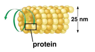
Microtubules
Straight hollow tubes composed of globular proteins called tubulins. Grow longer or shorter by addition/removal of tubulin proteins.
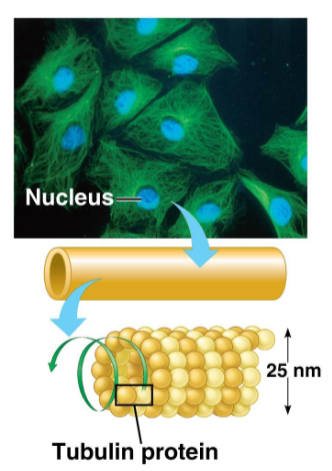
Tubulins
Globular proteins that make up microtubules. Each protein consists of two subunits.
Centrosome
Region in animal cells that microtubules grow out of
Microtubules function
Supports and shapes the cell
Track along which organelles can move with the help of motor proteins
Guides the movement of chromosomes when cells divide
Main component of flagella and cilia
Intermediate Filaments
Fibrous proteins that supercoil into cables
Permanent fixture in the cell (not made shorter or longer)
Outer layer of your skin is made of dead cells packed full of intermediate filaments.
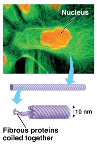
Function of Intermediate Filaments
Reinforce cell shape and anchor some organelles that should not move
Microfilaments (Actin Filaments)
Rods composed of globular actin proteins, arranged as a twisted double chain
Network in the cell membrane that supports the cell shape
Important in animal cells since they do not have a cell wall
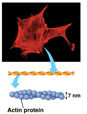
Function of Microfilaments (Actin Filaments)
Supports the cell shape
Involved in cell movement
Actin filaments and myosin motor proteins interact to make muscle cells contract and in amoeboid crawling movement
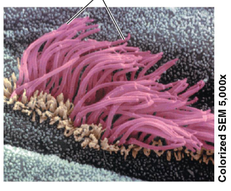
Cilia
Short numerous appendages protruding from the cell. Move together in a sweeping motion
Cilia Function
Propel single-celled organisms
Sweep other things, like line our trachea to sweep mucous out of our lungs
Antennae for signal reception (non-motile)
Flagella
Long tail-like appendage on cells (one to a few per cell)
Propels a cell using a undulating whip-like motion
Commonly found on animal sperm cells
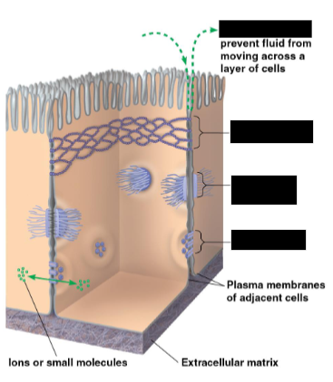
Extracellular Matrix
Holds cells together in tissues
Protects and supports the plasma membrane
Relays signals that affect gene expression across diff cells
Directs the movement of embryonic cells
Structure of Extracellular Matrix
Matrix of glycoproteins outside of the plasma membrane
Most abundant glycoprotein is collagen
Collagen fibers are embedded in a network of small glycoproteins and polysaccharides
Bound to the plasma membrane by proteins called integrins
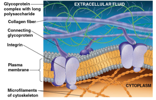
Integrins
Proteins that bind the extracellular matrix to the plasma membrane
Tight Junction
Plasma membranes of adjacent animal cells are knit tightly together. Used to prevents fluid leakages between layers
Anchoring junction
Intermediate filaments fasten animal cells together into strong sheets. For tissues susceptible to stretching and mechanical stress
Gap Junction
Channels of pores that allow the flow of small molecules from one animal cell to another (ie, nutrients)
Types of Junctions between animal cells
Tight, anchoring, gap.
Plasma Membrane (Cell membrane)
Flexible boundary between the living cell and its surrounding envrioment. Regulates the flow of material in and out of the cell.
Proteins embedded in the lipid bilayer
Hydrophobic regions embedded in the membrane
Hydrophilic regions protrude into the aqueous solutions inside or outside the membrane
Follows a fluid mosaic model structure and is selectively permeable
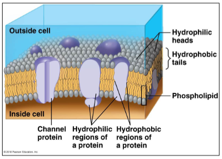
Fluid mosaic model
Diverse protein molecules suspended in a fluid phospholipid bilayer
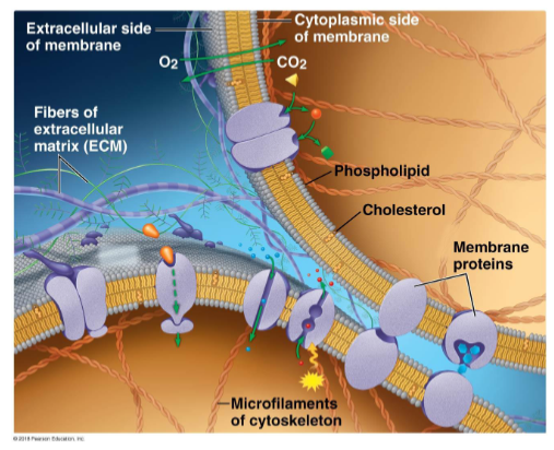
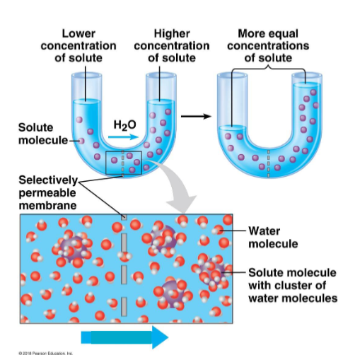
Selective permeability
Some substances can cross the plasma membrane more easily then others

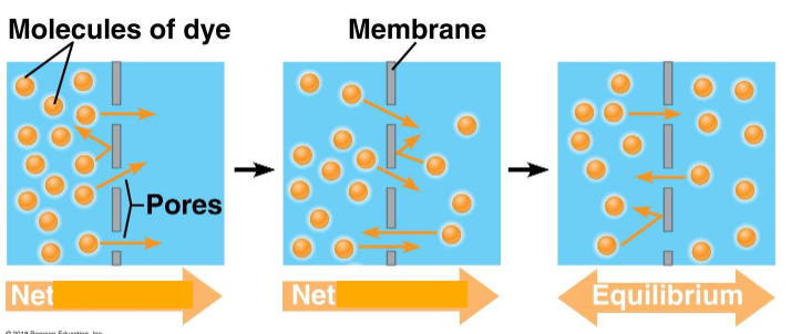
Diffusion
The tendency for particles of any substance to spread out into the available space. Net movement from a high concentration to low concentration until equilibrium is reached due to equal concentrations.Thermal energy allows movement. Particles move through air, water, and across membranes
Passive transport
Diffuse across a membrane with no energy investment. Molecules move down their concentration gradient. Substances move independently of each other
Substances that diffuse through cell membrane using passive transport
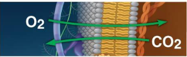
Small non-polar molecules (O2 enters, CO2 leaves ← animals, reverse for plants). Can move directly through phospholipid bilayer
Ions and polar molecules using simple transport proteins (can’t move through phospholipid bilayer). Must move down their concentration gradient
Osmosis
Diffusion of water across a membrane. Water passes through the membrane until the concentration of a solute is equal on both sides