H1 Toth- Endomembrane system I + II: key organelles, vesicles, signals and sorting

0.0(0)
Studied by 0 people
call kaiCall Kai
learnLearn
examPractice Test
spaced repetitionSpaced Repetition
heart puzzleMatch
flashcardsFlashcards
GameKnowt Play
Card Sorting

1/8

encourage image

There's no tags or description

Looks like no tags are added yet.

Last updated 4:19 PM on 1/23/26
Name
Mastery
Learn
Test
Matching
Spaced
Call with Kai

No analytics yet

Send a link to your students to track their progress

9 Terms

1
New cards

how are proteins secreted in eukaryotes?

  • the proteins are translocated co-translationally by the ribosomes of the rough ER, either integrating into the membrane or being released into the lumen

  • during this process, N-linked glycans are added- short oligosaccharides (sugar chains) are coupled to the protein via a nitrogen in an asparagine residue

  • this is used as a signal throughout the secretion process to determine whether the protein is folded and modified correctly (controlled by chaperone proteins), as the glycan chain is truncated and edited at different points, for quality control

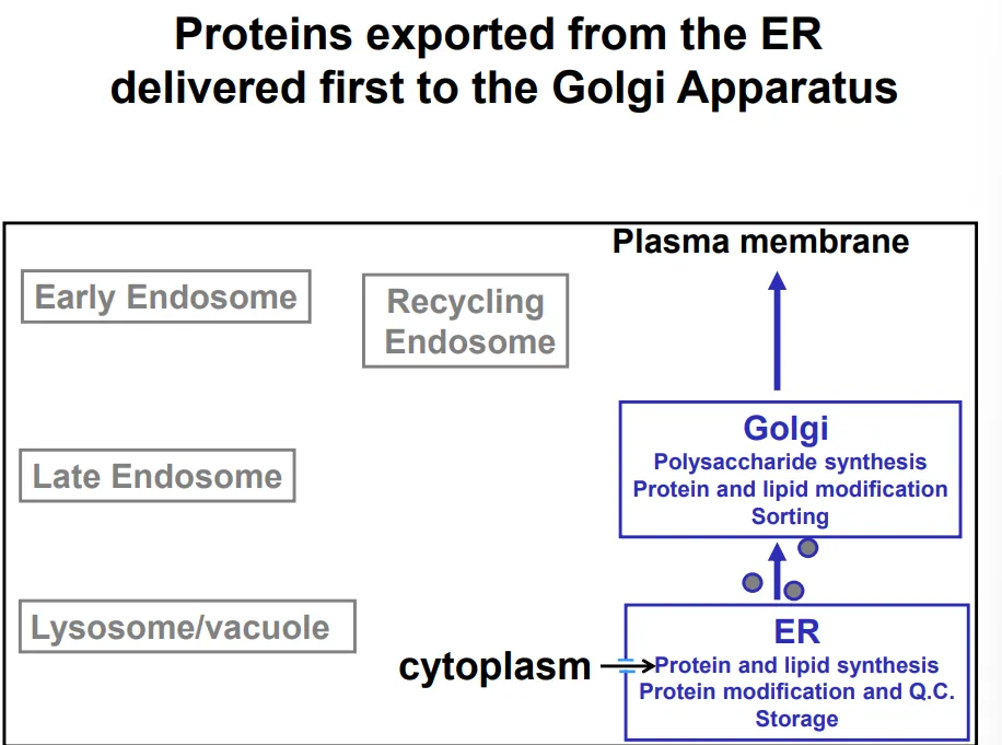
  • the proteins are transported from the ER to the golgi apparatus by the membrane budding into COP-II vesicles (so that they don’t need to cross membranes to be secreted)- this is also used to transport lipids produced by the smooth ER

  • the golgi apparatus (cis → medial → trans layers of cisternae) is a polysaccharide factory, which modifies the existing N-glycan chains for signalling and adds further polysaccharides of functional importance for glycoproteins and glycolipids

  • the vesicles bud off again and release the contents of the lumen (topologically equivalent to extracellular space) out of the cell, either immediately or following an external signal, in the case of regulated secretory vesicles

<ul><li><p>the proteins are <strong>translocated co-translationally</strong> by the ribosomes of the <strong>rough ER</strong>, either integrating into the membrane or being released into the lumen</p></li><li><p>during this process, <strong>N-linked glycans</strong> are added- short <strong>oligosaccharides </strong>(sugar chains) are <strong>coupled </strong>to the protein via a <strong>nitrogen </strong>in an <strong>asparagine </strong>residue</p></li><li><p>this is used as a <strong>signal </strong>throughout the secretion process to determine whether the protein is <strong>folded and modified correctly</strong> (controlled by chaperone proteins), as the glycan chain is <strong>truncated </strong>and <strong>edited </strong>at different points, for <strong>quality control</strong></p></li><li><p>the proteins are transported from the ER to the <strong>golgi</strong> <strong>apparatus </strong>by the membrane <strong>budding </strong>into<strong> COP-II vesicles </strong>(so that they don’t need to cross membranes to be secreted)- this is also used to transport lipids produced by the <strong>smooth ER</strong></p></li><li><p>the golgi apparatus (cis → medial → trans layers of cisternae) is a <strong>polysaccharide factory</strong>, which <strong>modifies </strong>the existing<strong> N-glycan chains</strong> for signalling and adds further polysaccharides of functional importance for <strong>glycoproteins </strong>and <strong>glycolipids</strong></p></li><li><p>the vesicles bud off again and <strong>release </strong>the contents of the <strong>lumen </strong>(topologically equivalent to extracellular space) out of the cell, either <strong>immediately </strong>or following an <strong>external signal</strong>, in the case of <strong>regulated </strong>secretory vesicles</p></li></ul><p></p>
2
New cards

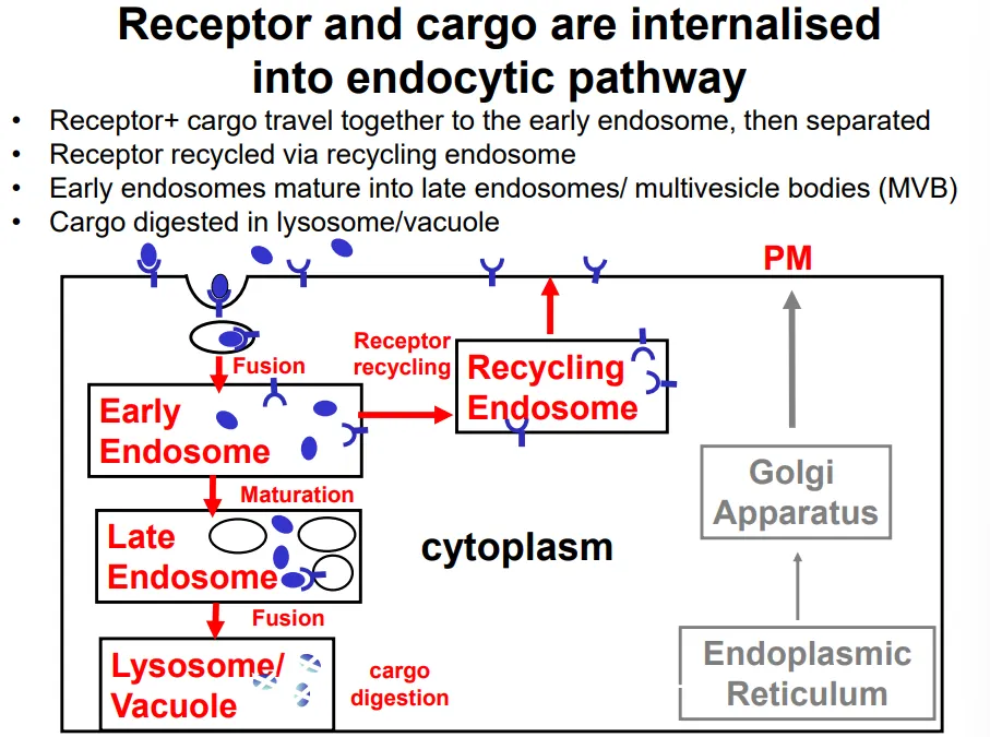
what is the endocytic pathway in eukaryotes?

  • endocytosis and phagocytosis are initiated by specific receptor proteins, which form a receptor-cargo complex

  • clathrin-coated vesicles bud off the plasma membrane into the cell, producing an endosome

  • the receptor and cargo dissociate in the early endosome, so that the receptors can be re-inserted into the plasma membrane via a recycling endosome

  • the cargo remains in the endosome, which matures into a late endosome upon receiving additional contents (eg. enzymes) from the golgi apparatus

  • exisiting lysosomes fuse to this, releasing further digestive enzymes that degrade the cargo (nutrients, pathogens and signalling molecules)

  • during maturation, the endosome becomes increasingly acidic until it becomes a lysosome, where the pH is low enough for the digestive enzymes to function (acidic optimum so that they don’t damage the cell earlier)

<ul><li><p>endocytosis and phagocytosis are initiated by <strong>specific receptor proteins</strong>, which form a receptor-cargo complex</p></li><li><p><strong>clathrin-coated vesicles bud </strong>off the plasma membrane into the cell, producing an <strong>endosome</strong></p></li><li><p>the receptor and cargo <strong>dissociate </strong>in the <strong>early endosome</strong>, so that the receptors can be re-inserted into the plasma membrane via a <strong>recycling endosome</strong></p></li><li><p>the cargo remains in the endosome, which <strong>matures </strong>into a<strong> late endosome </strong>upon receiving additional contents (eg. enzymes) from the<strong> golgi apparatus</strong></p></li><li><p>exisiting <strong>lysosomes </strong>fuse to this, releasing further <strong>digestive enzymes </strong>that degrade the cargo (nutrients, pathogens and signalling molecules)</p></li><li><p>during maturation, the endosome becomes increasingly <strong>acidic </strong>until it becomes a lysosome, where the pH is <strong>low </strong>enough for the digestive enzymes to function (<strong>acidic optimum</strong> so that they don’t damage the cell earlier)</p></li></ul><p></p>
3
New cards

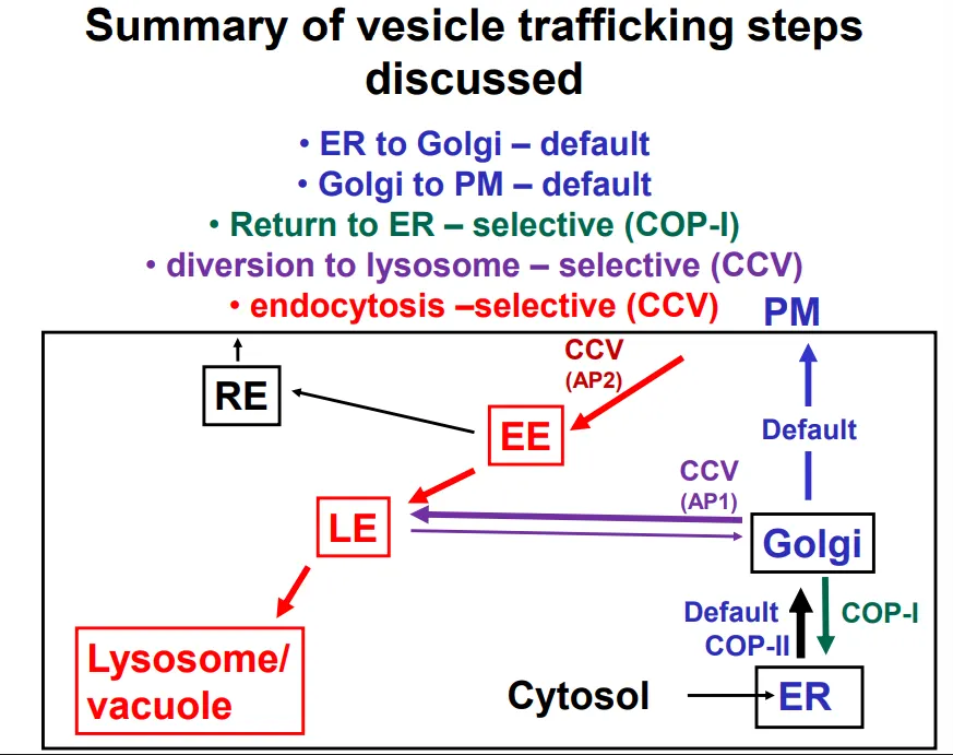
how are transport vesicles formed and what proteins do each kind of vesicle use in the process?

  • curvature of membranes is energetically unfavourable, so to form vesicles, they must be forced into deformation by a protein coat

  • protein coat subunits self-assemble on the cytoplasmic side of the membrane

  • these are also responsible for collecting and packaging the cargo into the vesicle

  • ER → golgi vesicles use COP-II coats

  • golgi → ER (retrieval) vesicles use COP-I coats

  • PM → early endosome vesicles use clathrin and adaptin 2 to form clathrin coated vesicles (AP2)

  • golgi → late endosome vesicles use clathrin and adaptin 1 to form clathrin coated vesicles (AP1)

<ul><li><p><strong>curvature </strong>of membranes is <strong>energetically unfavourable</strong>, so to form vesicles, they must be <strong>forced </strong>into deformation by a <strong>protein coat</strong></p></li><li><p>protein coat subunits<strong> self-assemble</strong> on the <strong>cytoplasmic </strong>side of the membrane</p></li><li><p>these are also responsible for collecting and <strong>packaging </strong>the <strong>cargo </strong>into the vesicle</p></li></ul><p></p><ul><li><p>ER → golgi vesicles use <strong>COP-II </strong>coats</p></li><li><p>golgi → ER (retrieval) vesicles use <strong>COP-I </strong>coats</p></li><li><p>PM → early endosome vesicles use <strong>clathrin </strong>and <strong>adaptin 2 </strong>to form <strong>clathrin coated </strong>vesicles (AP2)</p></li><li><p>golgi → late endosome vesicles use <strong>clathrin </strong>and <strong>adaptin 1</strong> to form <strong>clathrin coated</strong> vesicles (AP1)</p></li></ul><p></p>
4
New cards

how are proteins signalled to be retained or exported in the ER?

  • export from the ER doesn’t require a signal, it occurs by default ‘bulk flow’ (but some signals may accelerate export)

    • bacterial proteins that are inserted into eukaryotes (adding a signal for production in the ER, but not a signal for retention) are excreted

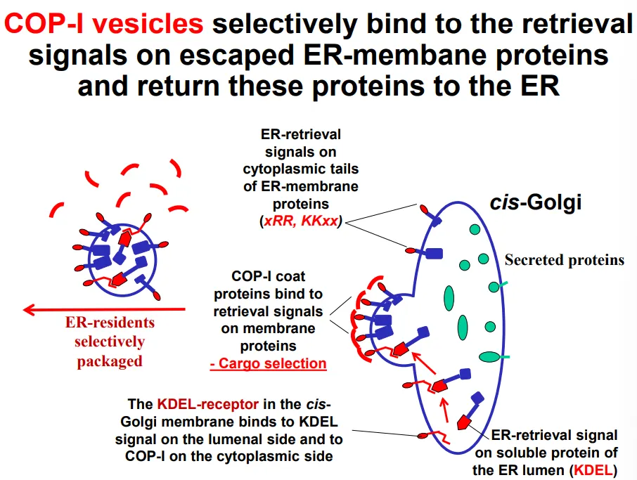
  • resident proteins aren’t prevented from leaving the ER, but they are continually retrieved from the golgi apparatus due to an ER-retrieval signal

    • these are necessary (deletion of the signal results in secretion instead of residency) and sufficient (the only sequence that is required to cause secretion)

  • COP-I vesicles selectively bind to retrieval signals (primary structure) for escaped ER resident proteins that are found in the cis-golgi:

    • diarginine (N terminus) and dilysine (C terminus) motifs on the cytoplasmic side of transmembrane ER resident proteins

    • KDEL receptor proteins in the cis-golgi membrane bind to the KDEL sequences (C-terminus) of luminal ER resident proteins

  • this is cargo selection, which then triggers vesicle formation

  • the KDEL receptor proteins dissociate from the cargo upon return to the ER due to pH differences between the ER and golgi lumen (the ER is less acidic, which discourages the association), so the receptors can be recycled to the golgi in COP-II vesicles

<ul><li><p><strong>export </strong>from the ER doesn’t require a signal, it occurs by <strong>default ‘bulk flow’ </strong>(but some signals may <strong>accelerate </strong>export)</p><ul><li><p>bacterial proteins that are inserted into eukaryotes (adding a signal for production in the ER, but not a signal for retention) are excreted</p></li></ul></li><li><p><strong>resident </strong>proteins <strong>aren’t prevented from leaving </strong>the ER, but they are <strong>continually retrieved</strong> from the golgi apparatus due to an<strong> ER-retrieval signal</strong></p><ul><li><p>these are <strong>necessary </strong>(deletion of the signal results in secretion instead of residency) and <strong>sufficient </strong>(the only sequence that is required to cause secretion)</p></li></ul></li></ul><p></p><ul><li><p><strong>COP-I</strong> vesicles<strong> selectively bind</strong> to retrieval signals (primary structure) for <strong>escaped ER resident proteins</strong> that are found in the <strong>cis-golgi</strong>:</p><ul><li><p><strong>diarginine</strong> (N terminus) and <strong>dilysine </strong>(C terminus) motifs on the cytoplasmic side of <strong>transmembrane </strong>ER resident proteins</p></li><li><p><strong>KDEL receptor proteins</strong> in the cis-golgi membrane bind to the <strong>KDEL sequences </strong>(C-terminus) of <strong>luminal </strong>ER resident proteins</p></li></ul></li><li><p>this is cargo selection, which then triggers vesicle formation</p></li><li><p>the KDEL receptor proteins <strong>dissociate </strong>from the cargo upon <strong>return </strong>to the ER due to <strong>pH differences </strong>between the ER and golgi <strong>lumen </strong>(the <strong>ER is less acidic</strong>, which discourages the association), so the receptors can be <strong>recycled </strong>to the golgi in<strong> COP-II</strong> vesicles</p></li></ul><p></p>
5
New cards

what is the structure of COP-I vesicles?

  • pre-assembled COP-I triads are present in the cytoplasm

  • these complexes have a natural curvature

  • multiple triads assemble to cause membrane bending, with the cargo-motif binding sites positioned against the membrane for cargo selection

  • these are flexibly bound together so that the size of the vesicles can change

<ul><li><p>pre-assembled <strong>COP-I triads</strong> are present in the cytoplasm</p></li><li><p>these complexes have a<strong> natural curvature</strong></p></li><li><p>multiple triads assemble to cause <strong>membrane bending</strong>, with the <strong>cargo-motif binding sites</strong> positioned against the membrane for cargo selection</p></li><li><p>these are <strong>flexibly </strong>bound together so that the <strong>size </strong>of the vesicles can change</p></li></ul><p></p>
6
New cards

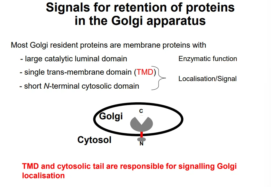
how are proteins signalled to be retained or exported in the golgi apparatus?

  • the large luminal domain of golgi resident proteins is functional enzymatically eg. to make polysaccharides

  • this is attached to a trans-membrane domain and a short N-terminal cytoplasmic domain, which act as a secondary structure signal

  • the different organelles in the secretion pathway have different lipid compositions, so the membrane thicknesses increase from ER → golgi → PM

  • this means that the TMDs of membrane proteins also increase in length, so they may be rejected if they are foreign to each organelle (eg. the hydrophobic region isn’t long enough, so charged residues clash with the fatty acid tails)

<ul><li><p>the large <strong>luminal domain</strong> of golgi resident proteins is functional <strong>enzymatically </strong>eg. to make polysaccharides </p></li><li><p>this is attached to a<strong> trans-membrane domain</strong> and a short <strong>N-terminal cytoplasmic domain</strong>, which act as a <strong>secondary structure </strong>signal</p></li></ul><p></p><ul><li><p>the different organelles in the secretion pathway have different <strong>lipid compositions</strong>, so the <strong>membrane thicknesses increase</strong> from ER → golgi → PM</p></li><li><p>this means that the <strong>TMDs </strong>of membrane proteins also <strong>increase </strong>in length, so they may be <strong>rejected </strong>if they are foreign to each organelle (eg. the hydrophobic region isn’t long enough, so charged residues clash with the fatty acid tails)</p></li></ul><p></p>
7
New cards

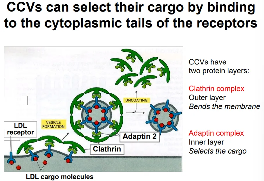
how does endocytosis occur selectively? use cholesterol as an example

  • cholesterol is transported through the blood as part of low density lipoprotein particles

  • these contain a large protein signal molecule, which binds to LDL receptors on the plasma membrane of target cells

  • these receptors bind to adaptin 2 complexes (cargo selection) in the cytoplasm, which in turn attach to clathrin triskelion complexes that cause membrane bending by spontaneous self-assembly, to produce a clathrin coated vesicle (CCV)

  • the clathrin and adaptin dissociate (using energy) from the vesicle to allow fusion to the early endosome, where the LDL receptors dissociate due to the lower pH so that they can be recycled back via recycling endosomes

<ul><li><p>cholesterol is transported through the blood as part of <strong>low density lipoprotein particles</strong></p></li><li><p>these contain a large <strong>protein signal </strong>molecule, which binds to<strong> LDL receptors</strong> on the <strong>plasma membrane</strong> of target cells</p></li><li><p>these receptors bind to <strong>adaptin 2 complexes</strong> (cargo selection) in the cytoplasm, which in turn attach to <strong>clathrin triskelion complexes</strong> that cause <strong>membrane bending </strong>by spontaneous self-assembly, to produce a <strong>clathrin coated vesicle</strong> (CCV)</p></li><li><p>the clathrin and adaptin <strong>dissociate </strong>(using energy) from the vesicle to allow <strong>fusion </strong>to the <strong>early endosom</strong>e, where the <strong>LDL receptors dissociate</strong> due to the <strong>lower pH</strong> so that they can be <strong>recycled </strong>back via recycling endosomes</p></li></ul><p></p>
8
New cards

how are proteins signalled to be transported from the golgi apparatus to the late endosome?

  • digestive hydrolases made in the ER have a signal patch in their tertiary structure, which is recognised by enzymes in the cis-golgi

  • these enzymes phosphorylate a mannose residue on the N-glycan chain of the protein

  • this modification is recognised by a mannose-6-P receptor in the trans-golgi, which binds to the glycan chain as well as adaptin 1 complexes from the cytoplasm

  • this causes the formation of clathrin coated vesicles, which transport the cargo to the late endosome

  • here the Man-6P receptor dissociates due to a decrease in pH, so that it can be recycled back to the trans-golgi

  • these hydrolase proteins are activated by proteases found in the lysosome, by proteolytic cleavage, so that they don’t cause damage to the cell earlier in the process

9
New cards

how are transport vesicles targeted to the correct destination?

  • the vesicle temporarily docks to a membrane

  • here its specific v-SNARE (vesicular) proteins test their interactions with the t-SNARE (target) proteins on the membrane

  • if the t and v-SNARE proteins are complementary to each other, they form a very tight coiled-coil structure, which brings the vesicle close to the target membrane so that fusion can happen

  • if not, they won’t coil and the temporary docking will release the vesicle to try elsewhere

  • after fusion, NSF complexes remove the v-SNARE proteins from the target membranes, using ATP, to maintain compartment identity