LAB QUIZ 2 part 1
1/34
There's no tags or description
Looks like no tags are added yet.
Name | Mastery | Learn | Test | Matching | Spaced | Call with Kai |
|---|
No analytics yet
Send a link to your students to track their progress
35 Terms
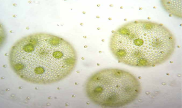
Colony Morphology
Only on isolated colonies [Physical appearance of colonies (shape, edge, elevation) on a plate]
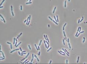
Bacterial Cell Morphology
Microscope observations of individual cells or how those cells cluster together- Bacillus, Streptococcus, Coccus, etc
[Shape and arrangement of individual cells under a microscope (SIZE, TEXTURE]
Difference between colony vs cell morphology
Colony morphology refers to the physical appearance of bacterial colonies on a plate, including their shape, edge, and elevation, while cell morphology describes the shape and arrangement of individual bacterial cells observed under a microscope.
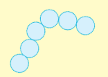
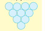
Coccus (Cocci)
Perfect Circle-shaped bacteria

Cocci/ Coccus
Bacilli/ Bacillus
Elongated rod (Also called Rods)

Bacilli/ Bacillus
prefixes
Diplo: groups of two
Strepto: cells in a chain
Staphylo: cells in a cluster (many togather)

diplococci
Streptococci
ci
Staphylococci
Gram stain
A staining method used to classify bacteria
Gram staining steps
Make smear
Stain with Crystal Violet: Stains Peptidoglycan
Iodine (mordant):Fixes stain to cells in a complex that cannot be removed
Alcohol (decolorization): removes non-binded crystal violet
Water rinse: remove alcohol
Stain with Safranin: Counter stain, stains everything:
Blot and observe:Phase contrast or Bright Field?
Mordant
A chemical that helps stain stick (iodine)
Counterstain
Second stain that colors remaining cells
Gram-positive
Purple (keeps crystal violet) because thick cell wall keeps stain
Gram-negative and why it color
Pink/red (loses purple, takes safranin) because thin cell wall loses stain
Bright Field Microscopy uses and best for
1.) uses
Uses transmission (light passing THROUGH something) of light through the objective lens
Image appears dark on a bright background
2.) best for:
Non living (Inanimate) objects
Stained or pigmented cells (mostly dead) Because staining adds contrast
⚠ Not good for live cells (they are too transparent)
Memory tip:
“Bright background → need stain to see things”

Bright Field Microscopy

Phase Contrast Microscopy uses and best for
1.) uses
Uses differences in refractive index
(how much light bends when it goes through something) between bacteria and environment
2.) best for
Living and Unstained cells
Shows internal details more clearly
Memory tip:
“No stain → still see live cells”
Phase Contrast Microscopy
Is colony morphology the same as cell morphology?
NO
Which step fixes stain to cells?1.)
1.)Iodine (mordant)
Which step removes stain
Alcohol
What does “staphylo” mean?
Clusters
What did the bacteria grow on? (plate, broth, slant)
Media
Which step acts as the mordant in Gram staining?” (Translation: Which step locks the stain?)
Iodine
What is the counterstain in Gram staining?
( Translation: What is the second stain?)
Safranin
The difference of Mordant vs Iodine AND Counterstain vs Safranin
Mordant And Counterstain → the job (Function: what it does)
Iodine And Safranin → the chemical (what it is, the actual substance)
What is the mordant in Gram staining?
Iodine
What is iodine’s role?
Mordant
What is the counterstain?
Safranin
What is the role of safranin?
Counterstain
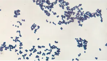
coccus
Staphylococcus
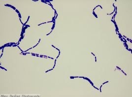
Streptobacillus