Back And Vertebral Column (EXAM 1)
1/80
There's no tags or description
Looks like no tags are added yet.
Name | Mastery | Learn | Test | Matching | Spaced | Call with Kai |
|---|
No analytics yet
Send a link to your students to track their progress
81 Terms
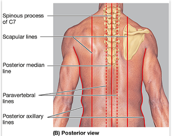
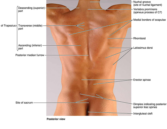
Where is the Mid-vertebral line?
Overlying the spinous processes of the vertebrae/ posterior median line
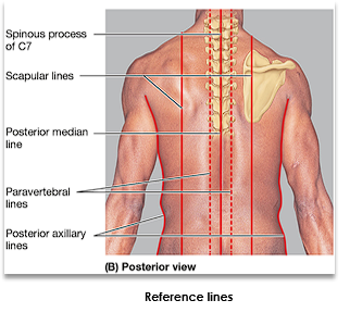
Where is the scapular line?
Passes through the inferior angle of the scapula.
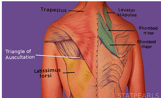
Where is the triangle of auscultation?
Boundaries include:
latissimus dorsi (inferior)
trapezius (medial)
scapula (lateral)
rhomboid major, facial sheet, 6th and 7th ribs, intercostal space (Anterior/ floor)
Where is the vertebral prominens?
The prominent spinous process of C7
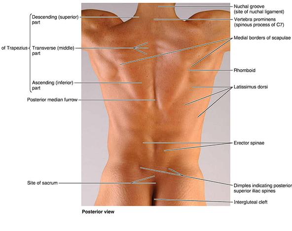
Where is the vertebral furrow?
landmark that indicates the location of the vertebral spines
erector spinae muscles lie on both sides
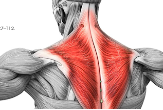
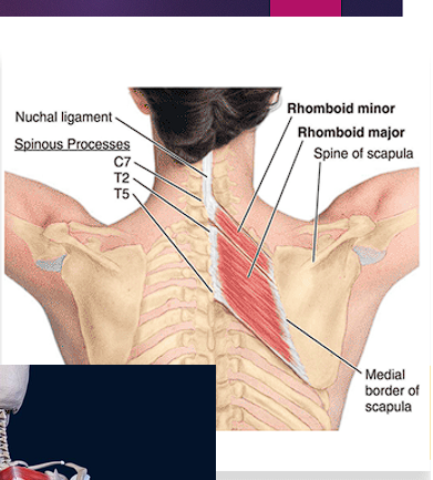
Describe the major muscles associated with the superficial muscles/ Extrinsic back muscles, what they contribute to, and nerve supply
Trapezius
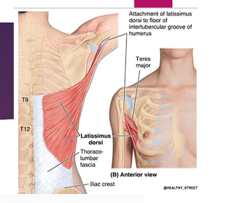
Latissimus dorsi
Levator scapulae
Rhomboid major
Rhomboid minor.
(superficial to deep)
Connect upper limbs to the trunk and assist in upper limb movement via scapula and humerus
Ventral rami of spinal nerves (brachial plexus branches). EXCEPTION: trapezius= spinal accessory nerve (CN XI)
Describe the Innervation and actions of the Trapezius
C7-T12 vertebrae
Action: elevates, retracts, depresses, and upwardly rotates scapula
innervation: Motor: spinal accessory nerve (CN XI)
Describe the Innervation and actions of the Levator scapulae muscle
C1-C4 vertebrae
Action: Elevation and downward rotation of the scapula
Innervation: Dorsal scapular nerve (C5), branches from C4-C5 ventral rami
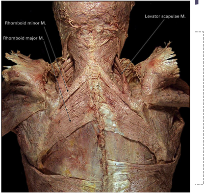
Describe the Innervation and actions of the Rhomboid major and minor
minor: C7-T1 vertebrae
Major: T2-T5
Action: retraction of the scapula
Innervation: Dorsal scapular nerve
Describe the Innervation and actions of the latissimus dorsi muscle
T7-sacrum
Action: Adduction, extension, and medial rotation of the humerus at the glenohumeral joint
Innervation: Thoracodorsal nerve (C6-C8)
List the scapular movements
Elevation
Depression
Protraction (abduction)
Retraction (adduction)
Rotation
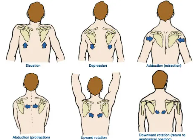
Describe scapular elevation
Scapula moves superiorly (as in shrugging the shoulder)

Describe Scapular depression
scapula moves inferiorly

Describe scapular protraction (abduction)
scapula moves away from the middle

Describe scapular retraction (adduction)
scapula moves away toward the midline

Describe scapular rotation
defined by the direction that the glenoid fossa faces (glenoid fossa faces superiorly for upward rotation and inferiorly for downward rotation)

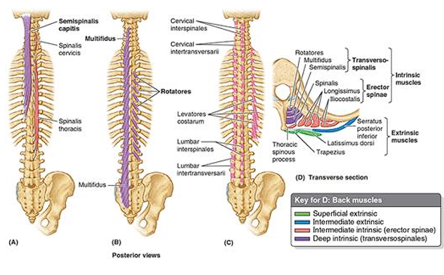
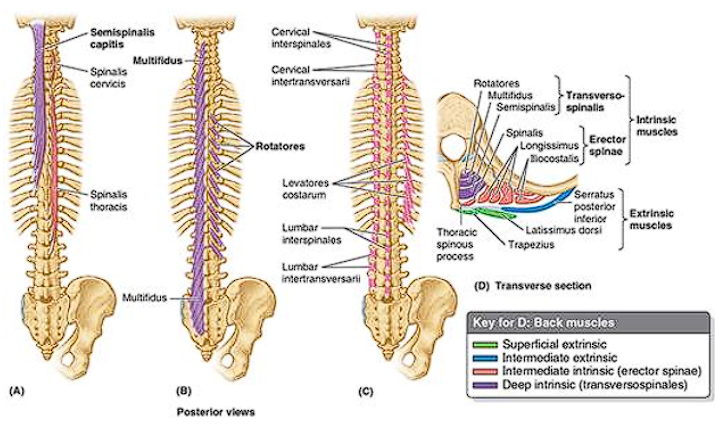
What muscles are considered the deep back muscles/ Intrinsic back muscles (superficial → intermediate → deep), what are they innervated by, and what are they responsible for?
Splenicus capitus and cervicis
Erector spinae
Transversospinalis, Suboccipital muscles
Innervated by dorsal (posterior) rami of spinal nerve at each vertebral level
Responsible for maintaining posture
Describe the location, Innervation, and actions of the splenius capitusa and cervicis muscle
Located deep to the Levator scapulae and rhomboid muscles, and superficial to erector spinae muscles
Action:
Bilateral contraction: extension of the head and neck
unilateral contraction: lateral flexion and rotation of the head and neck
Innervation:
segmentally innervated by dorsal rami

What are the major groups of deep back muscles of the splenius?
Splenius capitis and Splenius Cervicis
Describe the location, Innervation and actions of the Erector spinae muscles
Long muscles of the back, paraspinal muscles
Action:
Bilateral contraction: extension of the vertebral column and control of posture
Unilateral contraction: lateral flexion of vertebral column
Innervation:
segmentally innervated by dorsal rami
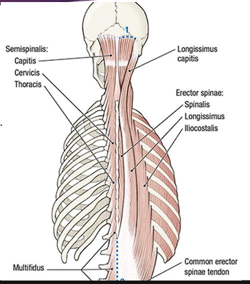
What are the major groups of deep muscles for the Erector spinae muscles?
From lateral to medial: Iliocostalis, Longissimus, Spinalis
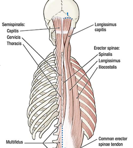
Describe the location, Innervation, and actions of the transversospinalis muscles
located deep to the erector spinae muscles
Action:
Stabilization
Bilateral contraction: extension of the vertebral column
Unilateral contraction: rotation of vertebral column to the contralateral side of the contracting transversospinalis muscle
Innervation:
segmentally innervated by dorsal rami
What are the major groups of deep muscles for the Transversospinalis muscles?
From superficial to deep: semispinalis, multifidus, and rotators
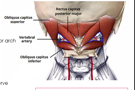
Describe the location, Innervation, and actions of the suboccipital muscles
Located inferior to the occipital bone and deep to the semispinalis capitis muscle
Action:
Mainly postural muscles
May contribute to extension and rotation of the head
Innervation:
Dorsal ramus of C1 spinal nerve (suboccipital nerve)
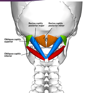
What are the major groups of deep muscles for the suboccipital muscles?
Rectus capitis posterior major and minor
Obliquus capitis superior and inferior

Explain the role of intrinsic back muscles in maintaining posture and facilitating movements of the vertebral column
Maintain posture, provide stabilization, extend the vertebral column/head/neck via bilateral contraction, and cause lateral flexion/rotation via unilateral contraction.
Describe the anatomical boundaries and contents of the suboccipital triangle
Superomedial- Rectus capitis posterior major
Superolateral- obliquus capitis superior
Inferior- obliquus capitis inferior
Floor- Posterior antlanto-occipital membrane and posterior arch of the C1 (atlas)
Roof- Semispinalis capitus
Vertebral artery, suboccipital nerve, greater occipital nerve
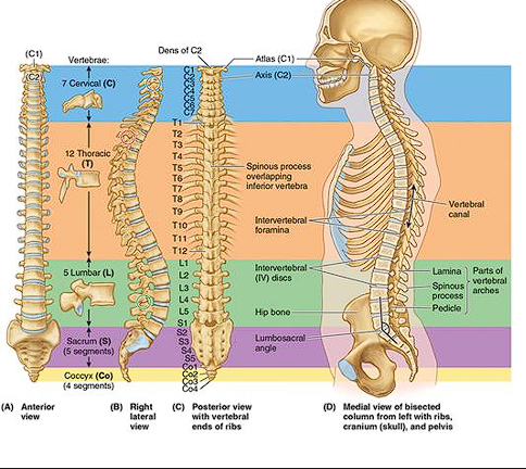
List the regions and number of vertebrae that compose the vertebral column
Cervical (7)
Thoracic (12)
Lumbar (5)
Sacral (5)
Coccygeal (4 segments that fuse into one solid bone)
33 vertebrae
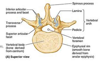
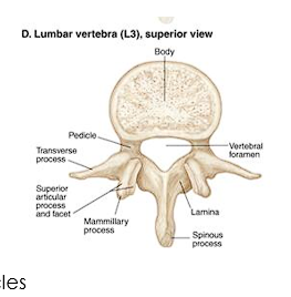
Describe the structural components of the vertebral body
Anterior more massive part
Gives strength to the vertebral column and support body weight
Size increase as column descends
Trabecular bone containing red marrow
Surround by a thin external layer of compact bone
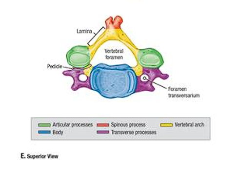
Describe the structural components of the vertebral arch
Posterior to the body
2 pedicles: Right and Left. Short, cylindrical process. Project posteriorly
2 Lamina: Broad, flat bones. Connect transverse and spinous processes
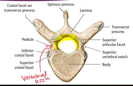
Describe the structural components of the vertebral processes
Arise from vertebral arch
Spinous process (1)- Posterior projecting tip, easily palpated
Transverse process (2)- lateral
Articular process (4)
2 superior and 2 inferior
Facet
Zygapophyseal (facet joint)
The transverse process and spinous process provide attachment to what muscles and what do they serve as ?
Deep muscles
Serve as a lever to facilitate the muscles to change position
Describe the structural components of the vertebral foramen
Formed from the vertebral arch and posterior surface of the vertebral body
contains Spinal cord
What anatomical features are unique in respect to the cervical spine?
Atlas (C1)
Axis (C2)
C7
What anatomical features are unique in respect to the thoracic spine?
T1
T2
What anatomical features are unique in respect to the Lumbar spine?
Accessory process
Mammillary process
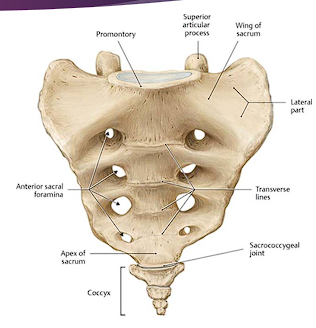
What anatomical features are unique in respect to the sacrum?
Sacral foramen
sacral promontory
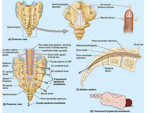
Sacral canal
Sacral hiatus
Describe the features of the Atlas (C1)
Atypical
Supports the skull, allows nodding movement
Articulates with the skull (occipital condyles)
Ring, kidney shaped
LACKS spinous process and body
Nod up and down
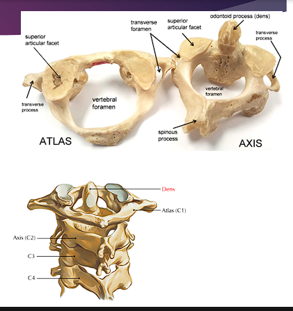
Describe the features of the Axis (C2)
Strongest cervical vertebrae- carries cranium
Dens (odontoid process)
Attachment for transverse, apical, and alar ligaments
Bifid spinous process
Provides rotational movement (through it articulates with C1)

Describe the features of C7
Long spinous process
Spinous process is NOT BIFED
Larger body
Smaller transverse foramen
Palpable bulge on back of neck
Transmits only small accessory veins
Describe the features of T1
Atypical
Long almost horizontal spinous process
Complete costal facet on the superior edge of its body for the 1st rib
Demifacet on its inferior edge that contributes to the articular surface of 2nd rib
Spinous process is elongated and resembles that a cervical vertebrae
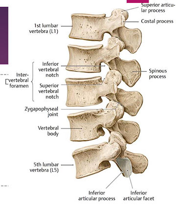
Describe the features of T12
Mammillary process (small tubercle)
Transitions in characteristics
Superior half-thoracic characteristics
Rotary movement
Inferior half-lumbar characteristics
Permit flexion and extension
Most commonly fractured vertebrae
Describe the accessory process of the lumbar spine
Attachment for the intertransversarii muscles
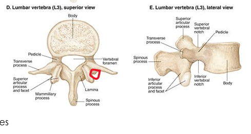
Describe the Mammillary process of the lumbar spine
Posteriorly located
Attachment of Multifidus and intertransversarii muscles
Describe the features of the sacral foramen
4 pairs
Exit of the posterior and anterior rami of the spinal nerves
Anterior (pelvic) are larger than posterior (dorsal) ones
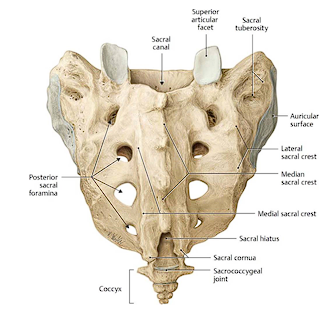
Describe the features of the sacral promontroy
Anterior projecting edge of the body of S1
L. mountain ridge
Important OB landmark
Describe the features of the sacral canal
Continuation of the vertebral canal in the sacrum
Contains the Cauda equina
bundle of spinal nerve roots arising inferior to the L1 vertebrae
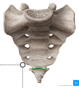
Describe the features of the sacral hiatus
U shaped
leads into the sacral canal
Between sacral cornua, inferior to median sacral crest
Provides access to the epidural space
Used in OB, pediatric procedure, and chronic pain management
Describe the features of the coccyx
Tailbone
Triangular bone
Remnant of the embryonic tail-like cauda eminence
Last 3 coccygeal vertebra fuse during midlife forms beak-like coccyx
Provides attachments for parts of the gluteus maximus and coccygeus muscles and anococcygeal ligament
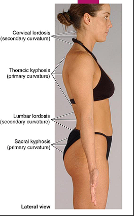
What is the functional significance of primary (Kyphotic) curvature
Thoracic and Sacral
Develop in the fetal period
Concave anteriorly
Retain through life
Abnormal curvature-kyphosis, excessive kyphosis
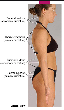
What is the functional significance of secondary (Lordotic) curvature
Cervical and Lumbar
Develop postnatal period
Concave posteriorly
Abnormal curvature-lordosis, excessive lordosis
Compare and contrast the characteristics of the five regions of the vertebral column
As the vertebral column descends, bodies increase in size to handle increased weight-bearing. The size of the vertebral canal changes relative to the diameter of the spinal cord.
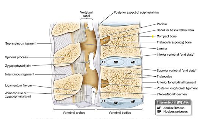
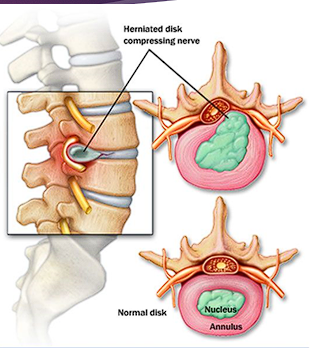
Describe the features of the annulus fibrosis of the disc
Bulging fibrous ring
Consists of concentric lamellae of fibrocartilage
Insert into smooth rounded epiphysial rims on the articular surface
Thinner posteriorly, may be incomplete in an adult in the cervical region
Decreasingly vascularized centrally
Outer third receives sensory innervation
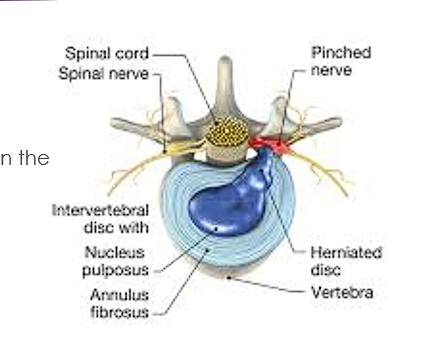
Describe the features of the nucleus pulposus of the disc
Central core of disc (jelly like center)
At birth-88% water, more cartilage than fiber
Responsible for the flexibility and resilience of the disc and column
Becomes broader when compressed
Thinner when tensed or stretched
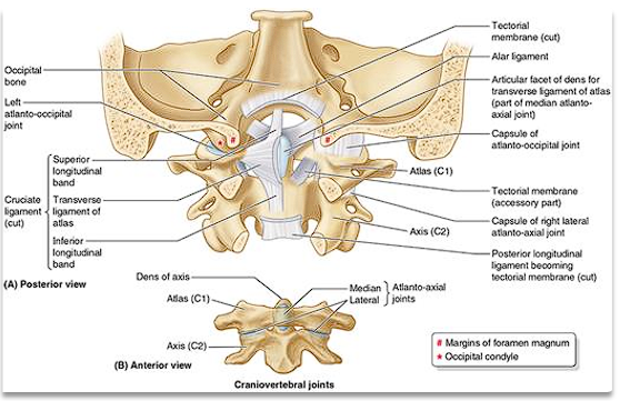
Describe the features of the craniovertebral joints
2 sets- atlanto-occipital joints and atlanto-axial joints
Synovial joints that have no IV discs
Wider range of movement than the rest of the vertebral column
Articulations involve the occipital condyles, atlas, axis

What joints are associated with Craniovertebral joints
Atlanto-Occipital joints
Atlanto-axial joints
What are the features of Atlanto-Occipital joints
Between atlas (C1) and occipital bone of the cranium
Synovial joint of condyloid type-thin, loose capsules
Permit nodding of the head (extension and flexion) the “YES” motion
Permit sideways tilting of the head (lateral flexion)

What are the features of the Atlanto-axial joints
Between the atlas (C1) and axis (C2)
3 articulations (2 lateral and 1 median)
Lateral- gliding type synovial joints
Median- pivotal joint
Permits rotating head side to side- saying “no” (rotation)
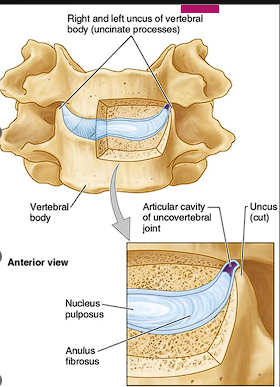
Describe the features of Uncovertebral Joints (joints of Luschka)
Between uncinate process of the lower vertebrae and the body of the vertebrae above
Found ONLY in the cervical spine (typically C3-7)
Lateral and posterolateral margins of IV discs
Considered synovial joint
Clefts (degenerative spaces)
Degenerative spaces in the discs occupied by extracellular fluid
Clinical correlation:
Sites of bone spur (osteophyte) formation neck pain
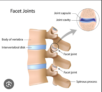
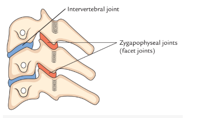
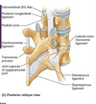
Describe the features of Zygapophysial joints (facets) (joints of vertebral arches)
Plane synovial joints between the superior and inferior articular processes of adjacent vertebrae
Surrounded by a thin joint capsule
Thin and loose in cervical region= movement
Attached to the margins of the articular surfaces of the articular processes
Accessory ligaments unite the laminae, transverse processes, spinous processes to help stabilize the joint
Permit gliding movements between articular processes
Enable vertebral flexion, extension and limit rotation
Innervated by articular branches from the medial branches of the posterior rami of the spinal nerves
Each joint is supplied by 2 nerves
Describe the features of intervertebral joints
Fibrocartilaginous symphysis (secondary cartilaginous joints)
Between vertebral bodies of adjacent vertebrae
The intervertebral disc forms the connection between the bodies
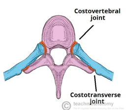
Describe the features of costovertebral joints
Joints of the head of the ribs
Supported by multiple ligaments
Synovial plane joint
Joints of Head of ribs
Slight gliding
Describe the features of Lumbosacral joint
Articulation between L5 and S1
Intervertebral symphysis and Zygapophyseal (facet) synovial joint
Highly weight bearing transition point between lumbar and sacrum
High stress at the lumbosacral angle
Clinical correlation:
Common site of herniated disc, spondylolisthesis, degenerative changes
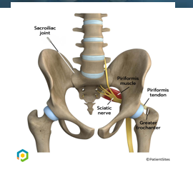
Describe the features of the Sacroiliac joints
Joint of the pelvic girdle
Between auricular surface of the sacrum and ilium
Strong, weight bearing joints
Anterior synovial joint-, covered with articular cartilage
Transfers weight from spine to the pelvis to the lower limbs
Provides stability
Very limited mobility
Slight gliding and rotary movement
Due to interlocking of articulating bones and ligaments
Clinical correlation
SI joint dysfunction
Sacroilitis
Describe the features of the Sacrococcygeal joints
Articulation between sacrum and coccyx
Symphysis
Become partially or completely fused with age (sometimes)
Allows very limited movement
Slight flexion/extension of the coccyx during sitting, defecation, childbirth
Describe the Anterior longitudinal ligament
Strong, broad fibrous band
On anterior surface of vertebral column
Attaches to the vertebral bodies and intervertebral discs
Extends from sacrum to C1 and occipital bone
Maintain stability of the joints between the vertebral bodies
Prevents hyperextension of vertebra column
ONLY ligament that limits extension
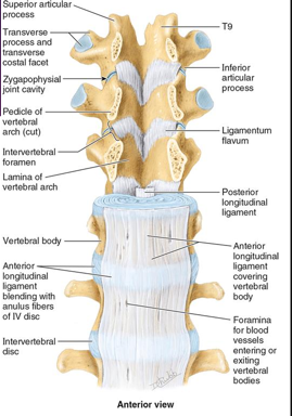
Describe the posterior longitudinal ligament
Narrow, weaker
Runs within vertebral canal along the posterior aspect of the vertebral bodies
Supports the IV discs posteriorly
Helps prevent/redirect posterior herniation of the nucleus pulposus
Weakly resists hyperflexion
Describe the ligamentum flavum (accessory ligaments of IV joints)
Broad, pale, yellow elastic fibrous tissue
Extends from lamina above to the lamina below
Forms the posterior wall of the vertebral canal
Limiting abrupt flexion of the vertebral column, prevents injury to the IV discs
Preserve normal curvature, Assist with straightening after flexing (standing up)
Cervical Region- thin
Thoracic Region Thicker
Lumbar- Thickest
Describe the Interspinous ligament (Accessory ligament of IV joints)
Weak, thin
Connect adjoining spinous processes
Attach to the root to the apex of each spinous process
Limits flexion
Describe the Supraspinous ligament (Accessory ligament of IV joints)
Strong, cord-like band
Connects tips of spinous process from C7 to sacrum
Limits flexion
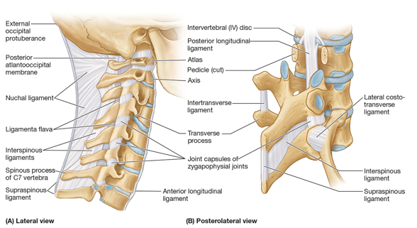
Describe the Nuchal ligament (Accessory ligament of IV joints) (ligaments of neck)
Merges with the supraspinous at the back of the neck
Strongest median ligament of the neck
Extends from external occipital proturbance and posterior border of the foramen magnum to the spinous process in the cervical vertebrae (C2 to C7)
Replaces the supraspinous ligament in the cervical region
Provides muscle attachment
Helps support the head and limit cervical flexion
Describe the structure of the cruciate ligament (Cruciform ligament) (ligaments of neck)
Transverse ligament of Atlas
Horizontal, strong band
Attaches to the medial tubercles on the lateral masses of C1
Passes posterior to the dens (odontoid process of C2)
Holds dens against anterior aspect of the atlas
Prevents anterior displacement of C1 on C2- stabilizes the atlanto-axial joint
Longitudinal bands (superior and inferior)
Vertical, Weaker
Pass from transverse ligament to occipital bone superiorly and to the body of C2 inferiorly
Stabilizes the atlanto-axial joint
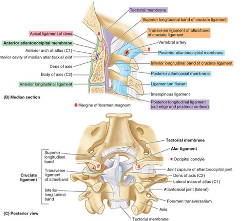
Describe the Alar ligament (ligaments of neck)
Short, rounded cords
Attach the cranium to C1
Extend from the sides of the dens to the lateral margins of foramen magnum
Prevent excessive rotation at the joint

Describe the Apical ligament of Dens (ligaments of neck)
small, fibrous cord-like
Extends from tip of dens to anterior edge of foramen magnum
Stabilizes the dens by attachment to the occipital bone
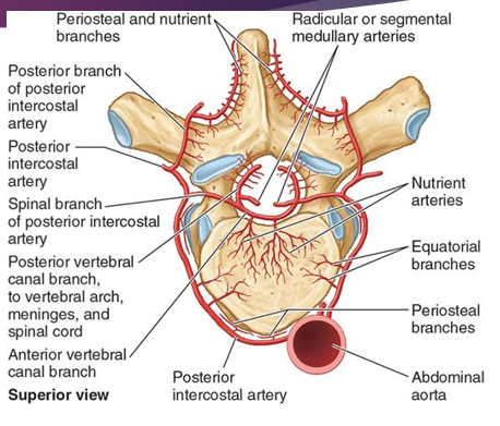
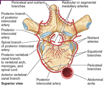
The Spine is supplied by the….
Vertebral artery system (longitudinal source)
Describe the segmental arteries
Enter through intervertebral foramen at each level and then splits into vertebral body supply and spinal cord branches
Intercostal branch
Lumbar branch
Sacral branch
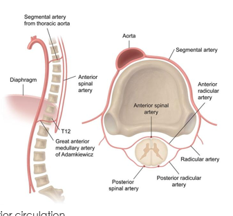
Describe the vertebral arteries of the Cervical spine
Arise from subclavian artery
Ascends through the transverse foramina (C6→C1)
Supplies cervical vertebrae, spinal cord (spinal branches), posterior circulation
Describe the vertebral arteries of the thoracic spine
Posterior intercostal arteries (same as segmental arteries)
From thoracic aortal
Each level gives
Vertebral body branches
Spinal branches (enter intervertebral foramina
Describe the vertebral arteries of the Lumbar spine
Lumbar arteries
Direct branches of abdominal aortal
Supplies vertebral bodies, discs, spinal canal
Describe the vertebral arteries of the Sacral spine
Lateral sacral arteries
From iliac artery
Supplies: sacrum, cauda equina region
Describe the costotransverse joint
Slight gliding
Rotation
Elevation and depression
Articulates with tubercle of the rib