609B Poll Questions/Forums
 Call Kai
 Learn
Learn Practice Test
 Spaced Repetition
 Match
Match Flashcards
 Knowt Play
1/44

Earn XP
Description and Tags
UT 609B - OB 2
Name | Mastery | Learn | Test | Matching | Spaced | Call with Kai |
|---|
No analytics yet
Send a link to your students to track their progress
45 Terms
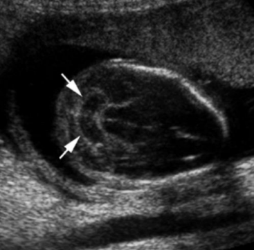
What sign is depicted by the arrows in this image?
a. Stargazer
b. Banana
c. Lemon
d. Strawberry
Banana
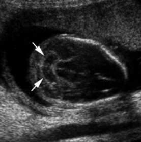
If you see this when scanning the fetal head, what else will you find?
a. Holoprosencephaly
b. Choroid plexus cyst
c. Spina bifida
d. Absent corpus callosum
Spina bifida
Which condition is associated with the stargazer sign?
a. Iniencephaly
b. Anencephaly
c. Chiari malformation
d. Exencephaly
Iniencephaly
A bony defect of the calvarium that allows herniation of meninges and/or brain is called:
a. Myelocele
b. Myelomeningocele
c. Rachischisis
d. Encephalocele
Encephalocele
The most common cranial neural tube defect is:
a. Spina bifida
b. Anencephaly
c. Acrania
d. Encephalocele
Anencephaly
What part of the brain does the rhombencephalon become?
a. Cerebellum
b. Lateral ventricle
c. Thalami
d. Third ventricle
Cerebellum
The network of arteries at the base of the fetal brain is:
a. Sagittal sinus
b. Circle of Willis
c. Vein of Galen
d. Ventricle system
Circle of Willis
Which of the following is not an ossified portion of the spine?
a. Spinous process
b. Vertebral body
c. Space between lamina & pedicle
d. All of the above are ossified
Spinous process
Partial or complete absence of the cerebral hemispheres is:
a. Hydrocephalus
b. Hydranencephaly
c. Holoprosencephaly
d. Dandy-Walker malformation
Hydranencephaly
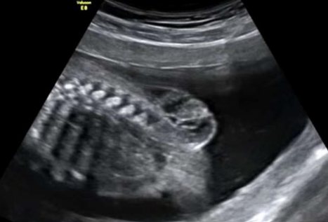
What is this pathology?
a. Brachial cleft cyst
b. Encephalocele
c. Cystic hygroma
d. Myelomeningocele
Myelomeningocele
A collection of CSF in the posterior fossa with an intact vermis is known as:
a. Dandy-Walker malformation
b. Vein of Galen aneurysm
c. Arachnoid cyst
d. Porencephaly
Arachnoid cyst
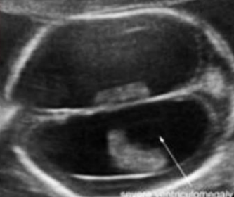
What is the anomaly visualized in this image?
a. Hydranencephaly
b. Dandy-Walker malformation
c. Hydrocephalus
d. Holoprosencephaly
Hydrocephalus
What is the structure of the brain that is highly vascular and produces CSF?
a. CSP
b. Corpus callosum
c. Choroid plexus
d. Thalami
Choroid plexus
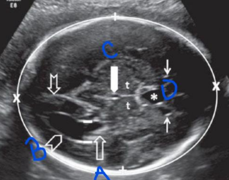
What structure is labeled A?
a. Third ventricle
b. CSP
c. Thalami
d. Choroid plexus
Choroid plexus
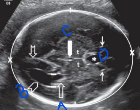
What structure is labeled C?
a. Third ventricle
b. CSP
c. Thalami
d. Choroid plexus
Third ventricle
The bony structures of the fetal face are typically first seen on ultrasound at ___ weeks.
a. 6-7
b. 8-9
c. 11-12
d. 14-16
11-12
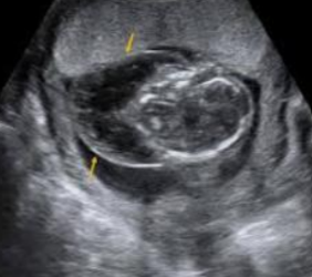
What is being depicted in this image?
a. Teratoma
b. Cystic hygroma
c. Thyroglossal duct cyst
d. Cyclopia
Cystic hygroma
A single midline orbit is known as:
a. Anopthalmia
b. Hypertelorism
c. Microphthalmia
d. Cyclopia
Cyclopia
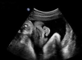
What is being depicted in this image?
a. Normal nose/lips
b. Cleft lip
c. Cleft palate
d. Proboscis
Normal nose/lips
Rostral means:
a. Toward the tail
b. Toward the head
c. Midline
d. Neurulation
Toward the head
The prosencephalon gives rise to which structure?
a. Thalamus
b. Cerebellum
c. Cerebral peduncles
d. Medulla oblongata
Thalamus
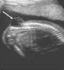
What are we seeing in this image?
a. Myelomeningocele
b. Teratoma
c. Cystic hygroma
d. Brachial cleft cyst
Myelomeningocele
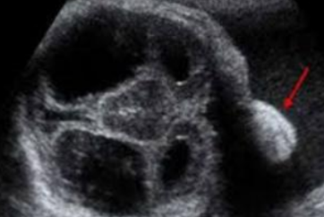
What is being depicted in this image?
a. Macroglossia
b. Micrognathia
c. Hypertelorism
d. Proboscis
Proboscis
Which of the following lies along the floor of the lateral ventricle and the roof of the third ventricle?
a. CSP
b. Thalami
c. Choroid plexus
d. Cisterna magna
Choroid plexus
The ________ is a nerve bundle that connects the right and left hemispheres of the brain.
a. Corpus callosum
b. CSP
c. Falx
d. Thalami
Corpus callosum
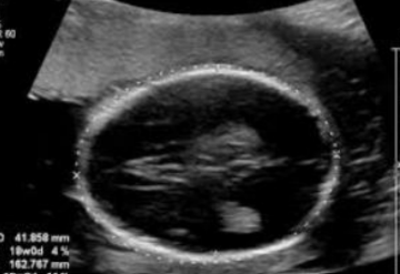
What is this ultrasound image depicting?
a. Normal anatomy
b. Enlarged third ventricle
c. Enlarged lateral ventricle
d. Agenesis of the corpus callosum
Agenesis of the corpus callosum
Which of the following is not an ossification center of the vertebrae?
a. Vertebral body
b. Right lateral process
c. Spinous process
d. Left lamina
Spinous process
A defect in the spine where a sac that contains spinal cord and meninges protrudes from the spinal defect is known as:
a. Meningocele
b. Myelomeningocele
c. Chiari malformation
d. Cystic hygroma
Myelomeningocele
Mandibular hypoplasia that causes a receding chin is known as:
a. Hypotelorism
b. Macroglossia
c. Micrognathia
d. Microcephaly
Micrognathia
Causes of microcephaly include all of the following except:
a. Craniosynostosis
b. Zika virus
c. Enlarged ventricles
d. Maternal drug use
Enlarged ventricles
Which type of holoprosencephaly is the most severe?
a. Alobar
b. Semilobar
c. Lobar
d. Hydranencephaly
Alobar
Which of the following is associated with cyclopia and proboscis?
a. Hydrocephalus
b. Hydranencephaly
c. Holoprosencephaly
d. Choroid plexus cyst
Holoprosencephaly
A missing vermis and a cystic filling of the posterior fossa is a sign of _______.
a. Choroid plexus cyst
b. Arachnoid cyst
c. Vein of Galen aneurysm
d. Dandy-Walker malformation
Dandy-Walker malformation
Partial or complete absence of the cerebral hemispheres is known as:
a. Hydrocephalus
b. Hydranencephaly
c. Holoprosencephaly
d. Dandy-Walker malformation
Hydranencephaly
The dilation of the ventricular system is:
a. Ventriculomegaly
b. Hydrocephalus
c. Dandy-Walker malformation
d. Holoprosencephaly
Hydrocephalus
Choroid plexus cyst can be associated with which of the following?
a. Holoprosencephaly
b. Dandy-Walker malformation
c. Arachnoid cyst
d. Trisomy 18
Trisomy 18
A collection of CSF in the posterior fossa with an intact vermis is known as:
a. Dandy-Walker malformation
b. Vein of Galen aneurysm
c. Arachnoid cyst
d. Porencephaly
Arachnoid cyst
A cyst filled with CSF that usually communicates with the ventricles, subarachnoid space or both is:
a. Dura sinus malformation
b. Schizencephaly
c. Porencephaly
d. Teratoma
Porencephaly
The most common intracranial solid tumor is:
a. Teratoma
b. Glioblastoma
c. Choroid plexus cyst
d. Arachnoid cyst
Teratoma
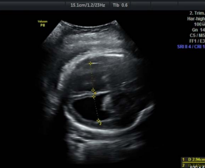
What is being depicted in this image?
a. Hydranencephaly
b. Dandy-Walker malformation
c. Porencephaly
d. Hydrocephalus
Hydrocephalus
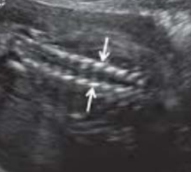
What are the arrows pointing to in this image?
a. Vertebral body ossification
b. Lateral ossification centers
c. Fetal cranium
d. Myelomeningocele
Lateral ossification centers
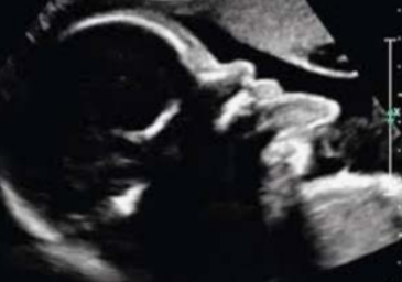
What is being depicted in this image?
a. Proboscis
b. Macroglossia
c. Absent nasal bone
d. Cleft palate
Macroglossia
Where is the Circle of Willis located in the brain?
a. Frontal lobe
b. Base
c. Cerebellum
d. Apex
Base
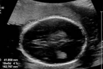
What is being depicted in this image?
a. Dandy-Walker malformation
b. Hydranencephaly
c. Normal fetal brain
d. Absence of the corpus callosum
Absence of the corpus callosum
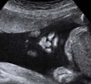
What is being depicted in this image?
a. Cleft lip
b. Cleft palate
c. Normal nose/lips
d. Proboscis
Cleft lip