1- Anatomy and physical examination of the auricle, external auditory canal and tympanic membrane
1/14
There's no tags or description
Looks like no tags are added yet.
Name | Mastery | Learn | Test | Matching | Spaced | Call with Kai |
|---|
No analytics yet
Send a link to your students to track their progress
15 Terms
Describe the auricle
composed of elastic cartilage covered with perichondrium and skin
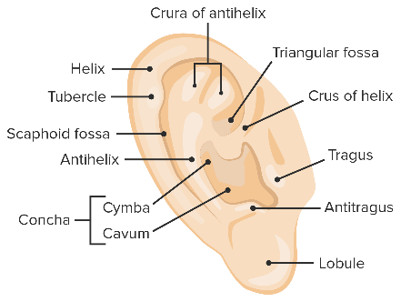
What is the function of the auricle
collect sound waves into the external ear canal → directional hearing
Describe the external auditory canal
consists of an outer cartilaginous and inner bony part
Canal curves and narrows to protect tympanic membrane
during otoscopy- ear is puleld upwards and back
What is the function of the external auditory canal
transmission of sound waves to the tympanic membrane
Describe the cartilaginous part of the external auditory canal
covered by skin containing hair follicles and ceruminous glands that produce cerumen/ ear wax
provides pathway for bacterial spread from parotid gland, infratemporal fossa and skull base
Describe the bony part of the external auditory canal
covered by thin layer of skin, tightly adhering to periosteum
glands and hair is absent
between cartilaginous and bony part- stricture where foreign bodies can lodge
What is the tympanic membrane
separates external ear from middle ear
3 layers- skin, fibrous tissue, mucosal layer
thin, cone shaped membrane
What is the function of the tympanic membrane
Sound waves cause vibration of the tympanic membrane
→ transmits these vibrations to the ossicles of the middle ear (malleus, incus, stapes)
What are the quadrants of the tympanic membrane
anterior/ posterior + superior/ inferior
2 parts
Pars flaccida
pars tensa
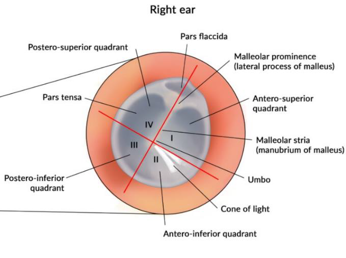
What is the pars flaccida of the tympanic membrane
has 2 layers- non fibrous tissue
fragile
associated with eustachian tube dysfunction and cholesteatomas
What is the pars tensa of the tympanic membrane
has 3 layers
comparatively robust
“cone of light”- cone shaped light reflection of the otoscope light in anterior inferior quarant helps with orientation
tympanic nerve passes in superior region
for intervention, posteroinferior quadrant is chosen
What are the steps to examine the ear
history
inspection and palpatiion of auricle, mastoid and tissues around ear
examination of external ear canal, first without speculum
examine ear canal with aural speculum with head mirror and light source
examine ear canal and tympanic membrane with otoscope
How do you examine the external ear canal without a speculum
pull ear upwards and back
straightens external ear canal
in infants- pull downwards
what are the types of otoscopy
battery powered
battery powered light source with magnification
pneumatic
detect perforation of tympanic membrane or fluid in middle ear
assess the mobility of tympanic membrane by applying positive and negative pressures with the rubber squeeze bulb
How do you clean the external ear
can be blocked by ear wax/ cerumen, skin debris, purulent discharge
wax removal and aural speculum + cerumen spoon or alligator forecepts
fluid removal with suction or cotton tipped metal applicator
if no perforation of TM, can be cleaned by irrigation with water at body temperature
too warm or cold can cause vertigo