cell bio lab
1/53
There's no tags or description
Looks like no tags are added yet.
Name | Mastery | Learn | Test | Matching | Spaced | Call with Kai |
|---|
No analytics yet
Send a link to your students to track their progress
54 Terms
B cell activation steps
Ag bings to BCR, activates B cell, proliferates, differentiates into plasma B cell, plasma cells produce IgM Abs
Ist Ab to be secreted during infection
IgM
Most abundant Ab
IgG
Dilution formula
C1v1=c2v2
AA with ring structures
Tyrosine, phenylalanine, tryptophan
cell culture medium examples
DMEM, TCM, RPMI 1640
what does cell culture contain
inorganic salts, vitamins, sugar/glucose, buffered pH, phenol red indicator, AA, hormones and growth factors, fetal bovine serum, sodium pyruvate, non essential AA, L glutamine, HEPES buffer
how do we feed cell cultures so they can proliferate and grow
cell culture medium
hormones in cell culture medium
insulin- for glucose
transferrin- for iron binding
growth factors
optimal cell culture growth conditions
37 degrees Celsius, humid atmosphere, proper CO2 content to maintain buffering system (5-8%)
two types of cell cultures
primary explants and continuous cell lines
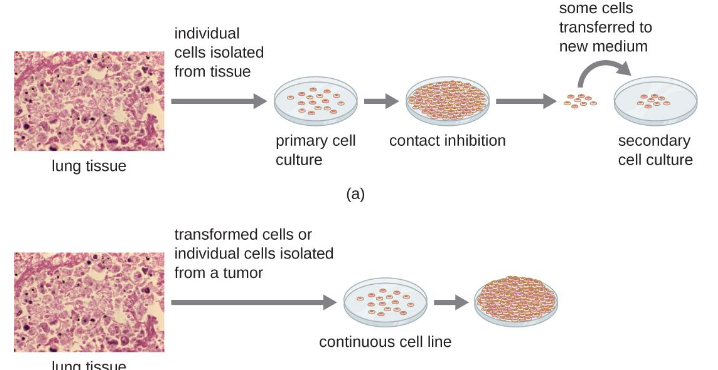
primary explants
cells derived from tissue or blood, the primary culture reaches a certain density then they have contact inhibition, and at that point you can transfer some to make a secondary culture
continuous cell lines
established cultures of cells essentially the same type, will not stop growing after contact inhibition, due to genetic modification using plasmid
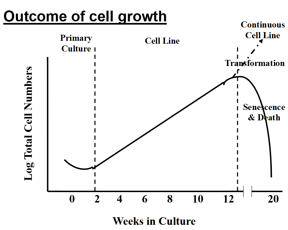
outcome of cell growth- somatic cells vs transformed cell line
primary somatic cells enter senescence after 12 weeks roughly (due to telomere shortening after many cell cycles), primary cell culture can last 12 weeks, so when you transform the cells into continuous cell line they can last many years
types of cell lines
nontransformed- normal, have limited lifespans, we won’t use in the lab
transformed- genetically modified to last but still has normal functions of a cell, altered to show continuous growth
cancer cell lines- derived from cancers, shows continuous growth, may have alter functions due to being a cancer cell
cell characteristics- adherence and examples
non adherent- like blood leukocytes
adherent- like epithelial, endothelial, fibroblast, neuronal
epithelial cell characteristics
skin, intestinal/colon mucosa, liver or other organs, usually grow flat and spread out
fibroblasts
connective tissue, grow as a spindle shape
endothelial
line the blood vessels, grow spreading and flat
3T3 cells
mouse fibroblast transformed cells, don’t have contact inhibition
HT29
human colon carcinoma, cancer cells, they don’t have contact inhibition but may have modified function
IEC-6
rat small intestinal epithelial cells, non transformed
Caco-2
human colon carcinoma cells isolated from cancer
how do we avoid contamination
sterilize solutions and equipment, aseptic or sterile techniques, laminar flow hood, antimicrobials
3 methods for sterilizing solutions and equipment
autoclave- use high pressure and temperature to kill microbes
filtration- pass solutions through a membrane with a small pore size
irradiation- X rays or gamma rays to kill microbes (not UV bc it will change the nutrients in the culture medium)
aseptic or sterile technique examples
gloves, lab coat, ethanol spray
laminar flow hood purpose
circulates the air, turn on the hoods before using them, must work in the center of the hood
antimicrobials purpose in cell cultures
to reduce bacterial load, not for sterilization
penicillin- kills gram pos bacteria
streptomycin- kills gram neg bacteria
gentamicin- broad spectrum
amphotericin B- anti fungal
none are antivirals, so virus and mycoplasm bacteria with no cell wall can be a problem
role of trypsin/EDTA
EDTA secretes metal ions to enhance the cleaving ability of trypsin, which breaks the connections between cells and adherent surface
how to determine if cells are trypsinized
cells become round and bright instead of flat
role of trypan blue
stains dead cells blue
hemocytometer and how to use
a dyed cell counter, use trypan blue, pipet cells into hemocytometer, 10 microliters each side, count cells in each quadrant up to 100 cells, finish the number of cells in the last quadrant
dilution for hemocytometer
10 microliters cells, 80 of medium, 10 trypan blue
formula for percent viable cells
percent viable cells = viable cells/all cells
formula for viable cells/mL
viable cells/mL = (total viable/number of quadrants counted) x dilution factor x 10^4
why do we need viable cells/mL
to set up new cell cultures, to prepare dilutions
unregulated cell death
necrosis
necrosis
energy independent cell death, cell is damaged and then swells and bursts, releases intracellular contents, enzymes, DNA, and this release can damage nearby cells and activate the inflammatory response
regulated cell death types
apoptosis, autophagy, necroptosis, pyropoptosis, ferroptosis
ferroptosis
iron dependent cell death, driven by lipid peroxidation, not mediated by caspases, ROS accumulate in the cell
pyropoptosis
highly inflammatory mode of cell death, common in immune cells, is activated by infection from microbe/virus inside the cell, caspases are activated and pores are formed on the cell membrane, IL-1B and IL-18 cytokines are released causing the inflammatory response
necroptosis
programmed form of necrosis, is triggered if the apoptosis system is blocked, causes membrane rupture and inflammation
autophagy
cell eats itself, lysosome dependent degradation, provides nutrients during stress, maintains cellular homeostasis, is normally a helpful system but can lead to cell death if excessive, GLP-1s slow down digestion so much that cells do this
apoptosis
cell death without the release of cellular contents, is energy dependent, no nearby inflammatory response, important in development bc its used to build the nervous system and shape body parts, removes old cells in the epithelium, removes cells after immune response, kills infected cells
morphological changes: chromatin condenses, DNA begins to fragment into 200 bp pieces, nuclear envelop disassembles, cytoskeleton breaks, cell blebs forming bodies that can be phagocytosed, once caspase activation begins, cell will die no matter what
quantifying apoptotic cells
run a gel to see how many fragments, does it form a ladder
look for the activation of apoptosis specific proteases/caspases
electron microscopy for cell blebbing
stain the cells for condensed DNA
Syto24 stain- binds to DNA, can see if its in tiny blebs or in a blebbing cell
inducing apoptosis in cells
serum starve the cells causes in 2-3 days
why do we favor serial dilutions
there is less error than direct dilutions
serial dilution
making a smaller dilution ratio and then using that to create your final dilution
calculating dilution factor
the reciprocal of the fraction of the dilution
cell confluence
the cells are touching each other, no gaps between cells
importance of standard deviation
values are significant if values do not overlap with mean +/- SD
how to determine apoptosis
Syto 24 dye and epifluorescent microscope- stains nuclei in cells to view blebbing