Unit 3 - Cell Structure, Adhesion & Replication
1/259
There's no tags or description
Looks like no tags are added yet.
Name | Mastery | Learn | Test | Matching | Spaced | Call with Kai |
|---|
No analytics yet
Send a link to your students to track their progress
260 Terms
UTIs are caused by
UPEC
UPEC Infection Process
UPEC adheres with pili
Takes and replicates
Burst cell to repeat cycle
Pili
Lectin proteins that interact with mannose-glycoprotein in epithelial
Lectins
Made of FimA, D, H and G
FimH
Adhesive on the tip, and necessary for assembly of type 1 fimbriae
FimF and G
Attachment proteins
FimA
Creates length
FimD
Anchor
Mannose-Containing Glycoproteins
Oligosaccharide units
Proanthocyanides
Polyphenol molecule in cranberries that decrease UPEC adherance
Inhibitor Cocktail
Combination of inhibitors that target many different bacteria adhesion molecule
Inner Cell Mass
Forms early embryo
Embryo Layers
Endoderm, Ectoderm, Mesoderm
HV Wilson Experiment
Separate 2 sponge cells with a mesh, then mixed them together which they eventually clump in their own species naturally
Johannes Holtfreder
Took cells from eggs ectodermal and mesodermal to separate them, but the cell aggregated by type
Cell Adhesion Molecules (CAMs)
Transmembrane proteins that help with clumping of like cells
Junctions
Formed after aggregation stabilize cell interactions and facilitate communications between cells
Epithelial Sheets
Form inner lining of digestive system and outer skin
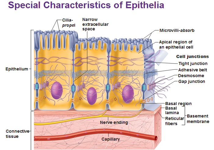
Basal Surface
Anchored to extracellular structures that give structure to sheets of cells, includes basal lamina, basement membrane
Hemidesmosomes
Anchors cells to extracellular matrix on basal region
Adhesion Complexes
Connects lateral surfaces
Types of Adhesion Complexes
Tight junctions
Adherence junctions
Gap junctions
Desmosomes
Tight Junctions (Zonula Occludens)
Below the apical surface of occludin and claudin to form continous seals to stop diffusion between apical and basolateral
Occludin and Clotin
Closely arrange between neighbouring cells
Gap Junctions
1-5-2 nm width that directly links cytosol of two cells, allowing for metabolic integration through ions (cAMP, Ca)
Hexagonal Connexin Hemichannel
6 individual nexin protein subunits
Hemichannels
Link up with other cell’s hemichannels to allow for rapid coordination of cardiac and uterine muscle contractions
Plasmodesmata
Like gap junctions in plant cells for phloem structure
Phloem
System of elongated tubes from linear arrays of connected cells, carrying nutrients like products of photosynthesis, from leaves to plants
Sieve Tube Elements
Phloem cells connected by menlarged plasmodesmata that form the sieve tube plate
Companion Cells
Associated with development and function of sieve tube elements by giving ATP, proteins and connects to the phloem cells by plasmodesmata
Communication through Plasmodesmata happens through
Transcription factors, gene transcripts, sRNAs
Viral Pathogens exploit
Gap junctions to facilitate cellular spread
Anchoring Junctions
Hemidesmosomes
Adherens junctions
Desmosomes
All associated with actin filaments
Desmosomes
Links two cells together
Adherens Junctions
Indirectly connect actin cytoskeleton between neighbour cells
Homophilic Interactions
Association of similar cells
Heterophilic Interactions
Connects different cells together
Families of CAMs
Cadherins (IG superfamily)
Integrins
Selectins
Cadherins
Ca dependent CAMs that mediate homophilic interactions
E-Cadherin (Epithelial Cadherin)
Mediates Ca dependent adhesion of epithelial cells
N-Cadherin
Neural cadherin
P-Cadherin
Placental cadherin
Adhesion controlled by
Transmembrane coherence and cytosolic co-factors
Catenin
Anchors cadherins to actin
Endothelial Cells
Specialized epithelial cells that form the walls of the blood vessels
Extravasation
Cells/fluids move from blood vessel/capillary into surrounding tissue
Types of Leukocytes
Granulocytes
Monocytes
Lymphocytes
Grandulocytes
Targets pathogens, include neutrophils, eosinphils, basophils
Neutrophils
Most numerous, primarily targets bacterial infections and trauma
Moncytes
Differentiates into macrophages
Natural Killer Cells (NK)
Lyse virally cells and tumour cells
T and B cells
Make antibodies as part of immune response
Capture
Injury signal, mediated with cytokines and basal receptors that triggers release of selectins to appear on apical layer
P Selectins
Interacts with selectin specific glycoprotein in leukocytes, held in secretory vesicles until cytokine signal
Platelet Activating Factor (PAF)
Membrane anchored signal interacts with neutrophil’s paf receptor
PAF Receptor
Only happens when slow rolling occurs with neutrophils and interacts with PAF
PAF Binding
Initiates changes in gene expression and activation of integrin adhesion molecules on the neutrophil
Firm Adhesion
Integrin interacts with icamps that further slows movement
Integrins
Dimeric protein with propeller and alpha-beta domains that form the ligand binding domain and folded down when inactivated
Activation of Integrins
Signals for reorganization of actin cytoskeleton
Transmigration
Protease breaks down the endothelial cell adhesion, causing swelling from leaked blood and migration of neutrophils
Transendothelial Migration
Complete migration of neutrophil through the vessel wall into surrounding connective tissue
Dynamic Instability
Both polymerization and depolymerization, making the spindle self organizing
Centrosome
Made up of mother and daughter centriole and PCM
Pericentrilar Material (PCM)
Has y-Turc where microtubules nucleates and polymerizes
CDK and PLK4 Kinase
Initiates centrosome duplication in G1/S phase
M Phase CDK
Initiates centrosome splitting
Spindle Poles
2 MTOC on opposite sides
Bipolar Mitotic Spindle
Microtubule based machine that segregates duplicated chromosomes
Astral Microtubules
Links to cell cortex to anchor spindles
Polar Microtubules
Projects to the centre and overlap each other to help with pushing microtubules apart
Kinesin 1 and 2
2 ATP heads and a cargo binding subunit
Kinesin 5
Has no motor activity but helps with end diassembly
Bipolar Attachment
Chromosome attaches to spindle microtubules from each poles
Spindle Assembly Checkpoint
Forces from each kinetochore must be equal in order for anaphase to occur
Prophase
Mitotic spindle assemble, centrosomes move to opposite poles
Prometaphase
Chromosomes attach to bipolar microtubule spindles, kinetochores assemble at centromeres
Metaphse
Bipolar attachment, tension from chromosomes being pulled causes it to line up on metaphase plate
Telophase
Chromosome decondenses, mitotic spindle disassembles and endomembrane and nuclear envelope reassemble
Cytokinesis
Cell pinches from actin filament and myosin
Cyclin CDK Complexes
Heterodimer protein complexes that facilitate regulated phosphorylation
A-Cyclin
Regulates CDKs
E3 Ligase Complexes
Target specific protein for degradation in proteasome to turn off kinases or cell cycle inhibitors
Skip Cullen F box (SCF) Complexes
Releases cell from G1 and allows transition into S phase
Anaphase Promoting Complex (APC)
Has different target proteins depending on its accessory protein (CDC20 or CDH1)
APC CDC20
Regulates transition from metaphase to anaphase
APC CDH1
Regulates Exit from Mitosis
G1 Cyclin CDK
Targets phosphorylation of APC CDH1 to stop mitotic breaks, and inhibitors, and prepare S phase transcription
G1 S phase Cyclin CDK
Prepares for S and M phase by activating E2F and transcription of mitotic regulators
G1 S Phase Cyclin CDK phosphorylate
Activates DNA replication genes, and phosphorylates proteins to only fire once
M Phase Cyclin CDK Complex phosphorylates
Chromosomal proteins
Nuclear lamina
Microtubule proteins
Kinetochore proteins
APC Complex
Phosphorylation of Condensins and Histones
Chromosomes condense
Phosphorylation of Nuclear Lamina
Breakdown envelope
Phosphorylation Microtubule Proteins
Formation of mitotic spindle and centrosome separation
Phosphorylation of Kinetochore Proteins
Chromosome spindle association
Phosphorylation of APC Complex
Cell progression
Metaphase to Anaphase Transition (MAT)
Anaphase inhibitors degrade
Mitotic Exit Network (MEN)
Mitotic cyclin degrades to exit mitosis
Mitosis Promoter Factor (MPF)
Cyclin B + CDC2 that regulates phosphorylation