Unit 5 Body Systems ( Full )
1/185
There's no tags or description
Looks like no tags are added yet.
Name | Mastery | Learn | Test | Matching | Spaced | Call with Kai |
|---|
No analytics yet
Send a link to your students to track their progress
186 Terms
Definitons
Ventilation: The inhalation and exhalation of air using the ventilation system
Gas exchange: The diffusion of gases across the alveoli
Cell respiration: A controlled release of energy from organic substance inside the cell.
Properties of gas-exchange surfaces
Thin – Short diffusion distance
Moist – Dissolve respiratory gases
Large Surface area – Maximize diffusion
Permeable to respiratory gases – allow oxygen and carbon dioxide to pass through
E.g. Lung, Gills
Alveoli - Adaptations
It is the site for gas exchange
Large surface area:
There are many spherical shape alveoli
Maintain concentration gradient:
Surrounded by rich blood capillaries
Short diffusion distance:
Single-cell thick wall of type 1 pneumocytes
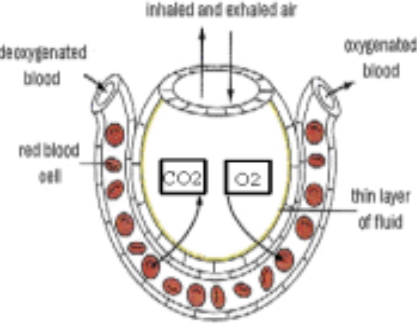
Alveoli - pneumocytes
Type 1 pneumocytes :
Extremely thin alveolar cells that are adapted to carry out gas exchange
Type 2 pneumocytes :
Secrete a fluid to keep the inner surface moist and allow gases to dissolve
Secrete surfactant to reduce surface tension.
Lung ventilation
Inspiration ( Inhalation ) Expiration ( Exhalation )
|
|
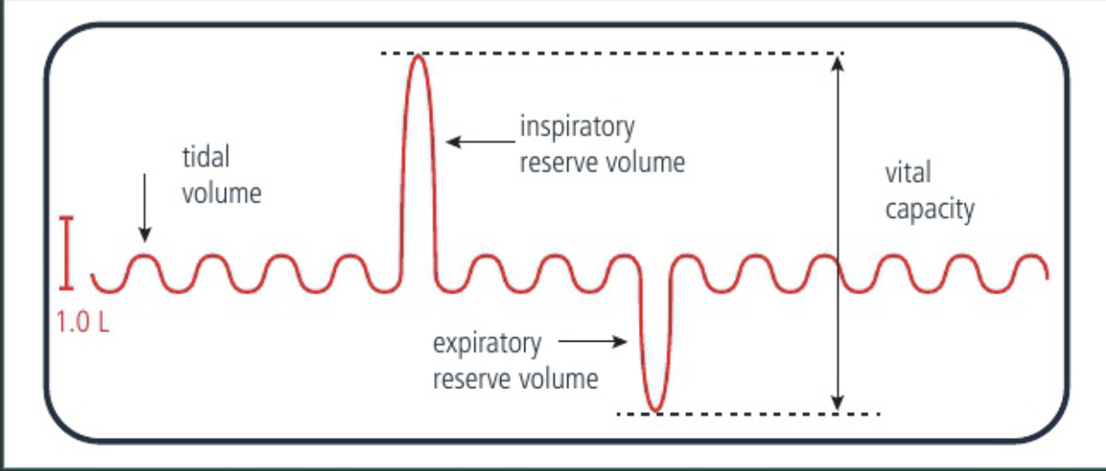
Lung volume measurement
Spirometer is used to measure lung volume.
Tidal volume: the volume of air that is breathed in or out when a person is at rest.
Inspiratory reserve volume: the maximum volume of air that a person can breathe in.
Expiratory reserve volume: the maximum volume of air that a person can breathe out.
Vital capacity: the sum of the inspiratory reserve volume, the tidal volume and the expiratory reserve volume.
Leaf structure – cross section of leaves
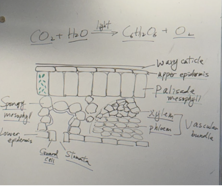
Leaves are organs that are responsible for photosynthesis.
Waxy cuticle – Prevent water loss from evaporation
Upper epidermis – Clear layer, allows light to pass through
Palisade mesophyll – Regular cells, contains a lot of chloroplasts
Spongy mesophyll – Irregular cells with a lot of air space, increases surface area for gas exchange
Guard cells – regulate the opening and closing of stomata
Stomata – site of gas exchange [in human it is alveoli]
Xylem – transport of water and mineral ions (from roots to leaves)
Phloem – transport of sucrose and amino acids (from source to sink)
Factors that affect Transpiration
Definition: Evaporation of water through the stomata.
Factors that affect transpiration rate | Reasons |
Light intensity
| 1. Increase light intensity, increase transpiration 2. Photosynthesis occur under light, when there is light, more stomata open for gas exchange, increase evaporation. |
Temperature
| 1. Increase temperature, increase transpiration 2. Increase kinetic energy in water molecules, so water evaporate faster. |
Air movement
| 1. Increase air movement, increase transpiration 2. Removal of the humid air, increase water potential gradient between inside and outside of the leaf. Thus water vapor diffuse faster |
Humidity
| 1. Increase humidity, decrease transpiration 2. Less water potential gradient differences. Less water vapor diffuse out |
Haemoglobin and oxygen transport
Haemoglobin → iron-rich protein in RBC that transports oxygen from the lungs to tissues, carbon dioxide from tissue to lungs
It consists of heme (iron) and globin, which form a complex structure enabling oxygen binding
Oxygen is bound to haemoglobin and carried in red blood cells.
Haemoglobin molecule consists of four polypeptide chains, with a haem prosthetic group at the centre of each chain.
Each haem group contains one iron atom, and one oxygen molecule binds to each iron atom.
So one haemoglobin molecule can bind up to four oxygen molecules.
Cooperative binding and Allosteric binding of haemoglobin and O2
Cooperative binding | Allosteric binding |
|
|
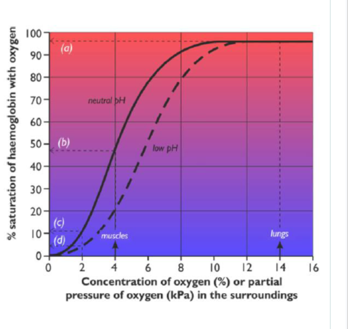
Oxygen dissociation curve
The more oxygen there is in the surroundings (high partial pressure), the more saturated the haemoglobin will be.
The concentration of oxygen in the surroundings can be measured as a percentage or measure it as a partial pressure (PO2, kPa)
Oxygen can only diffuse in and out of the blood from capillaries.
Oxygen dissociation curve Graph
(a) In the alveoli of the lungs
Oxygen is constantly being brought in by ventilation
Partial pressure of oxygen is kept high, at around 14 kPa.
As blood passes through the capillaries surrounding the alveoli, oxygen is loaded on to haemoglobin and become almost 100% saturated
(b) In tissues
e.g. liver or brain
Oxygen is used by respiration, so its partial pressure is low, about 4 kPa.
At this PO2 the haemoglobin is only 50% saturated
It unloads about half its oxygen to the cells, which use it for respiration.
(c) In tissues that are respiring quickly
e.g. contracting muscle cells
PO2 drops even lower, to about 2 kPa
haemoglobin saturation drops to about 10%
almost 90% of the oxygen is unloaded, providing more oxygen for the muscle cells.
(d) Actively-respiring tissues
A lot of CO2 is produced
CO2 dissolves in blood or tissue fluid to make carbonic acid and so lowers the pH
H+ ions leads to a decrease of affinity for O2 , therefore reduces the % saturation of haemoglobin at any PO2.
This right-hand shift is called the Bohr shift.
So at a PO2 of 2kPa, the % saturation is nearer 5%
95% of the oxygen are unloaded in respiring tissues.
*decrease affinity, release more O2.
blood vessels
The circulatory system of the human body contains several different types of blood vessel:
Arteries → away from heart
Arterioles
Capillaries → site of exchange
Venules
Veins → to heart
adaptations of capliiaries, arteries and vein
| Artery | Capillary | Vein |
Thickness of walls | Thick | Extremely thin - only one cell thick | Thin |
Function | Carries blood away from heart | Site of materials exchange between blood and tissue | Carries blood into heart |
Elasticity | Greater than vein | n/a | Less than artery |
Muscularity | Greater than vein | n/a | Less than artery |
Diameter of lumen | Narrower than vein | Extremely narrow (fits single RBC) | Wider than artery |
Valves? | no | no | yes |
Pressure of blood | high | low | low |
Capillaries:
Adaptations of capillaries for exchange of materials
One cell thick → reduces the diffusion distance for oxygen and carbon dioxide between the blood and the tissues of the body
The thin endothelium cells of some capillaries have gaps between them called fenestrations which allow blood plasma to leak out and form tissue fluid
Capillaries form branches in between the cells → increase the surface area for diffusion + substances to and from the cell
Capillaries have a lumen with a small diameter →
Red blood cells squeeze through capillaries in single-file
This forces the blood to travel slowly which provides more opportunity for diffusion to occur
It also reduces the diffusion distance as red blood cells are brought in close contact with the capillary wall
arteries:
narrow lumen : high pressure blood
thick muscle fibre : prevent rupture
thick elastic fibre : pulse.
elastc fibre allwos arteries to stretch - presure exerted on the artieral walls - elastic recoil - pushes blood forward. Contraction of artieries = One pulse
vein
thick lumen : maintain low pressure blood
valve : prevent backflow
thin layer of muscles and elastic fibres , srrounded by skeletal muscle.
skeletal muscle contract, squeeze vein , opens valve - blood move forward. Relaxes, valve close - blood trapped in vein
measurement of heart rate
radial pulse in wrisk
carotid pulse in neck
The rate is the number of beats per minute (bpm).
Heart rate depends on the body’s demand for oxygen, glucose and for removal of carbon dioxide. There is a positive correlation between intensity of physical exercise and heart rate.
DVT
Deep vein thrombosis (DVT) can occur after a long period of being stationary
for example on a long haul flight or in jobs which require a lot of standing.
conorary occultion
Coronary arteries supply the cardiac muscle with oxygen and nutrients
Plaque buildup: build up of fatty plaque in coronary artery walls narrows the lumen, reducing blood flow to heart muscle.
Wall damage and stiffening: Higher pressure damages walls; inelastic fibrous tissue repairs it, hardening (sclerosis) the artery.
Plaque rupture: Damaged plaque breaks open, triggering thrombus (clot) formation.
Occlusion: Thrombus restricts or fully blocks (occludes) the coronary artery; dislodged pieces block smaller arterioles downstream.
consequence
Heart attack
cardiac tissue requires oxygen and nutrients via conorary artieries to function
conorary artery blocked - result into heart attack
Treatment
bypass surgery
blood content (4)
Plasma (55%) - yellow fluid contain: blood cell, nutrient, Co2, O2, hormones, antibodies, urea, heat
RBC(45%) - carry oxygen
WBC + Platelets - (less than 1%)
WBC: body immunity - phagocytes, lymphocytes
platelets: clot blood, prevent futher entry of pathogen, excessive blood loss
Tissue Fluid
A solution that bath all cells.
Substances do not move directly between the blood and the cell
They first diffuse into the tissue fluid that surrounds all cells
Then diffuse from there to the cells
Tissue Fluid - At the arterial end of the capillary bed
Blood is at high pressure
Blood plasma is forced out through the permeable walls
Cells and proteins are too big to leave, so they remain in the blood
Tissue fluid is formed by pressure filtration
Tissue Fluid - At the venous end of the capillary bed
Blood is at low pressure
Blood and tissue fluid are now at around the same pressure
Tissue fluid returns by the methods below
Solutes enter the blood by diffusion
Water returns to the blood by osmosis
Tissue Fluid - Excess tissue fluid
Not all the fluid that left the blood returns to it
Excess tissue fluid will be drained into lymph vessels, which are found in all capillary beds
Lymph vessels have thin walls like capillaries, tissue fluid can easily diffuse inside forming lymp
what is lymphatic system
Consists of a network of lymph vessels flowing alongside the veins
The vessels lead towards the heart, where the lymph drains back into the blood system near the vena cava.
There is no pump, but there are numerous valves, and lymph is helped along by contraction of skeletal muscles.
collect waste and tissue from the tissue to the bloodstream
Lymphatic System Function
The lymphatic system has three different functions:
It drains excess tissue fluid
It absorbs fats from the small intestine.
It is part of the immune system.
There are networks of lymph vessels at various places called lymph nodes
White blood cells are developed in lymph nodes
They become swollen if more white blood cells are required to fight an infection.
lymph vessel
Lymphatic vessels (or ducts) have the following features:
Thin walls with gaps
Valves to prevent backflow
After filtration in the lymph nodes, lymph returns to the blood circulation
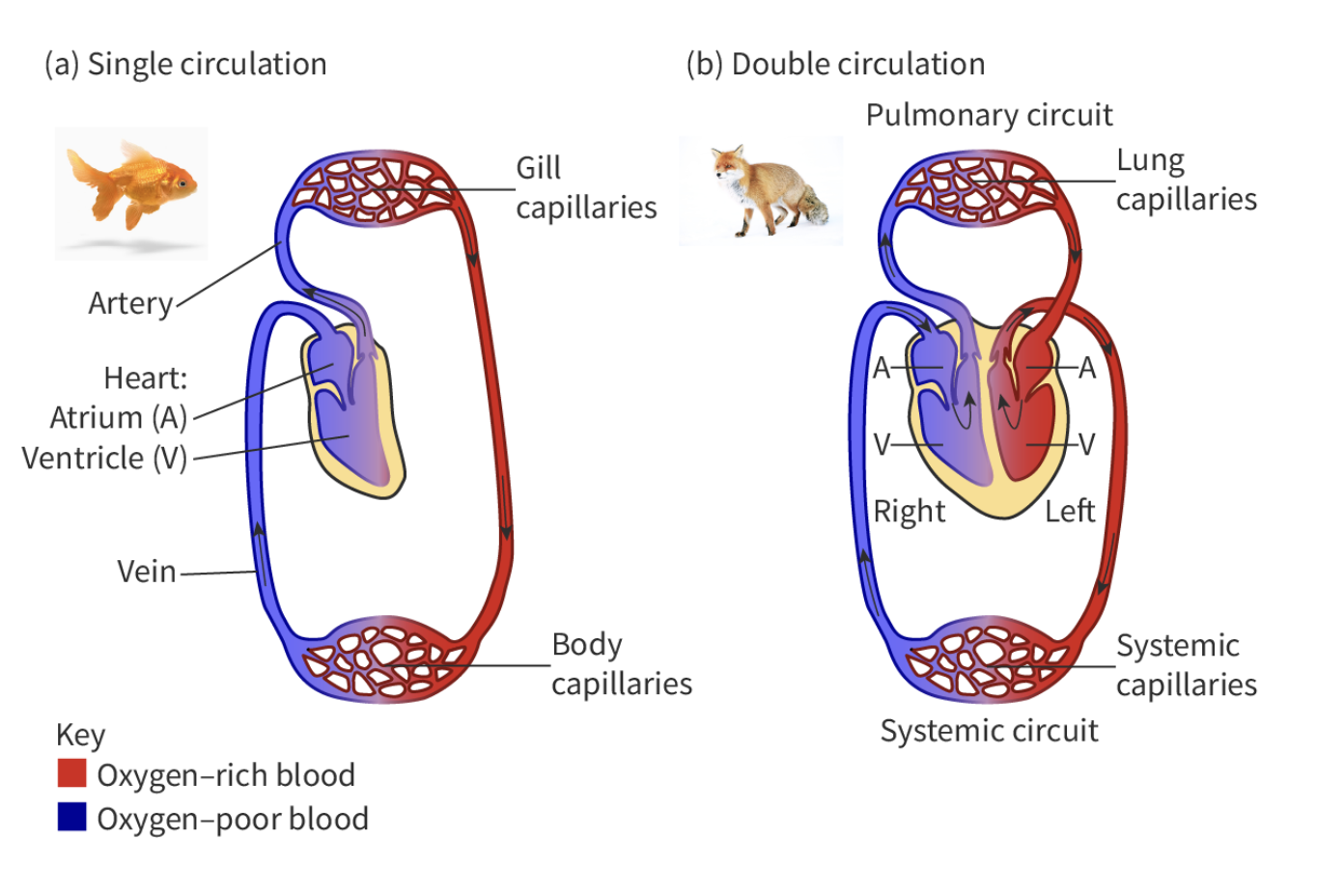
Differences between the single circulation of bony fish and the double circulation of mammals
Mammals
four chambers , two circuits
left: oxygenated blood enters the left side of the heart before being pumped to the body (systematic)
right: deoxygenated blood returns to right side of the heart before going to lungs ( plumonary)
separated by a septum
Fish
2 chambers, 1 circuit
mammal heart adaptation (8)
Double circulatory system Maintain a high concentration gradient → high metabolic needs | made out of the myogenic cardiac muscle generate own electrical contractions | Sinoatrial Node (SA node) pace maker initiate heartbeat | Atrioventricular and semilunar valves ensure one-way blood flow |
Four chambers (atria,ventricle) Thin musice atria - receive low-pressure blood Thick walls ventricles - generate high pumping pressure | Thicker muscle in left side of heart pump blood at high blood pressure | Coronary arteries surrounding heart Cardiac muscle is supplied with nutrients and able to remove waste | Septum separates right and left sides separate oxygenated and deoxygenated blood |
Blood flow through mammalian heart
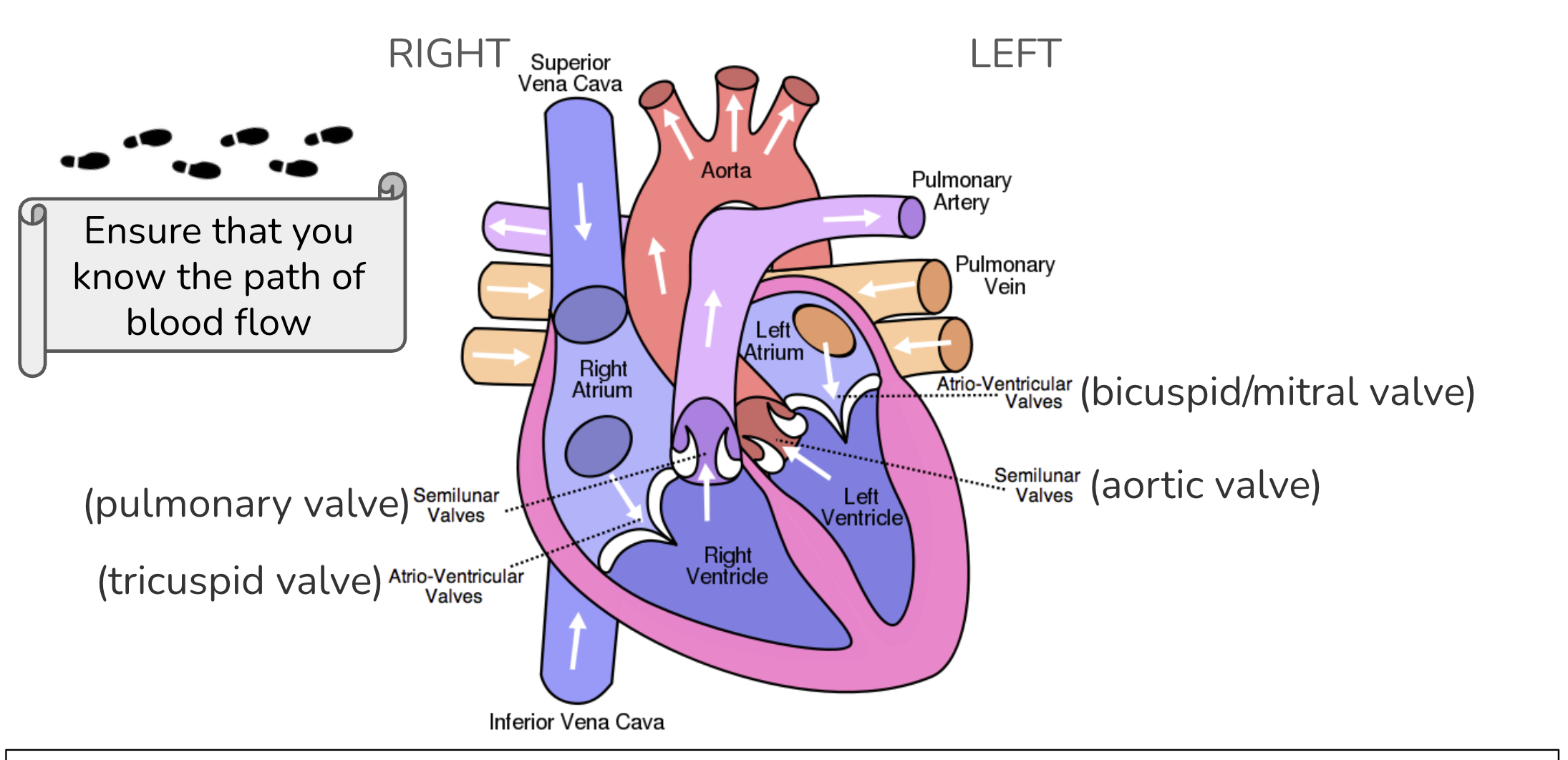
Deoxygenated
deoxygenated blood returns from body → Vena cava (superior/interior) → right atrium → tricuspid valve → Right ventricle → pulmnary valve → Pulmonary arteries → Lungs
Oxygenated
in the lungs , blood release Co2, Absorb O2
Pulmonary Veins → Left atrium → bicuspid valve → Left ventricle → Aortic valve → Aorta → Body tissues
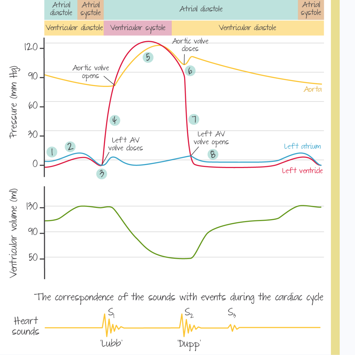
stages in cardiac cycle (3)
Atrial Systole:
Electrical impulse initiated by the Sino-atrial node (SA node) [also known as the pacemaker]
The electrical impulse are sent through the wall of the atrium and cause atrial contraction.
Volume of the atrium decreases and increases the pressure.
Pressure in the atrium is higher than the pressure in ventricle.
AV valves are open , Blood is pumped into the ventricle
Ventricle systole
Electrical impulse pass from SA node to Atrio-ventricular node (AVN).
The delay allows time for blood to transport from the atrium to the ventricles
AVN pass electrical impulse to Bundle of His, then spread through the Purkinje fibers in the ventricular wall
This cause ventricles contraction from bottom to top
Volume of the ventricle decreases and increases the pressure.
Pressure in the ventricle is higher than the pressure in the aorta and pulmonary artery.
Semi-lunar valves are forced open, and blood are pumped out through the aorta and pulmonary artery.
Pressure in the ventricle is higher than the pressure in atrium.
Atrio-ventricular valves are forced to close to prevent the backflow of blood.
Diastole
Ventricles are relaxed to allow blood to enter the atrium (Blood returns to the heart via the vena cava and pulmonary vein)
Pressure in the ventricle is lower than pressure in the aorta and pulmonary artery
Semi-lunar valves are closed to prevent the backflow of blood
Pressure in the ventricle is lower than pressure in the atrium
Atrio-ventricular valves are open
Blood flows passively into the ventricles
pressure of atrium > ventricular - Av valve opens
pressure of ventricular > pulmonary artery and aorta - SL valve open
Pressure changes during the cardiac cycle
1. Atrial contraction begins.
2. Atria eject blood into ventricles (atrial systole) .
3. Atrial systole ends; AV valves close ('lubb’).
4. Contraction of the ventricles occurs (ventricular systole) .
5. Ventricular ejection occurs.
6. Semilunar valves close ('dupp').
7. Relaxation of the ventricles occurs (ventricular diastole) .
8. AV valves open; passive ventricular filling occurs.
vascular bundle
Xylem → water+ minerals
Pholem → carbon compounds - sucrose+amino acid
xylem adaptations
Lignified walls: lignin strengthens cell walls against tension. Lignin is waterproof → walls are impermeable to water
Pits: pores where water easily moves in and out of xylem
No cell contents: allow unimpeded flow of xylem sap with minimal resistance
No End walls: allow unimpeded upwards flow of water
Transpiration
water vapour lost via stomata
facilitates
temperature regulation
absorption of water and minerals from soil
When water evaporates from cells wall during transpiration, more water is drawn from the xylem vessels to replace the loss.
Adhesion causes water molecules to stick to the cell walls, allowing them to move through the walls of the xylem and into leaf cells.
This movement due to adhesion in narrow tubes is called capillary action.
As water leaves the xylem, it creates tension / negative pressure potential within the xylem.
This tension produces a transpiration pull, draws water upwards through xylem from roots to leaves
Water is absorbed by the roots through osmosis to replace → generating higher hydrostatic pressure at the root which moves water up the xylem
Explain the process of transpiration and how xylem vessels are adapted for the
transport of water from roots to leaves. (7)
MAX 5 for transpiration (marking points a,b,c,d,e,f,g,h)
a. transpiration is the loss of water from (the surface of) the leaf / through stomata;
b. loss of water by evaporation from cell walls in leaf cells causes water to be drawn from
neighbouring/other cells;
c. lost water drawn out of the xylem/creates transpirational pull;
d. transpiration pull/tension draws water up the xylem;
e. cohesion is hydrogen bonding between water molecules;
f. cohesion (of water molecules) ensures a continuous column of water;
g. adhesion of water is (hydrogen bonding) between water and other polar molecules;
h. (adhesion is involved) in capillary action in soil/in plant cell walls/lignin;
Max 3 for xylem adaptation (for marking points i,j,k,l)
i. xylem (vessels) lack cell contents for unimpeded flow;
j. xylem (vessels) have lignified walls to withstand tensions;
k. xylem (vessels) have incomplete or absent end walls for unimpeded flow;
l. xylem (vessels) have pits/pores/gaps for entry and exit of water;
Generation of root pressure in xylem vessels by active transport of mineral ions
plants can also push water up from the roots by generating root pressure
Generated to cause water movement in roots and stems when transport in xylem due to transpiration is insufficient
For example when high humidity prevents transpiration, or before the leaves of deciduous plants develop in spring
__
Root pressure occurs as minerals are actively transported from the soil into root cells, which lowers the water potential of these cells (a lower water potential means a higher solute concentration).
As minerals enter, the water potential in the xylem decreases, and water follows by osmosis.
The entry of water into the xylem generates a positive pressure potential / hydrostatic pressure which pushes the column of water upwards—this is known as root pressure.
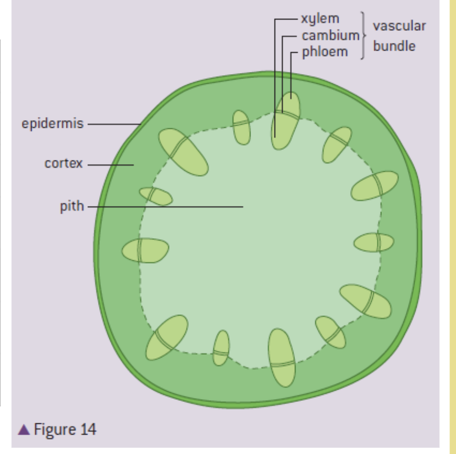
stem - draw and label and function
support, elevate leaves for seed dispersal and photosynthesis
epidermis - protection and waterproof
cortex - support and photosynthesis
pith - packing tissue ( bulking out the stem)
xylem - transport of water from roots - leaves
cambium - production of xylem&phloem tissue
phloem - transport of sugar from source to sink
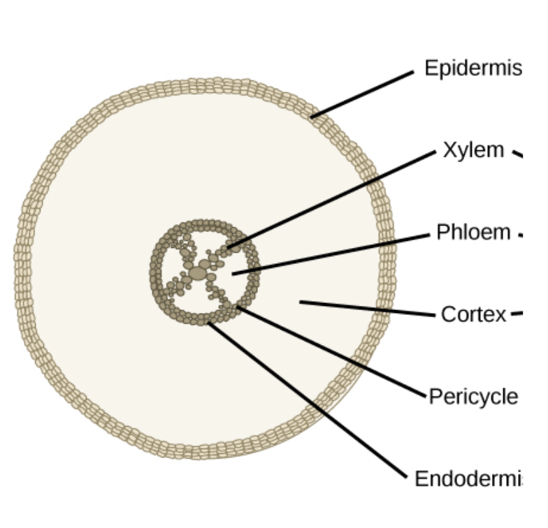
root - draw label and function
endodermis: layers of cell water pass through reach xylem
cortex: cells loosely packed - enable movement of water
epidermis: has root hair to increase water and mineral absorption
xylem: transport of water from roots to leaves
phloem: transport of sugar from source to sink
measure rate of transpiration
potometer
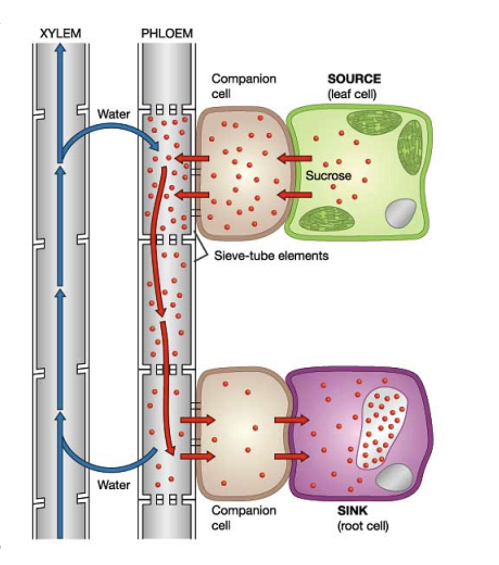
Phloem
Phloem is responsible for transporting sucrose & amino acid
Phloem is made up of the following of live cells: Companion cell and Sieve tubes.
Connections between companion cells and sieve tubes are called plasmodesmata.
As the cells are alive, it is possible for active transport to take place for translocation of nutrient to occur.
Adaptations of phloem sieve tubes and companion cells for translocation of sap
Sieve tube elements
pores in sieve plate → allows sap to flow between sieve tubes
no nucleus, reduced cytoplasm and organelles (ribosome,vacuole..) → maximises space for sap transport
Companion cell
Presence of many mitochondria → to synthesis ATP for active transport for phloem loading / unloading
Plasmodesmata
between companion cell and phloem sieve tube → exchange of material and communication between cells
Adaptations ease the flow of sap and enhance loading and unloading of carbon compounds into phloem sieve tubes
translocation
Active transport by phloem: Translocation (movement of sap)
Source: where carbon compounds are produced ; Sink: where carbon compounds are consumed
At the source, nutrients are actively transported into the companion cell, and flow through passively through plasmodesmata to the phloem sieve tube cells
This increases the solute potential, thereby causing water to enter from the xylem into the phloem sieve tube cells through osmosis (low to high solute potential)
This increases the hydrostatic pressure, thereby pushing the sap towards the sink
At the sink, nutrients actively/passively unload and reduces the solute potential, hence water returns back to the xylem, lowering hydrostatic pressure
skeleton (2)
exoskeleton → Arthropods such as spiders and insects have exoskeletons consisting of chitin that cover most of their body
endoskeleton (Vertebrates have endoskeletons consisting of bones )
Provide anchorage for muscles
act as levers for movement.
joint (2)
hinge joint ( elbow and knee)
one plant of movement
bend & straight
ball and socket joint ( hips, shoulder)
large range of movement
protraction, retraction , abduction, adduction , rotation
measure joint
goniometer
most allowing movement joint
synovial joint
ex. human hip joint
Movement at a synovial joint
Bone (Femur & Pelvis) | Cartilage | Synovial fluid | Ligaments | Muscles | Tendons |
Provide anchorage for muscles and ligaments. Guide the types of movements that can occur at a joint. | Tough, smooth tissue that covers a bone at the joint. Helps to prevent friction by preventing contact between regions of bone that might rub together. Absorbs shock. | Fills a cavity in the joint between the cartilages on the ends of the bones. Lubricates the joint, and helps prevent friction. | Connect bone to bone. Tough cords of tissue containing large quantities of collagen (protein). Prevent movements that would cause dislocation. | Provide forces that cause movement at the joint. | Attach muscle to bone. Composed of living tissue with large quantities of collagen. Allow forces to be transmitted between muscle and bone. |
Anagonistic muscle action to facilitate internal body movements
External and internal intercostal muscle fibres oriented differently - meaning contractions pull the rib cage in opposite directions
External intercostals
Contraction pulls the rib cage up and out - aiding inhalation - and stretches the internal intercostal muscles
Internal intercostals
Contraction pulls rib cage in and down - aiding exhalation - and stretches the external intercostal muscles
skeletal muscle
attach bones - cause movement of animal body
It consists of large multinucleated cells called muscle fibers.
There are also mitochondria between the myofibrils.
level of organisation
muscle fibres → myofibris → microfillaments → sacromere
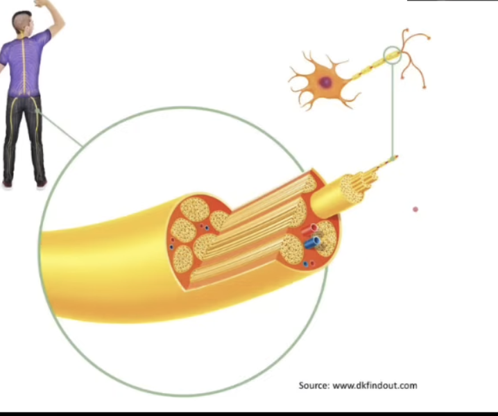
Structure and function of motor units in skeletal muscle
Around the myofibrils is a specialized type of endoplasmic reticulum – the sarcoplasmic reticulum.
Skeletal muscles are voluntary muscles that requires electrical impulse from the brain.
Electrical impulse are sent from the brain through the motor neuron to the neuromuscular junction.
Each motor neuron has a set number of muscle fibers that it control called a motor unit.
Motor units
Contraction of skeletal muscle is coordinated by motor units
A motor unit comprises a single motor neuron
and all of the muscle fibers that it stimulates via neuromuscular junctions
The muscle fibres contract when stimulated by the motor neuron
The stimulus passes from the neuron to the muscle fibre at a synapse called the neuromuscular junction
require neurotransmitter: acetylcholine
Sarcomere and Muscle contraction
A sarcomere is a subunit of a myofibril.
Between two Z lines is one unit of sarcomere.
two protein filaments : Myosin & Actin
myosin
Light bands are represented by thin actin filaments only, which are attached to either end of the Z lines.
actin
Dark band represent the region containing thick myosin filament, which contains heads that form cross-bridges by binding to the actin.
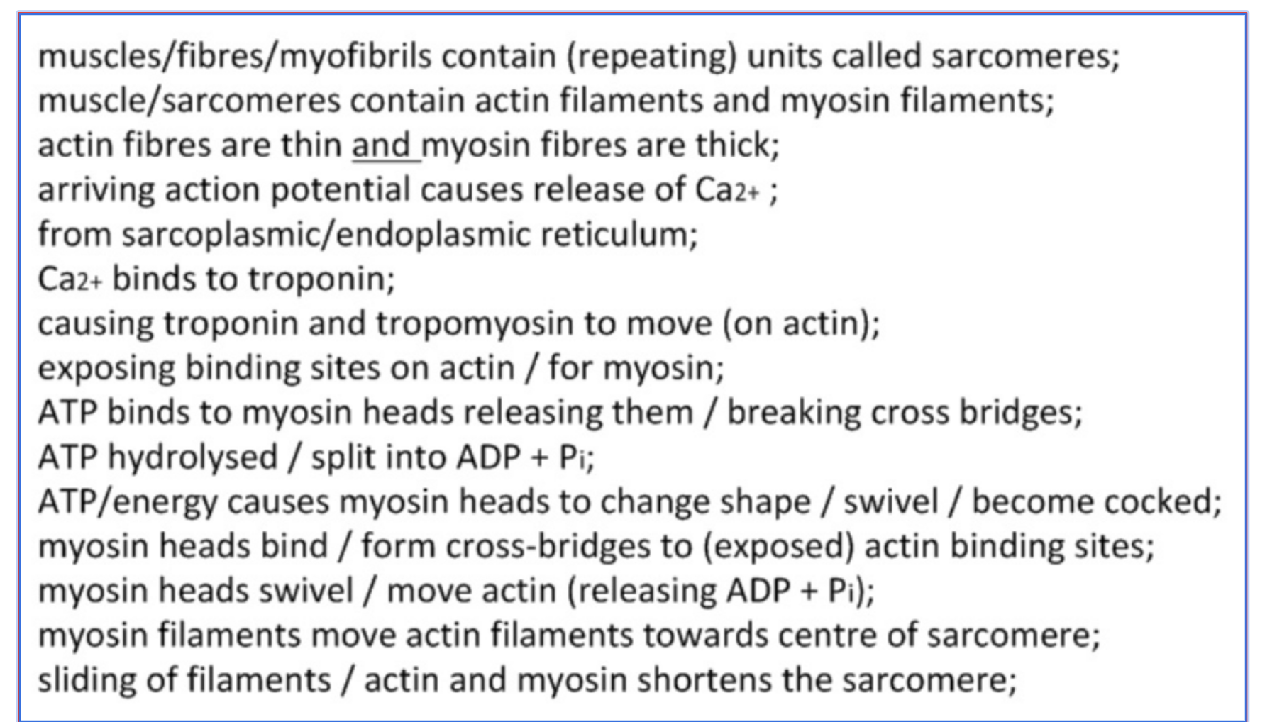
crose bridge cycle
When a nerve impulse arrives at the neuromuscular junction, Ca2+ ions are released from the sarcoplasmic reticulum
Ca2+ ions bind with troponin, causing it to change shape, moving tropomyosin to expose the myosin-binding site on actin
Myosin heads( hydrolyses ATP to ADP + Pi and stored that energy in its conformation, The myosin head is cocked) → bind to actin, forming crossbridges
Myosin releases ADP and Pi, causing the power stroke which pulls the actin filament towards the centre of the sarcomere
ATP binds to myosin → myosin detach from actin → breaking the crossbridges
ATP is hydrolysed to ADP and Pi → The energy released from hydrolysis is used to “recock” the myosin head (into its high‑energy conformation), ready to bind further along the actin filament toward the Z line.
Myosin heads bind to actin at a new binding site further along the sarcomere
The cycle continues until Ca2+ is pumped back into the sarcoplasmic reticulum, or there is no ATP available
titin
contraction of antagonistic muscle → creates energy
Energy is needed to lengthen a muscle, which stretches titin
Titin then releases energy as it recoils, adding to the force of contraction in that muscle
prevent overstretching of sacromere
holds myosin filaments in place
Explain how a skeletal muscle contracts
pathway of nerve impulse
Stimulus (e.g., heat/pain) is detected by a receptor (e.g., thermoreceptor)
which generates a nerve impulse;
The impulse travels along the sensory neuron (afferent) to the spinal cord/inter (relay) neuron/Central Nervous System (CNS);
The interneuron synapses with a motor neuron (efferent) which carries the impulse away from the CNS;
The motor neuron transmits the impulse to the effector muscle (e.g., biceps), causing it to contract/bring about the response;
Definitions
Tropism: the turning of all or part of an organism in a particular direction in response to an external stimulus.
Synergism - work together to stimulate a process
Antagonism - have opposing effects to regulate a process
Positive feedback: the amplification of a body’s response to a stimulus
Circadian rhythm: Pattern of sleep cycles that organisms are adapted to
Peristalsis: Muscle contraction that moves food through the digestive tract
System integration
This is a necessary process in living systems. Coordination is needed for component parts of a system to collectively perform an overall function
responsible for emergent properties.
communication/coordination 1. hormone 2. nervous signalling
Transport of materials
emergent properties
a property that is only present when parts of a system work together
ex. Cheetah as predators
Flexible spine: acts as spring during running; increases stride length
Longer hind limb bones - increases stride length
Grooves on claw pads to aid grip
All of these adaptations together make the cheetah a great predator!
Integration in terms of communication
Nervous system | Hormones: |
Nervous Systems Transmitted through neurons | Endocrine system Transmitted through the bloodstream |
Electrical impulse as messenger | Hormone as a messenger |
Quick in conduction | Slow in conduction(binds on receptor in target cells) |
Short duration of effect | Long duration of effect |
Controls both voluntary and involuntary functions (autonomic nervous system) | Controls involuntary functions |
E.g. Sympathetic(SNS) and Parasympathetic(PNS) nervous system → to control heart rate, adrenaline to control fight-or-flight response.
The brain as a central information integration organ
Receive info
Inputs (Afferent Neurons eg. sensory): The brain receives data from the outside world (sensory) and from inside the body (pain, temperature).
Stores info + Processes info
Process & Store: The brain interprets these signals, makes decisions, and stores memories.
Send signal → makes a decision + coordinates life processes
Outputs (Efferent Neurons eg. motor neurons): The brain sends commands back to Effector organs eg. muscles or glands to take action
Afferent and Efferent
Afferent : Carry signal towards CNS
Efferent : Carry signal away from CNS → initiate actions
Brain structure
Cerebrum is divided into two hemispheres.
The largest part of the brain composed of two halves known as the cerebral hemispheres.
Involved in controlling vision, thinking, learning, emotions as well as voluntary control of the body–collectively referred to as → advanced mental activity.
Cerebellum is divided into two hemispheres
and is responsible for: voluntary movement, coordination, and balance → movement
Brainstem is divided into
midbrain, pons, and medulla (involuntary activities)
Parts of cerebrum
The cerebrum contains many different parts:
Corpus callosum - a band that connects the two cerebral hemispheres
Frontal lobe - important for expressive language and higher-level functions such as learning.
Temporal lobe - this processes auditory information
Parietal lobe – important for processing somatosensory input (e.g. touch)
Occipital lobe - located at the back of the cerebrum this is known as the visual cortex
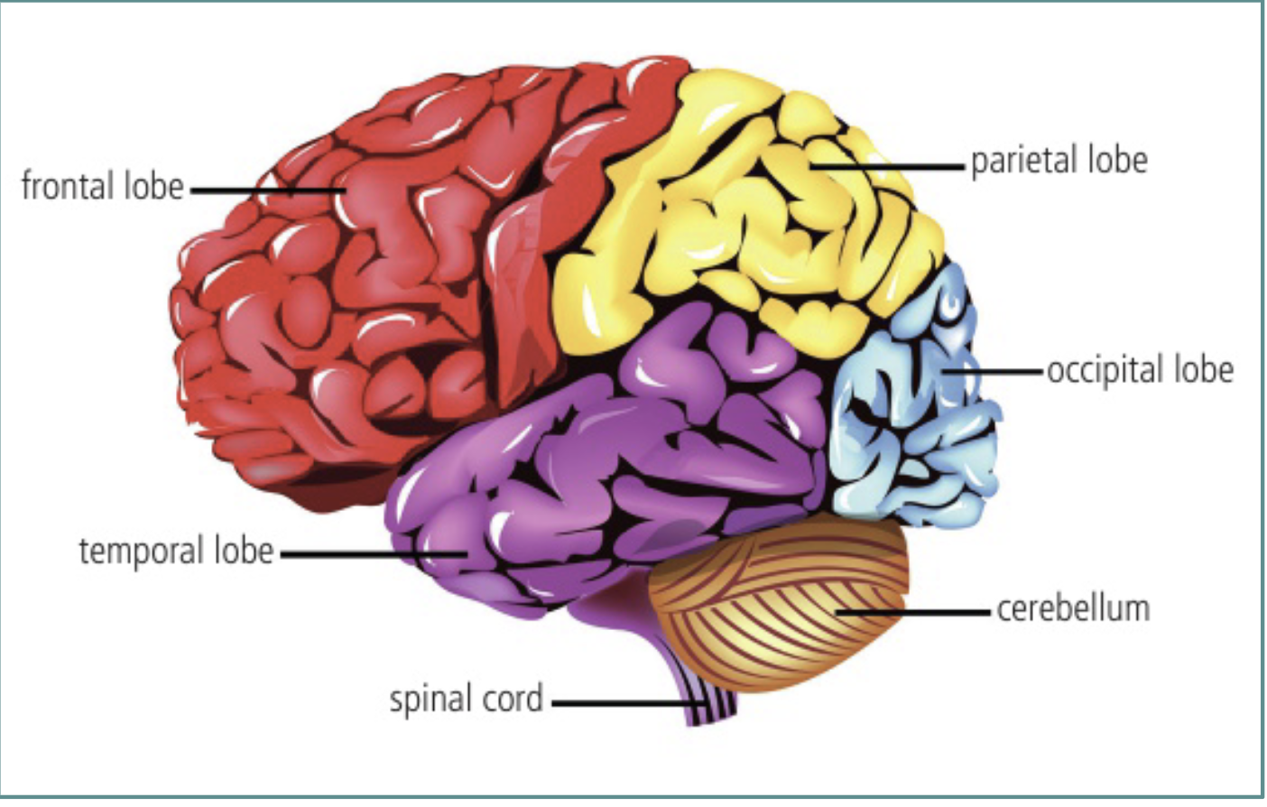
The brain and information processing - Cerebellum & Brain stem with medulla oblongata & hypothalamus & Pituitary gland
Cerebellum - located underneath the cerebrum, this plays an important role in coordinating muscle movements as well as balance.
—
Brainstem containing Medulla oblongata - located at the base of the brain, this controls many vital body processes such as breathing, heart rate, and blood pressure. → involuntary
—
Hypothalamus - found just beneath the middle part of the brain, this is involved in thermoregulation as well as the production of hormones that are involved in the control of the pituitary gland. ex. dopamine, ADH, oxytocin, GnRH
—
Pituitary gland - located on the underside of the brain and attached to the hypothalamus to secrete various hormones, such as oxytocin and FSH.
Nervous system includes
CNS
Neuron
→ CNS ( central nervous system )
brain → process complex sensory input, initiate motor actions, handle high order thinking(thoughts, emotion)
spinal cord → transmits signal between brain → body + involuntary action
→ PNS (peripheral nervous system)
Nerves
The spinal cord as an integrating centre for unconscious processes
Spinal cord as part of the central nervous system (CNS)
controls some of the unconscious reflexes associated with balance and other skeletal muscle functions that are not controlled by the brain.
The spinal cord mediates information between the brain and PNS.
It integrates information from unconscious processes only.
____
Two main tissues :
1. white matter
Transmit signal (transport)
Receive from sensory receptors ( Sensory receptors > Brain)
Transmit to other organs ( Brain > Organs )
composed on bundle of myelinated axons → carries electrical impulse to and from the brain.
Grey matter (neurons, unconscoscious processing)
contains cell body of motor neurons, relay neurons, and synapses
Process information + decision making for UNCONSCIOUS PROCESSING
ex. movement of digestive system
Input to the CNS through sensory neurons
Sensory receptors (detect external/internal environement) → Sensory neurons → CNS( process information) → Motor neuron → Effector organs
Sensory receptors
External
Touch, heat, light
Internal
stretch receptors, chemoreceptors
Output from the cerebral hemispheres to muscles through motor neurons
in betweem cerebral hemispheres → Motor cortex
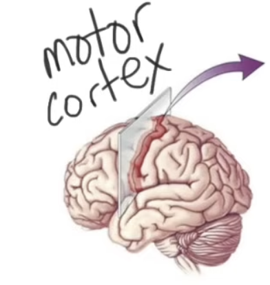
contains cell body of motor neuron → axon and terminal extend to different effector organs
Spinal cord as part of the central nervous system (CNS) controls some of the unconscious reflexes associated with balance
and other skeletal muscle functions that are not controlled by the brain.
Nerves & Neuron
Neurons are the nerve cells
They have long nerve fibres (axons) which may be myelinated or unmyelinated
Nerves are bundle of nerve fibre surrounded in a sheath
Most nerves contain fibres of both sensory and motor neurons
Pain reflex arcs as an example of involuntary responses with skeletal muscle as the effector
stimulus → sensory receptor → sensory neuron → CNS → motor neuron → Effector organs → Response
Receptors: Carries out transduction (the conversion of physical stimulus into electrical signal)
Sensory neuron: Carries electrical impulse to CNS
CNS - interneuron (relay neuron): Carries an electrical impulse to a specific motor neuron.
Motor neuron: Carries an electrical impulse to the effector organ
Effector: Can be either a muscle or a gland → carry out a response
Role of the cerebellum in
→ coordinating skeletal muscle contraction and balance
The cerebellum receives information from the cerebrum, brainstem and spinal cord.
The initiation of body movement is by the motor cortex of the cerebrum.
Movement begins : cerebellum receives feedback impulses from various area of the body.
Then sent out impulses to coordinate the movement and the timing.
Movement include
coordinate muscle contraction timing
balance
posture
things that require muscle memory
Hormones
Endocrine system
Endocrine glands secrete hormones directly into the bloodstream to cause changes in the body
Control of the endocrine system by the hypothalamus and pituitary gland
→ Hypothalamus attach to pituitary gland
→ links nervous system to hormonal system
The hypothalamus can respond to input signals by inhibiting or stimulating the pituitary gland.
The hypothalamus has many specialized groups of cells called nuclei.
contain sensors for blood temperature, osmolarity, or receive information from sense organs, e.g. the eyes
The nuclei in the hypothalamus control the release of hormones from the pituitary gland.
eg. Osmoregulation
Hypothalamus detects dehydration → promopts pituitary to releases ADH→ stimulates reabsorption of water
eg. Puberty
Hypothalamus releases GnRH (hormone) → stimulate pituitary gland releases LH, FSH
Modulation of sleep patterns by melatonin secretion as a part of circadian rhythms
Circadian rhythm: Pattern of sleep cycles that organisms are adapted to
Melatonin → secreted by pineal glands
Circadian rhythm set by special groups of cells in the hypothalamus called the suprachiasmatic nuclei (SCN)
control the secretion of the hormone melatonin from the pineal gland
Modulation of sleep patterns by melatonin secretion as a part of circadian rhythms
Light inhibits the production of melatonin.
Light receptor → CNS → Pineal gland
integrated by sensory neuron in eyes
Causes drop in temperature, drowsiness, sleep
Melatonin secretion decreases with age → sleep patterns become more irregular as we grow older.
The body’s circadian rhythms are disrupted by traveling rapidly between time zones → Jet lag
Epinephrine (adrenaline) secretion by the adrenal glands to prepare the body for vigorous activity
Adrenaline is responsible for flight-or-fight response / vigorous activity
Increase glucose and oxygen supply to skeletal muscle
Increase heart rate ( SA Node ) and blood pressure
Increase blood flow to liver and muscles (vasodilation)
Decrease blood flow to guts and kidneys (vasoconstriction) → not essential during emergency
Pupil dilation
Prepare body for vigorous, immediate response with intense muscle contractions.
Secreted by the adrenal glands.
Feedback control
Feedback control of heart rate - sensory inputs
The heart rate can be affected by hormones (e.g. adrenaline) and nervous control:
The medulla ( cardiovascular control centre) controls the sino-atrial node (SAN) via nerves.
The sympathetic nerve speeds up the heart rate in response to a decrease in pH in the blood due to CO2 rising
Impulses carried by the vagus (parasympathetic) nerve slow down the heart rate when the concentration of CO2 decreases, and pH increases
__
Nervous control of HR is coordinated by the medulla
Actioned by the vagus nerve (parasympathetic) and the sympathetic nerve.
Can be overridden by Adrenaline → stimulates the sinoatrial node to increase heart rate.
Feedback control of heart rate - baroreceptors and chemoreceptors
The structure that sets your heart rate → the pacemaker of the heart
This structure can adjust heart rate based on conditions within the body, which are sensed by
Baroreceptors: sense changes in blood pressure in aorta
Chemoreceptors: sense changes in pH (Co2, O2)
Located in the aortic arch and the branches of the carotid arteries.
Nerves carry signals from these receptors to the medulla in the brain
Receptors in vessels → sensory nerves → medulla → sympathetic/parasympathetic nerves → SAN and heart.
Feedback control of heart rate and stroke volume - increase in heart rate
During exercise, respiration rate and blood pressure increases, the wall of the artery is stretched and detected by the baroreceptors.
O2 decrease, CO2 increase, pH decrease, these are detected by the chemoreceptors.
These result in an increased rate of action potential sent to the medulla.
Medulla will respond by sending more impulses, through the sympathetic nerve, to the SAN → increase the heart rate and the force of contraction.
This will increase the cardiac output.
Feedback control of heart rate and stroke volume - decrease in heart rate
After exercise, respiration rate and blood pressure decrease, the wall of the artery is recoiled and detected by the baroreceptors.
O2 increase, CO2 decrease, pH increase, these are detected by the chemoreceptors.
These result in an decrease rate of action potential sent to the medulla.
Medulla will respond → sending more impulses to the SAN through the parasympathetic nerve to decrease the heart rate and the force of contraction.
This will decrease the cardiac output.
Feedback control of ventilation rate
During exercise, chemoreceptors in the brainstem detect a drop in pH, caused by increased CO2 level in the blood. (refer to transport in blood)
Chemoreceptor → send nerve impulses → respiratory center in the medulla.
Respiratory center then sends impulses intercostal muscles and diaphragm, causing them to contract harder and faster.
Control of peristalsis in the digestive system by the central nervous system and enteric nervous system
Peristalsis: Muscle contraction that moves food through the digestive tract
Peristalsis include voluntary and involuntary movement
voluntary: CNS(brain&spinal cord)
Inititation of swallowing
Egestion
Involuntary : ENS( enteric nervous system)
coordinating movement in the gut
HL
Tropism:
the turning of all or part of an organism in a particular direction in response to an external stimulus.
eg. phototrophism , hydrotrophism , gravitropism
Positive: Towards stimulus
Negative: Away stimulus
Phototrophism
Shoot bends towards light and shows positive phototropism.
As for roots, it bends away from light and shows negative phototropism.
Plants require sunlight and water to carry out photosynthesis
Tropic movements mean plants are able to meet these requirements
Stem of the plant has positive phototropism
Roots of the plant have positive hydrotropism
Phytochromes
Phytochromes are plant hormones that regulate physiological processes in plants.
Transported in the xylem and phloem to specific regions
Functions:
signal molecules to control growth
development of flowers, fruits and seeds
help the plant to respond to environmental stimuli.
Phytohormones
Auxins | Cytokinins | Gibberellins | Abscisic acid(ABA) | Ethylene |
Growth hormone Produced in shoot apical meristem (tip) Cell elongation for tropic movements Inhibit growth of lateral buds (causes vertical elongation) | Promote cell division Abundant in growing tissues Produced in the roots and pass to leaves and fruits Promote cell division and differentiation of the meristem | Group of hormones Plant growth Produced in apical meristem of roots and shoots Elongation of shoot Seed germination Flower maturation Breaking seed dormancy Delaying ageing | Inhibits elongation of stems Induces dormancy in seeds (seeds fail to germate even in ideal conditions) Involved in the dropping of leaves* | Gas produced by ageing tissues Causes leaves, fruits and flowers to drop Role in fruit ripening |
ABA: When water is scarce, plants synthesize more ABA, which travels to leaf guard cells to induce stomatal closure, significantly reducing transpirational water loss.
Auxin
grow at the tip of stem
stimulate cell elongation
stimulate growth of plant
→ negative phototropism, auxin move away from light stimulus
Auxin : Light over head & Light on one side
Light overhead
Auxin produced at the tip diffuses down the stem evenly
Auxin evenly distributed
All cells grow at the same rate
Shoot grows The shootvertically upwards
Light source to one side
Auxin molecules move towards the shaded side of the shoot, away from the light
Increased concentration of auxin on the shaded side
causes rapid cell elongation and growth on that side
Uneven growth causes the stem to bend towards the light source
Polar auxin transport
Transport of auxin is directional
Active directional cell-to-cell movement
Entry into a cell: Passively or via auxin influx carriers (proteins in membrane)
Exit from a cell: Auxin efflux pumps (proteins in membrane)
Phototropism controlled by Auxin – Auxin gradient
Auxin is produced at the apex (tips) of the shoot.
When light in the shoot is detected → they trigger movements of auxins by active transport carried out by auxin efflux pumps (carriers).
Efflux pump pumps auxin from the cytoplasm out into the cell wall, then diffuses to the next cell.
Once it enters the cell, the auxin is trapped inside the cytoplasm until the efflux pump pumps it out again.
Auxin efflux pumps are moved in response to the differences in light intensity, creating a concentration gradient of auxin from:
lower on the lighted side and higher in the shaded side.
Phototropism controlled by Auxin – Elongation of cell
Plant cells contain auxin receptor, when auxin binds, transcription of the genes for proton pump is promoted.
The expression of these genes causes the secretion of hydrogen ions into the cell wall.
The hydrogen bonds between the cellulose will be weakened and loosens the cell wall.
Allowing expansion of cell due the increase water uptake and higher turgor pressure.
Integration of root and shoot growth
Auxin is produced in the shoot and cytokinin is produced in the root.
Both areas are growing regions of the plant.
Auxin is responsible for cell elongation and cytokinin is responsible for cell division.
Both phytochromes needs to be transported to the opposing growth regions to regulate the growth of all parts of the plant and integrate both signals.
Cytokinin is transported through xylem up the plant and auxin is transported through phloem down the plant.
Root and shoot growth work together for cell growth
Together, the phytohormones work on meristems ( rapidly growing tissues made of undifferentiated cells) to integrate cell growth
The ratio of the two determines whether it results in:
Synergism - work together to stimulate a process
Antagonism - have opposing effects to regulate a process
Feedback control of fruit ripening
Positive feedback: the amplification of a body’s response to a stimulus
Ethylene (Ethene) is produced in ripping fruits.
Ones ripping process started, the fruit will produce more ethene.
When one fruit started to produce ethene, it will cause the surrounding fruit to ripen and produce even more ethene.
This helps fruits to become more attractive to herbivores therefore increasing the seed dispersal rate in their corresponding reproductive season.
Describe how an impulse passes from the relay neuron to the motor neuron. [3]
A. impulse causes relay neuron to release of neurotransmitter into synapse;
B. neurotransmitter diffuses across the synapse and binds to its receptor on the motor neuron;
C. causing Na+ (voltage-gated) channels to open;
D. new impulse generated in motor neuron;
E. if threshold is reached;
Definitions
Ligand: Molecules that bind reversibly to specific proteins.
Receptor: The protein to which a ligand binds.
Signal transduction pathways: A series of binding between various ligands and receptors that helps transducing the signals over varying distance between or within a cell and end with a response.
Quorum sensing - a mechanism by which bacteria can alter group behavior depending on population density.
Cytokines - small proteins involved in immune response
Transmembrane receptors: receptors that are embedded in the cell membrane.
Intracellular receptors: receptors that are within the cell cytoplasm
GPCR : Multi-pass transmembrane protein receptor
Acetylcholine receptor : Example of chemically gated ion channel receptor & Multi-pass protein: Composed of many domains that thread back and forth across the cell membrane several times
Tyrosine kinase ( insulin receptor) : A pair of single pass proteins with 3 domains
Kinases : enzymes that use a phosphate group from ATP to phosphorylate a specific molecule
Positive feedback : Amplifies cell signalling to enhance or reinforce a response
Negative feedback : Dampens cell signalling to prevent over-activation of a pathway
Ligand in Chemical Signalling
Ligand: Molecules that bind reversibly to specific proteins.
Receptor: The protein to which a ligand binds.
Signal transduction pathways: A series of binding between various ligands and receptors that helps transducing the signals over varying distance between or within a cell & end with a response.