Head & Neck IX: Ear
1/44
There's no tags or description
Looks like no tags are added yet.
Name | Mastery | Learn | Test | Matching | Spaced | Call with Kai |
|---|
No analytics yet
Send a link to your students to track their progress
45 Terms
inner ear: components/function
contains the apparatus for both auditory and vestibular functions
middle ear: function
enhance the auditory functions of the inner ear by aiding in the transfer of sound waves to the inner ear
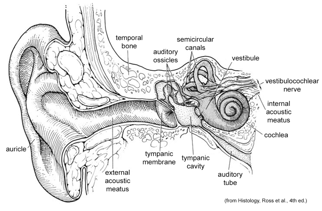
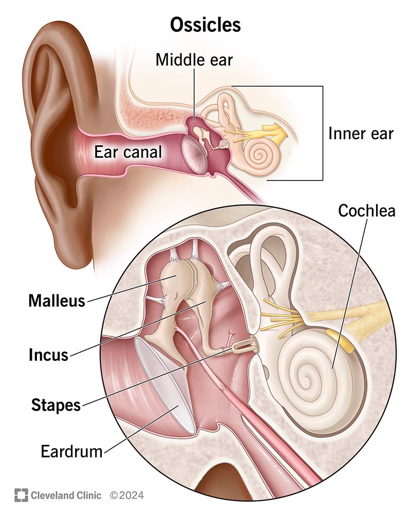
external ear: main components
auricle (pinna)
external acoustic meatus
where are the specialized receptors for sound located?
in the (fluid-filled) inner ear
airborne sound waves must compensate in the process for the loss in sound energy that occurs as sound waves pass from air into water. How is this done?
the apparatus of the external ear concentrates the energy of the airborne sound wave so that the liquid of the inner ear can be set in motion
auricle (pinna): description + function
elastic cartilage plate continuous with the wall of the external acoustic meatus. functions to collect sound waves and channels them down the external ear canal, concentrating high frequencies and localizing sound (via phase and intensity differences)
external acoustic meatus: description + function
lateral 1/3: cartilaginous and contains glands. functions to maintain constant temperature and humidity + prevent airborne particles from injuring tympanic membrane
medial 2/3: bony, has skin covering tympanic membrane. narrowest component of the external ear canal. functions as sound resonator for speech frequencies

tympanic membrane: function/how it works
closes the medial end of the external acoustic meatus; moves in response to air vibrations that pass through the external acoustic meatus.

why are surgical incisions of the tympanic membrane made in the posteroinferior portion?
to avoid the chorda tympani nerve, auditory ossicles, and blood vessels present in the superior portion
middle ear (tympanic cavity): location
located in the petrous portion of the temporal bone
middle ear (tympanic cavity): components & relationships
contains air, nerves, ossicles, and two muscles. the auditory tube connects the middle ear anteriorly with the nasopharynx.

roof of the tympanic cavity
separate the tympanic cavity from the dura mater and temporal lobe of the brain

floor of the tympanic cavity
separates tympanic cavity from the internal jugular vein

lateral wall of the tympanic cavity
tympanic membrane

medial wall of the tympanic cavity
formed by the basal coil of the cochlea
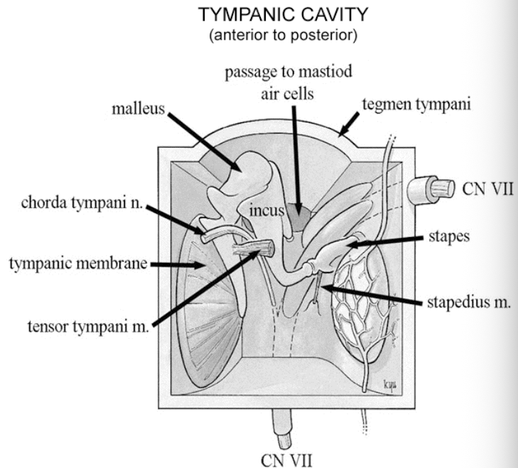
anterior wall of the tympanic cavity
contains opening for tensor tympani muscle, auditory tube, and exit of the chorda tympani nerve

posterior wall of tympanic cavity
contains openings to mastoid air cells and an aperture for the entrance of the chorda tympani nerve. forms attachment for stapedius muscle and opening for the nerve to stapedius (CN VII).

stapedius: function
dampens down the movement of the stapes in response to loud sounds
what must occur for the membrane to be free to move as sound waves strike it?
the resting air pressure on both sides of the tympanic membrane must be equal
the outside of the eardrum is exposed to atmospheric pressure that reaches it through the ___. the inside of the membrane is exposed to the atmospheric pressure via the ___, which connects the ____ to the ___.
ear canal, auditory tube, middle ear, nasopharynx
the cartilaginous portion of the tube normally remains (open/closed) except during ___.
closed; swallowing or yawning
opening the auditory tube involves which muscles?
tensor veli palatini and salpingopharyngeous muscles
what 3 small bones form a system of levers, which facilitate the transfer of vibratory movements of the tympanic membrane to the fluid of the inner ear at the oval window?
what are these collectively called?
auditory ossicles: malleus, incus, and stapes
the head of this bone articulates with the body of the incus
malleus
incus
stapes
malleus
what bone attaches to the posterior wall of the tympanic cavity and articulates with the head of the stapes
malleus
incus
stapes
incus

this bone fits into the oval window on the medial wall of the tympanic cavity
malleus
incus
stapes
stapes
what 2 mechanisms amplify the pressure of the airborne sound waves?
surface area of the tympanic membrane is larger than that of the oval window
the lever action of the ossicles provides additional mechanical advantage
surface area of tympanic membrane and lever action of ossicles provide an overall increase in force by approximately __ fold and
20; decreases amplitude
what two muscles reduce the movements of the auditory ossicles and tympanic membrane?
stapedius muscle (stapes) and tensor tympani muscle (malleus)
how do the stapedius muscle and tensor tympani muscle reduce loud sounds?
they decrease the range of movement of the tympanic membrane
the membranous labyrinth contains () and the end organs for (). the space between the bony membranous labyrinths contains ().
endolymph; hearing and balance; perilymph

this structure is the part of the labyrinth (of the inner ear) that is specialized for transducing sound waves into neural activity
cochlea
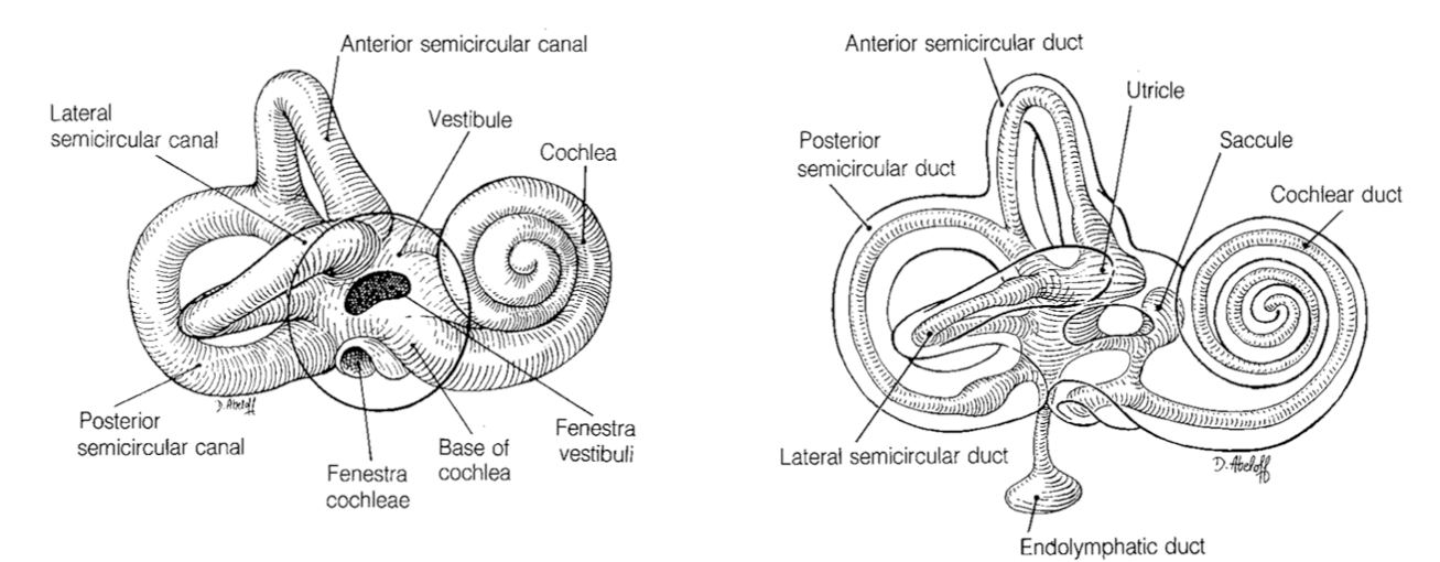

the cochlea is further divided by the membranous labyrinth into what three chambers?
scala vestibuli, scala media, and scala tympani


this bony chamber is filled with perilymph and is continuous with the scala tympani through the heliotrema at the apex of the cohlea.
scala vestibuli
where does the scala vestibuli open into?
into the vestibule of the bony labyrinth, where perilymph is freely exchanged between teh vesibule.
the stapes closes
the oval window (opening into the middle ear cavity)

this chamber froms a spiral, blind tube filled with endolymph and is firmly fixed to the internal and external walls of the cochlear canal.
scala media

what forms the roof and floor of the scala media?
vestibular membrane and basilar membrane

what organ contains the receptors for hearing? where is it situated?
Organ of Corti (spiral organ); situated on the basilar membrane
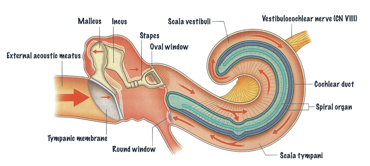
this bony chamber is filled with perilymph and is continuous with the scala vestibuli.
scala tympani
the secondary tympanic membrane closes
the round window (opening into the middle ear cavity)

significance of basilar membrane
when deflected by vibrations produced by the movement of the stapes, the hair-like projections of the sensory hair cells against the tectorial membrane bend. This causes the hair cells of the organ of Corti to be depolarized.
innervation of hair cells in organ of Corti
facial nerve
the central processes of the sensory neurons of the hair cells pass into the brain as the cochlear part of what nerve?
vestibulocochlear nerve
what results in sensorineural deafness?
damage of the hair cells in organ of Corti, spiral organ neurons, or CN VIII