Exam 2 BIOS 252 Chamberlain University
1/87
There's no tags or description
Looks like no tags are added yet.
Name | Mastery | Learn | Test | Matching | Spaced | Call with Kai |
|---|
No analytics yet
Send a link to your students to track their progress
88 Terms
Rostral
towards the forehead
Caudal
toward the tail or spinal cord
reticular formation
a nerve network in the brainstem that plays an important role in controlling arousal
reticular activating system
Located in the upper brain stem; responsible for maintenance of consciousness, specifically one's level of arousal.
Cerebellum function
the "little brain" at the rear of the brainstem; functions include processing sensory input and coordinating movement output and balance BALANCE
Hypothalamus function
A neural structure lying below the thalamus; it directs several maintenance activities (eating, drinking, body temperature), helps govern the endocrine system via the pituitary gland, and is linked to emotion and reward.
4 regions of hypothalamus
mammillary, tuberal, supraoptic, preoptic
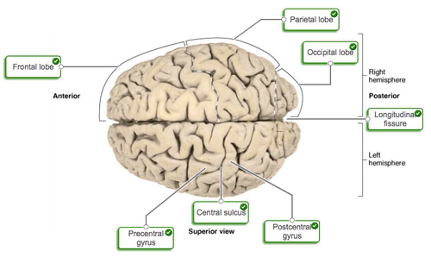
frontal lobe
The lobe at the front of the brain associated with movement, speech, and impulsive behavior. (primary motor lobe)
parietal lobe
receives sensory input for touch and body position (primary sensory area)
occipital lobe
visual cortex
insula
regions of cortex located at the junction of the frontal and temporal lobes
Thalamus
the brain's sensory control center, located on top of the brainstem; it directs messages to the sensory receiving areas in the cortex and transmits replies to the cerebellum and medulla (RELAY STATION BY OCCIPITAL LOBE)
brain
gray matter on the outside, white matter on the inside
spinal cord
gray matter on the inside, white matter on the outside
Dreams
a sequence of images, emotions, and thoughts passing through a sleeping person's mind (REM SLEEP)
Wernicke's area
controls language reception - a brain area involved in language comprehension and expression; usually in the left temporal lobe. Transmits speech to Broca's Area
Broca's area
Controls language expression - an area of the frontal lobe, usually in the left hemisphere, that directs the muscle movements involved in speech.
12 pair of cranial nerves
Attach directly to the brain and can be motor, sensory, or both
sensory cranial nerves
1, 2, 8 (I,II,VII)
motor cranial nerves
3, 4, 6, 11, 12 (III,IV,VI,XI,XII)
oculomotor, trochlear, abducens, accessory, hypoglossal
mixed cranial nerves
5, 7, 9, 10 (VVII,IX,X)
5 branches of facial nerves
temporal, zygomatic, buccal, mandibular, cervical
cranial nerve goes to innervate abdomen and thorax
visceral nerve
Divisions of the ANS
sympathetic and parasympathetic
sympathetic nervous system
the division of the autonomic nervous system that arouses the body, mobilizing its energy in stressful situations, fight or flight
parasympathetic nervous system
rest and digest; the division of the autonomic nervous system that calms the body, conserving its energy
adrenal cortex
the outer portion of the adrenal glands
adrenal medulla
secretes epinephrine and norepinephrine
Acetylcholine (ACh)
enables muscle action, learning, and memory
secreted by all preganglionic neurons in both divisions and by postganglionic parasympathetic nervous system.
Norepinephrine
secreted by all sympathetic postganglionic neurons
Alpha
Excitatory
Beta
inhibitory
Central control of autonomic functions
ANS regulated by levels of CNS
cerebral cortex
fear and anxiety
Hypothalamus
A neural structure lying below the thalamus; it directs several maintenance activities (eating, drinking, body temperature), helps govern the endocrine system via the pituitary gland, and is linked to emotion and reward.
spinal cord functions
defecation and micturation
cervical enlargement of spinal cord
Responsible for supplying nerves to the upper limb
lumbarenlargement
nerves to pelvic region and lower limbs
medullary cone
cord tapers to a point inferior to lumbar enlargement
cauda equina
collection of spinal nerves below the end of the spinal cord (L2 to S5)
Meninges
three protective membranes that surround the brain and spinal cord
Meninges layers (from superficial to deep)
dura mater, arachnoid mater, pia mater
cerebrospinal fluid
Fluid in the space between the meninges that acts as a shock absorber that protects the central nervous system. (in the subarachnoid space b/w arachnoid and pia layers)
gray matter
Brain and spinal cord tissue that appears gray with the naked eye; consists mainly of neuronal cell bodies (nuclei) and lacks myelinated axons.
gray matter function
site of information processing, synaptic integration
white matter function
abundantly myelinated axons that course up and down the cord providing communication between different levels of the CNS
funiculi
white matter columns on each side of the cord; 3 on each side
ascending tracts
carry sensory information up the spinal cord
descending tracts
carry motor commands down the spinal cord
Endoneurium
connective tissue external to neurilemma
perineurium
layers of squamous cells that wrap fascicles
Epineurium
connective tissue surrounding the entire nerve
Ganglion
A cluster of nerve cell bodies, often of similar function, located in the PNS.
spinal nerves
31 pairs of nerves arising from the spinal cord
cervical nerves
C1-C8 (8 pairs)
thoracic nerves
T1-T12 (12 pairs)
lumbar nerves
L1-L5 (5 pairs)
sacral nerves
S1-S5 (5 pairs)
coccygeal nerve
Co1 (1 pair)
dermatome
Area of skin supplied by a single spinal nerve
dermatome map
a diagram of the cutaneous regions innervated by each spinal nerve
Reflexes
quick, involuntary, stereotyped reactions of glands or muscle to stimulation
somatic receptors
in skin, muscles, or tendons
afferent nerve fibers
carry information from receptors to posterior horn of spinal cord or the brainstem
integrating center
A point of synaptic contact between neurons in gray matter of spinal cord or brainstem
Determines whether efferent neurons issue signal to muscles
efferent nerve fibers
carry motor impulses to skeletal muscle
Effectors
muscles or glands that carry out the response
stretch reflex
the contraction of a muscle in response to stretch of that muscle
patellar reflex
a reflex extension of the leg resulting from a sharp tap on the patellar tendon; one synapse between the afferent and efferent neurons
flexor reflex
the quick contraction of flexor muscles resulting in the withdrawal of a limb from an injurious stimulus
polysynaptic reflex
signals travel over many synapses on their way to the muscle
presynaptic neurotransmitter
acetylcholine
postsynaptic neurotransmitter
GABA
longitudinal fissure
separates cerebral hemispheres
Gyri
ridges of the brain
corpus callosum
a broad band of nerve fibers joining the two hemispheres of the brain.
Cerebrum (cerebral hemispheres)
83% of brain volume, important features and landmarks: gyri and sulci, longitudinal cerebral fissure, corpus callosum
Cerebellum (little brain)
second largest part of the brain; 50% of the neurons and 10% of brain volume
Brain stem
includes diencephalon, midbrain, pons, medulla oblongata
White matter (nickname)
bundles of myelinated axons; called tracts in the "CNS", "nerves" in the PNS.
central sulcus
separates frontal and parietal lobes

Parietal Lobe
A region of the cerebral cortex whose functions include processing information about touch.
Blood Capillaries
the smallest blood vessels in the body; protected by blood brain barrier
Brain protected by
blood; CSF barrier; forms tight junctions between the ependymal cells
medulla oblongata
Part of the brainstem that controls vital life-sustaining functions such as heartbeat, breathing, blood pressure, and digestion.
medial lemniscus
sensory tract to thalamus
corticospinal tracts
descending motor tracts to skeletal muscles
inferior olivary nucleus
relay center for signals to cerebellum