UNCW BIO 240 Lab Practical 2
1/86
There's no tags or description
Looks like no tags are added yet.
Name | Mastery | Learn | Test | Matching | Spaced | Call with Kai |
|---|
No analytics yet
Send a link to your students to track their progress
87 Terms
What conduction velocity? What does your value tell you about the diameter/myelination of nerve fibers involved in the reflex?
Spinal reflexes are very quick autonomic responses because they only involve one or two synapses in the spinal cord. Conduction velocity should be fast and one would expect the nerve fibers involved in the response to be myelinated and large in diameter.
If you increased the stimulus strength, would you expect the subject's reflex time to change with different stimulus strengths? Why does it or doesn't it change?
No, increasing the stimulus strength would not affect the subject's reflex time because the reflex occurs along the same path length every single time. With an increased stimulus however, you would expect an increased reflex response. This is due to the activation of more muscle spindle receptors and more neurons.
Compare the average reflex times between the Achilles and patellar tendon reflexes. What factors contribute to the difference between the two times?
One would expect that the shorter the distance the faster the response time. However, some groups data supported a faster reflex time for the Achilles reflex which was unexpected give that the length of the reflex pathway is greater for the Achilles reflex compared to the Patellar reflex. Some explanations for this phenomenon include: fibers may be later in diameter and more myelinated for the Achilles reflex. Fewer neuron fibers need to be activated for the Achilles reflex as compared to the Patellar reflex. The number of muscle fibers that need to be activated in the patellar reflex to create a response is greater and therefore the response may be slower that the Achilles reflex.
Describe Endomysium
-serves to insulate each fiber from the others
-innermost layer
-
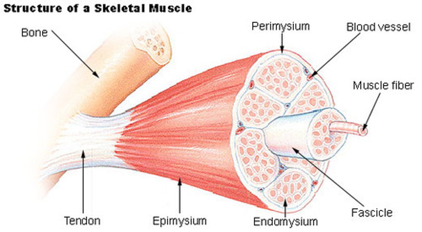
Describe perimysium
-several sheathed muscle fibers are wrapped in a collagen rich dense connective tissue
-formes a fascicle

Describe fascicle
-bundle of fibers
-makes up the perimysium
Describe the eipmysium
-a large number of fascicles are then bound together with connective tissue
-surrounds the entire muscle
-outermost layer
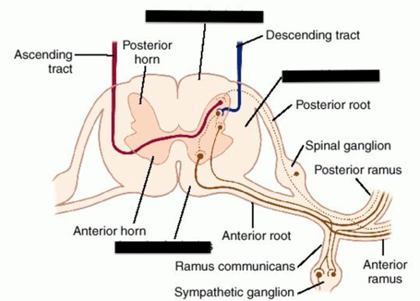
Label the dorsal/ posterior gray horns of spinal cord
(d)

Label the ventral/ anterior gray horns of spinal cord
(e)
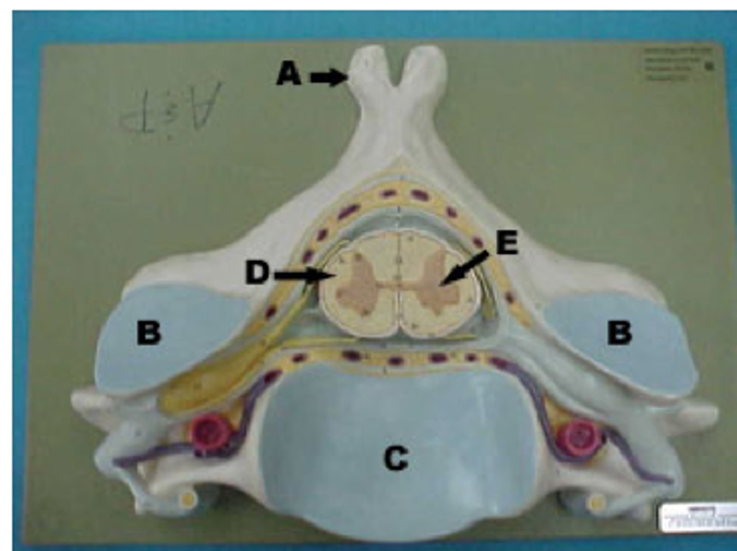
Label the gray commissure and central canal
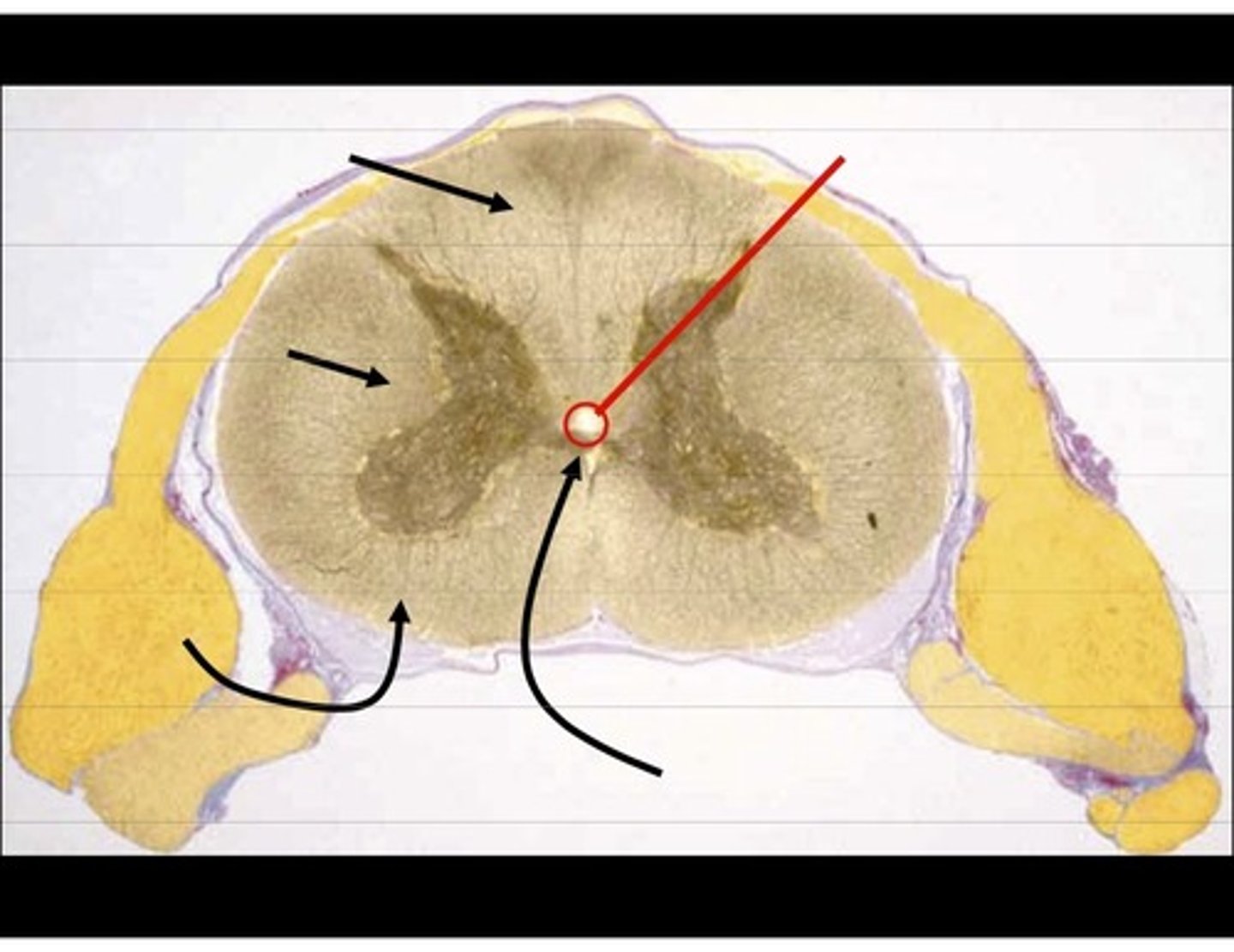
Label the dorsal root ganglion
Multipolar nuerons
-motor
-most neurons this type
Unipolar nuerons
-sensory
-most sensory neurons this type
Bipolar nuerons
-special senses
-vision
-hearing
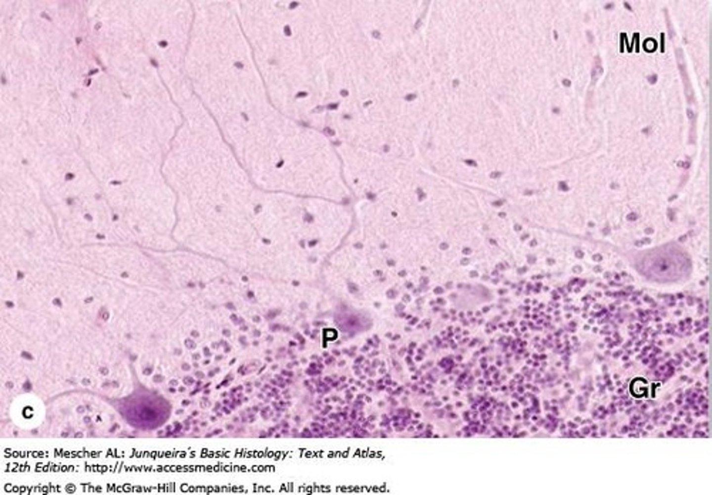
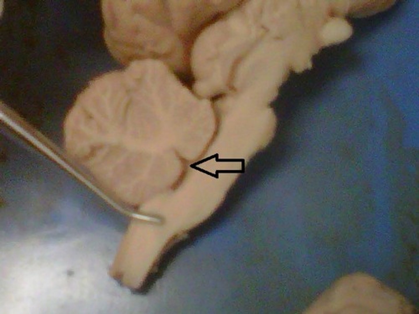
Identify Purkinje cells of the cerebellum
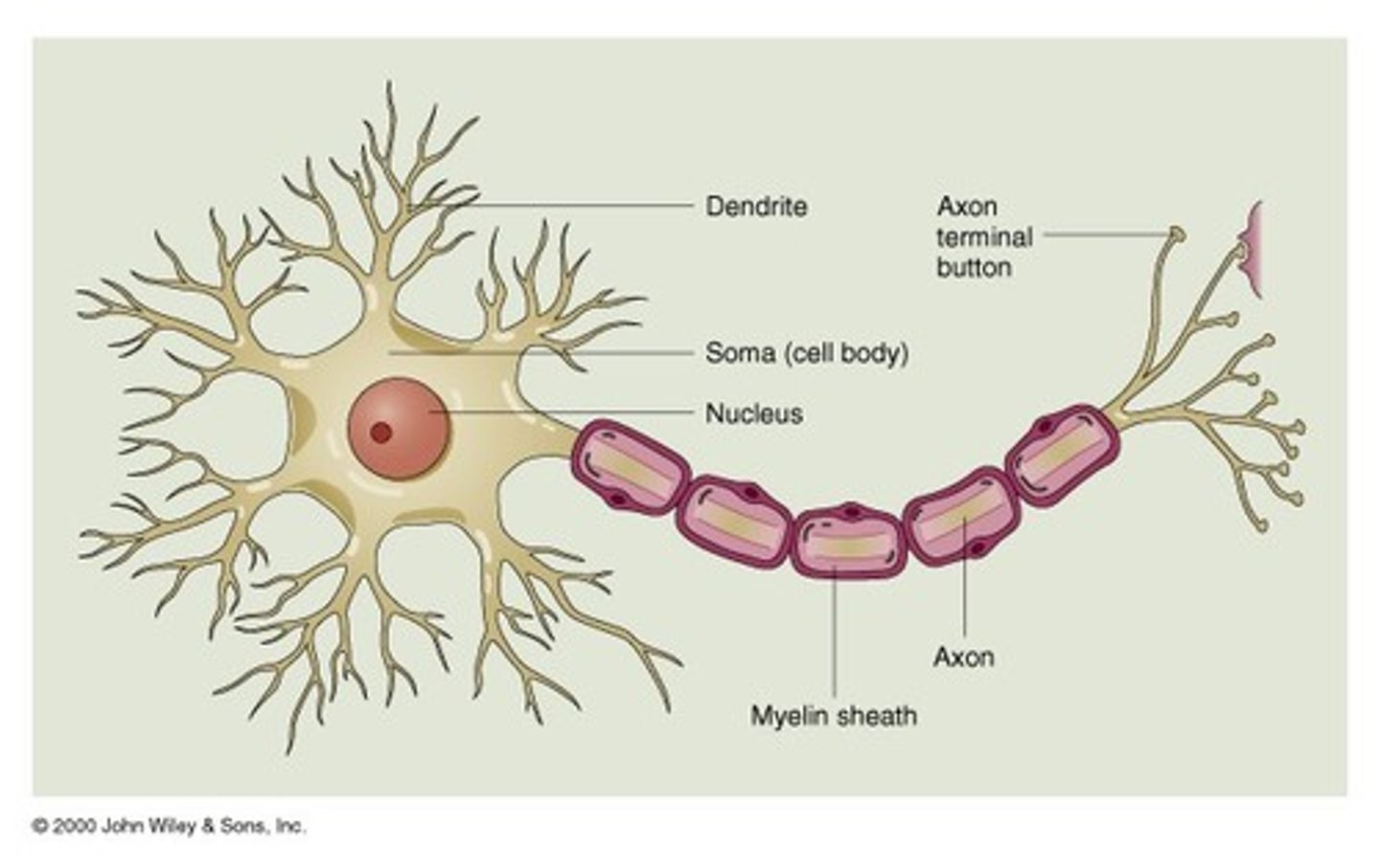
Identify the Nodes of Ranvier and the myelin sheath
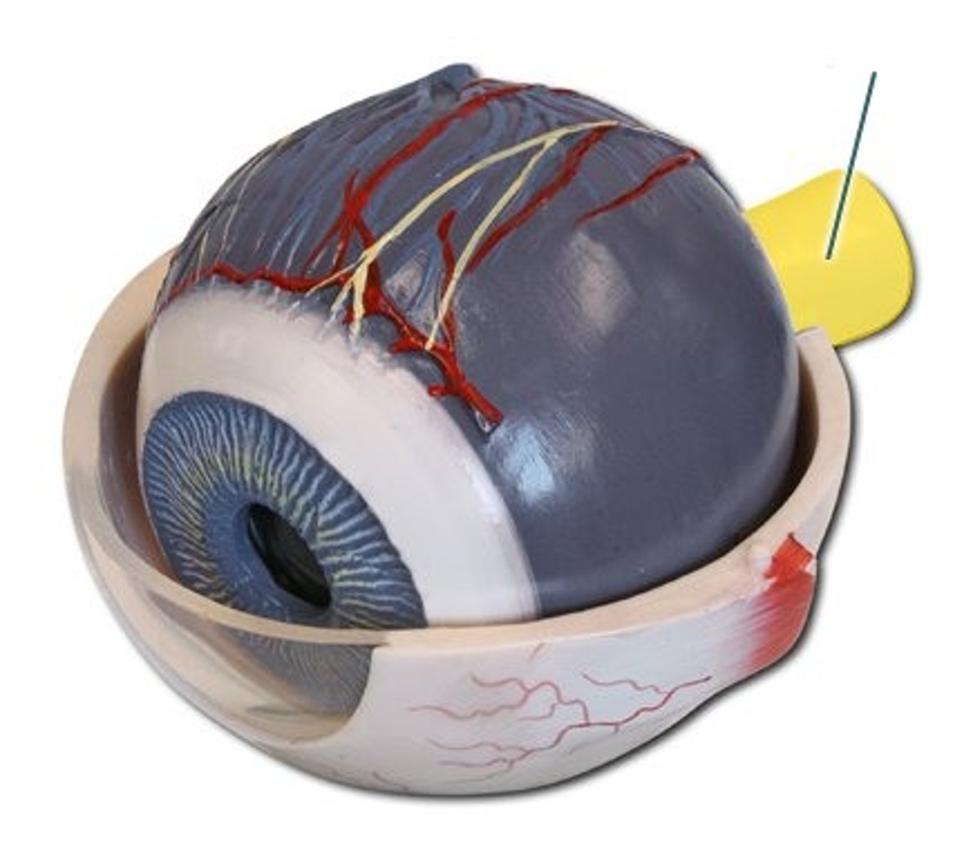
Dura mater

Arachnoid mater
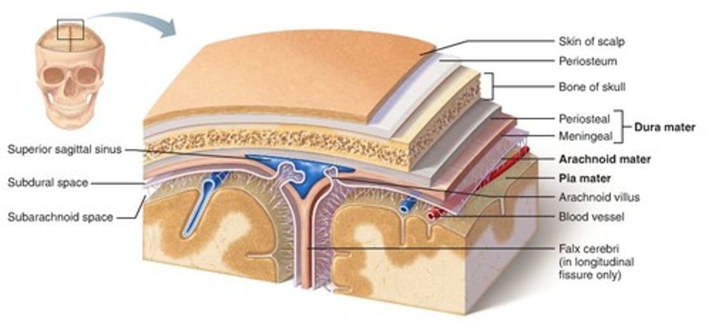
Pia mater
-could only see in sheep brains

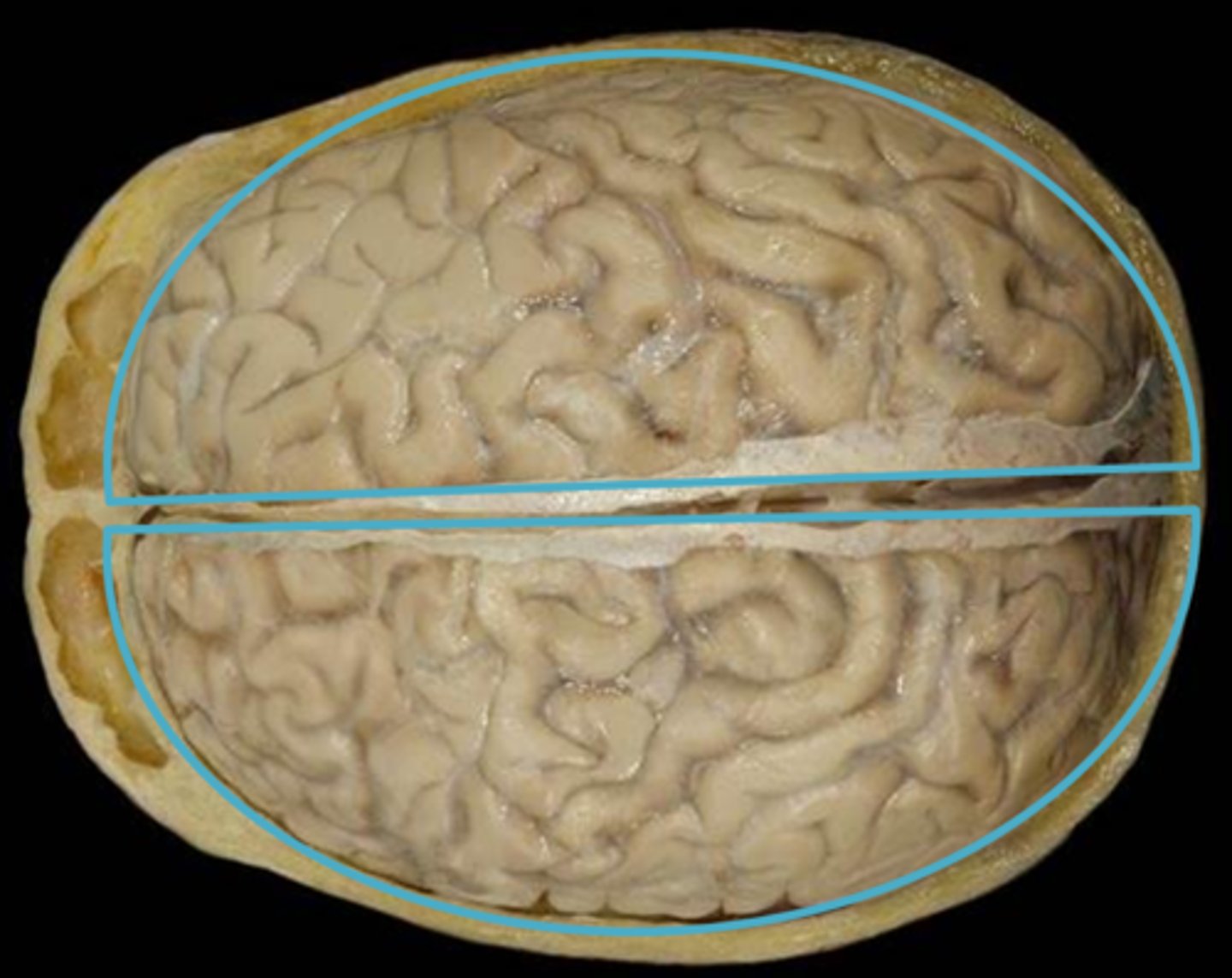
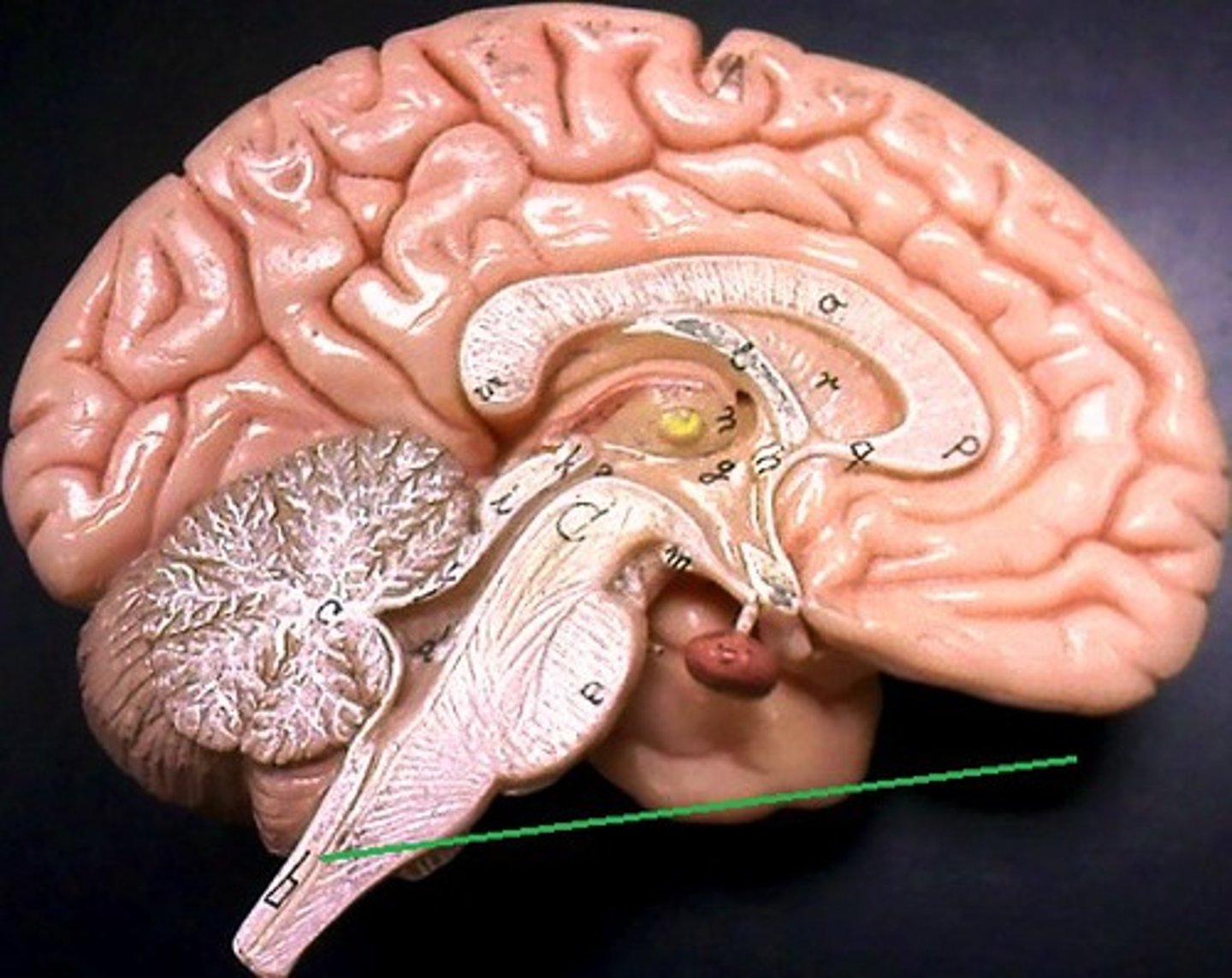
Cerebral hemispheres
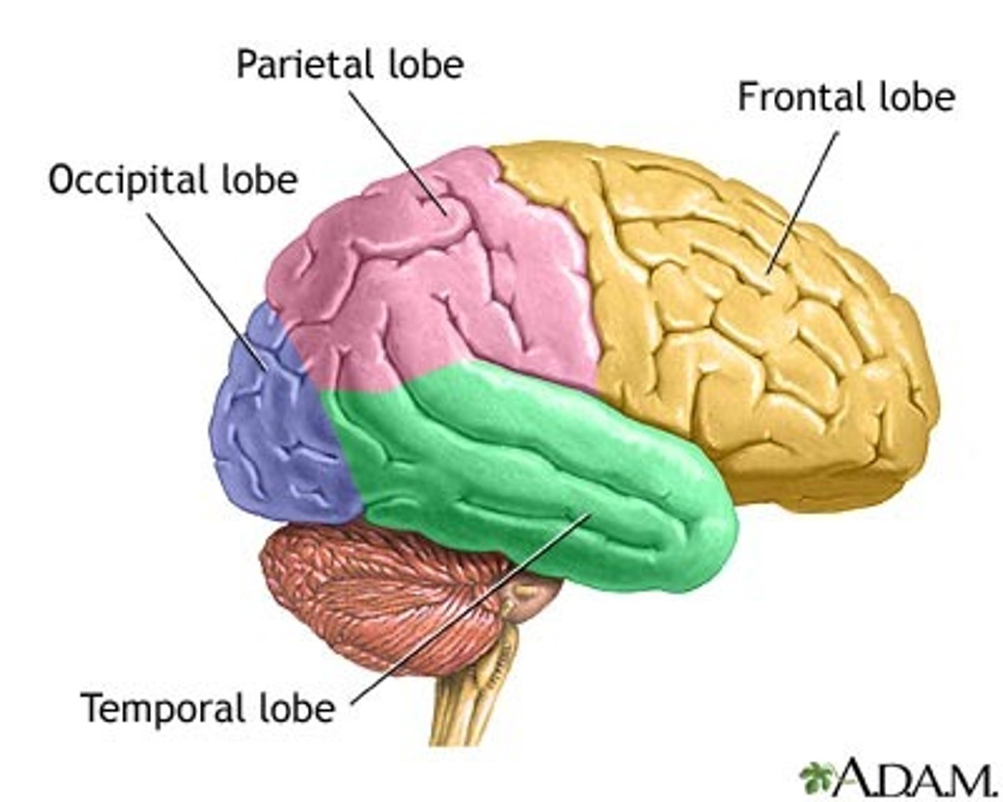
Cerebral lobes
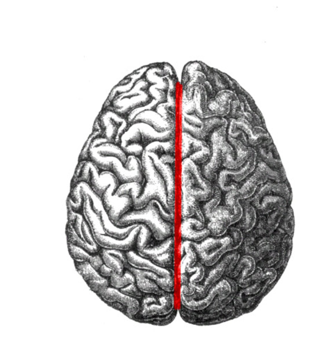
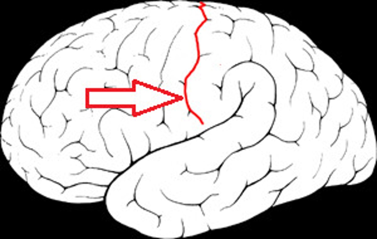
Longitudinal fissure
Transverse fissure
gyrus
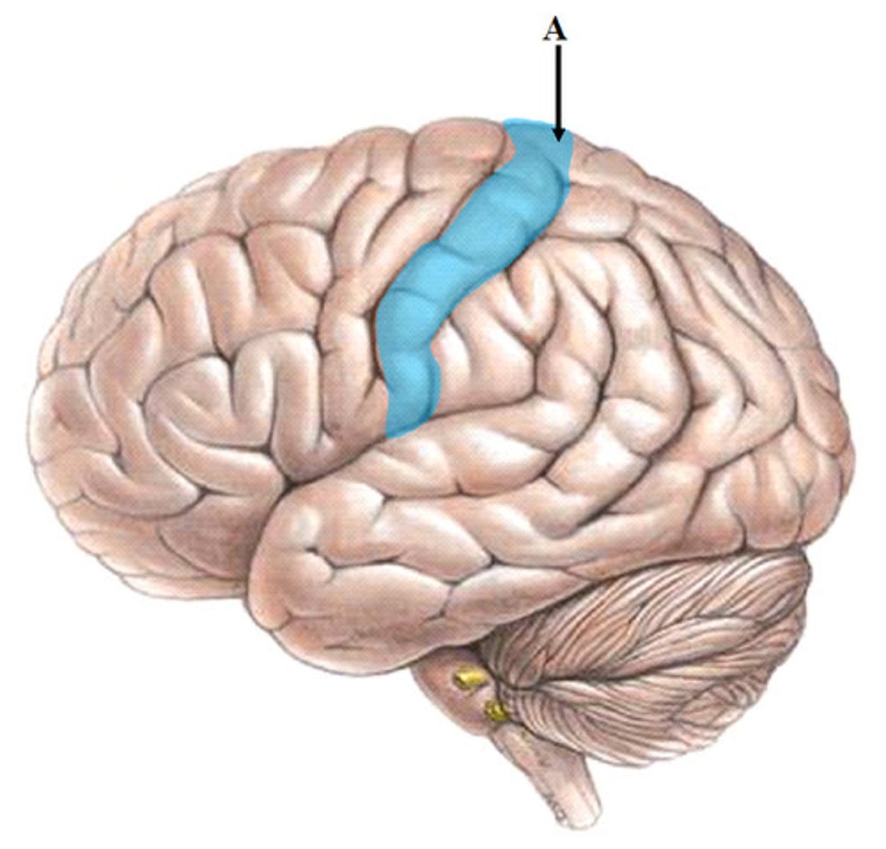
sulcus
corpus callosum
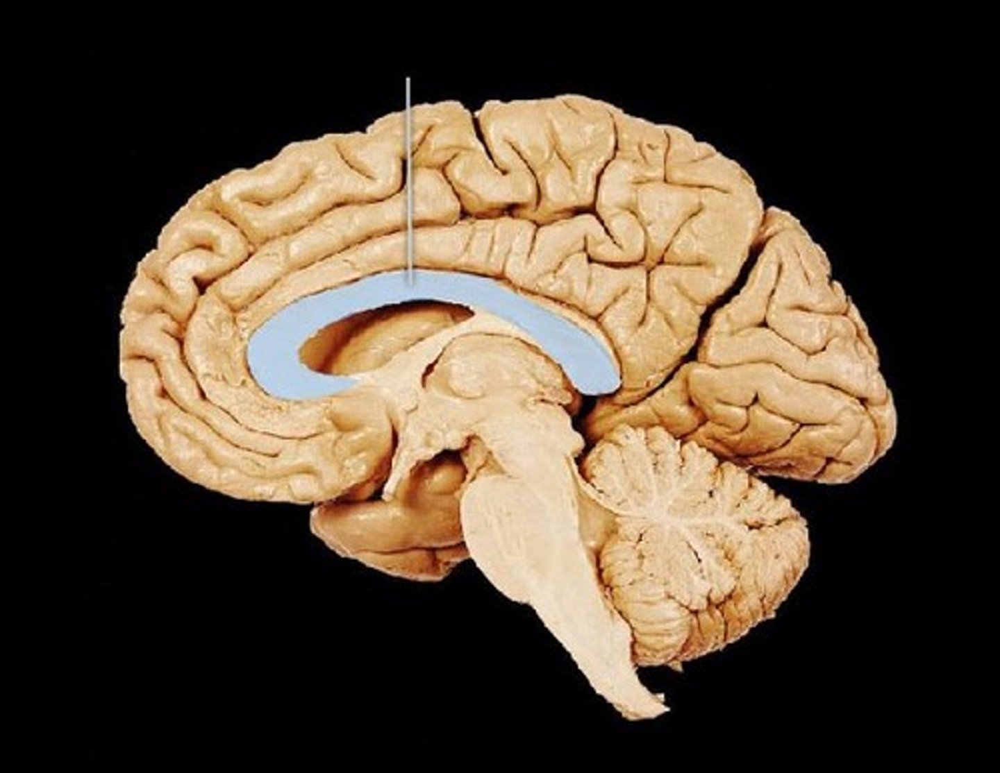
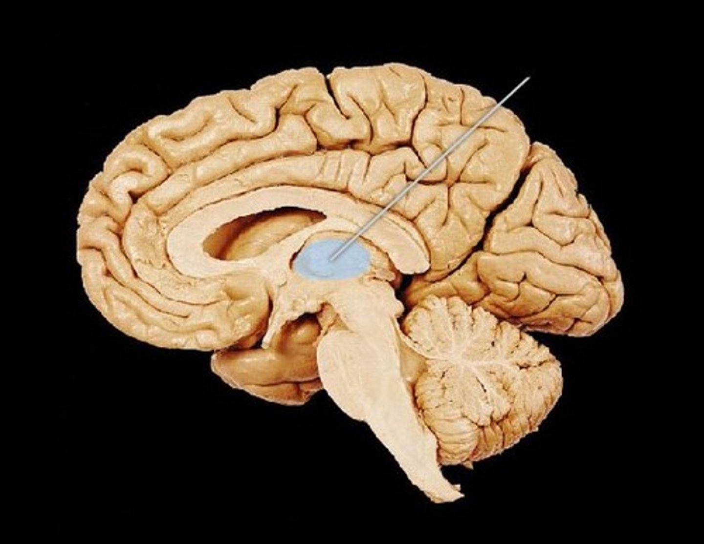
fornix lateral ventricles
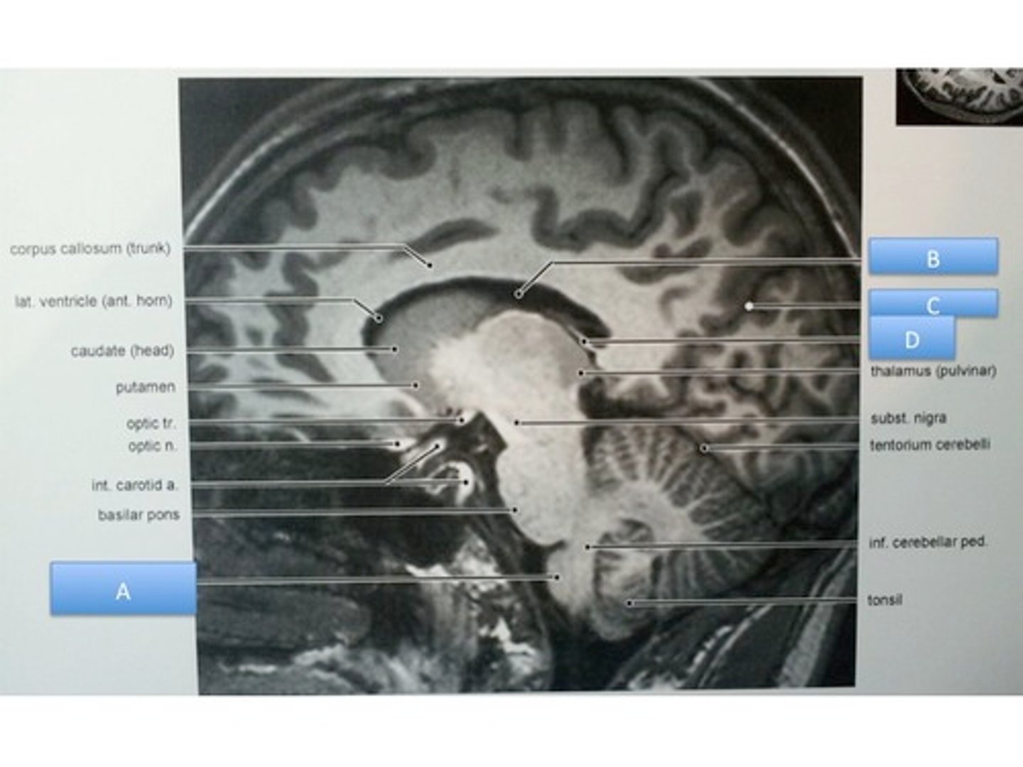
third ventricle
fourth ventricle
central canal
olfactory bulbs
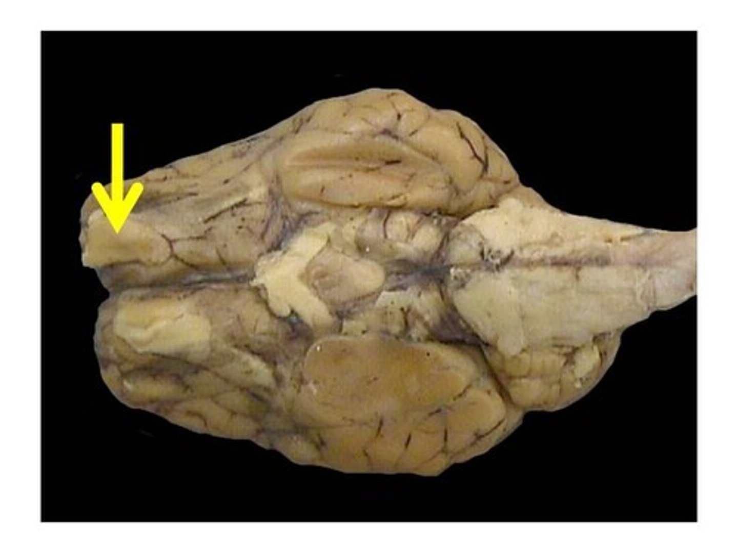
optic nerves
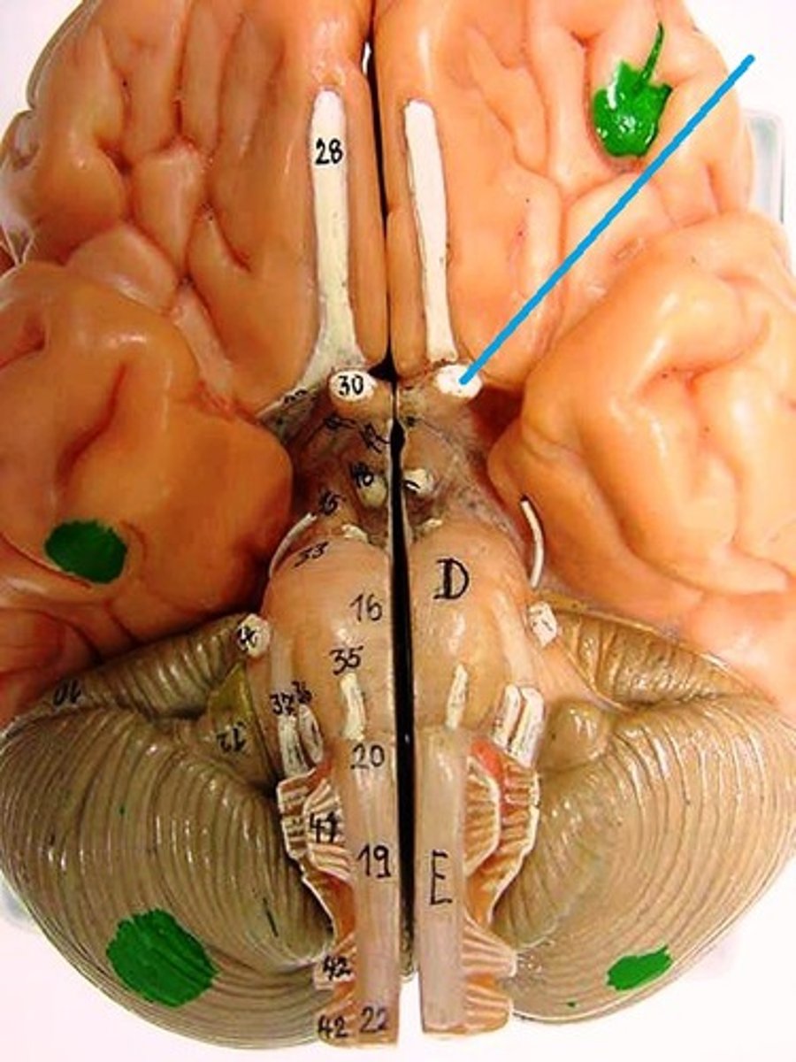
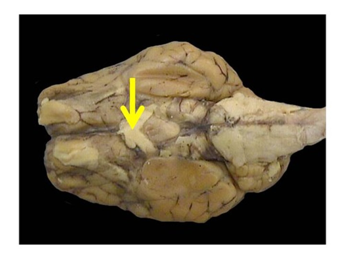
optic chiasma
medulla oblongata
pyramids of the brain
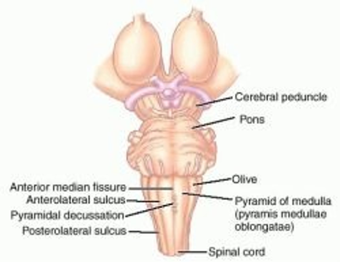
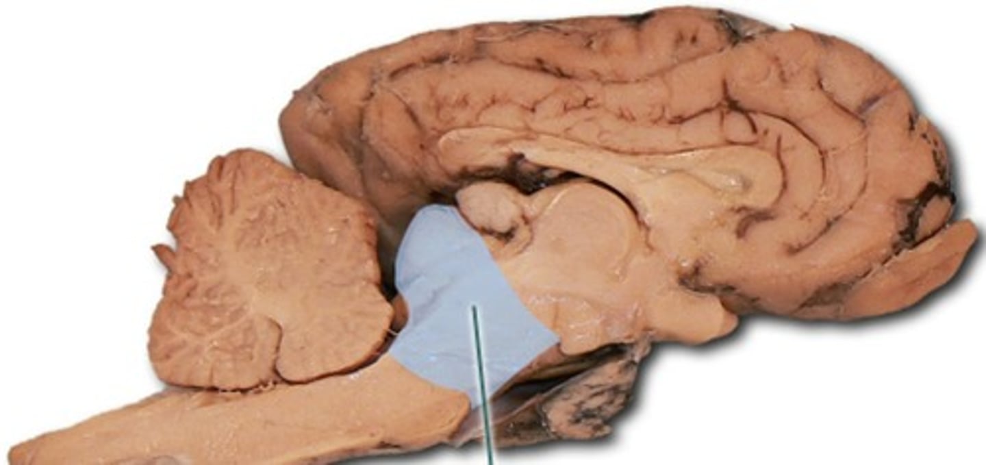
pons
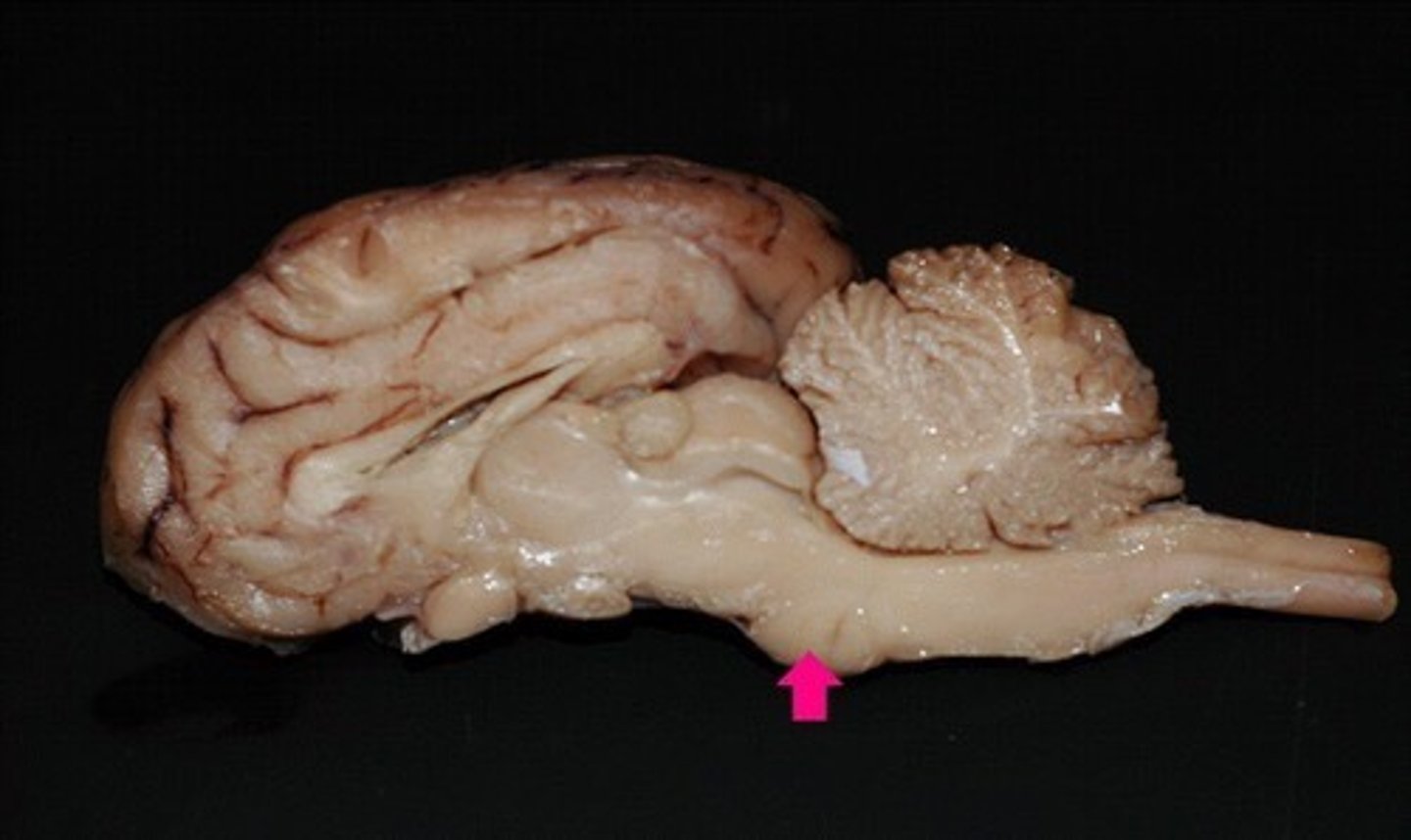
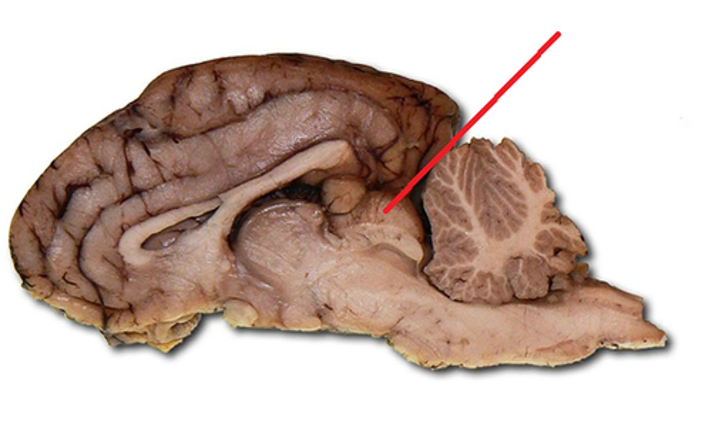
midbrain
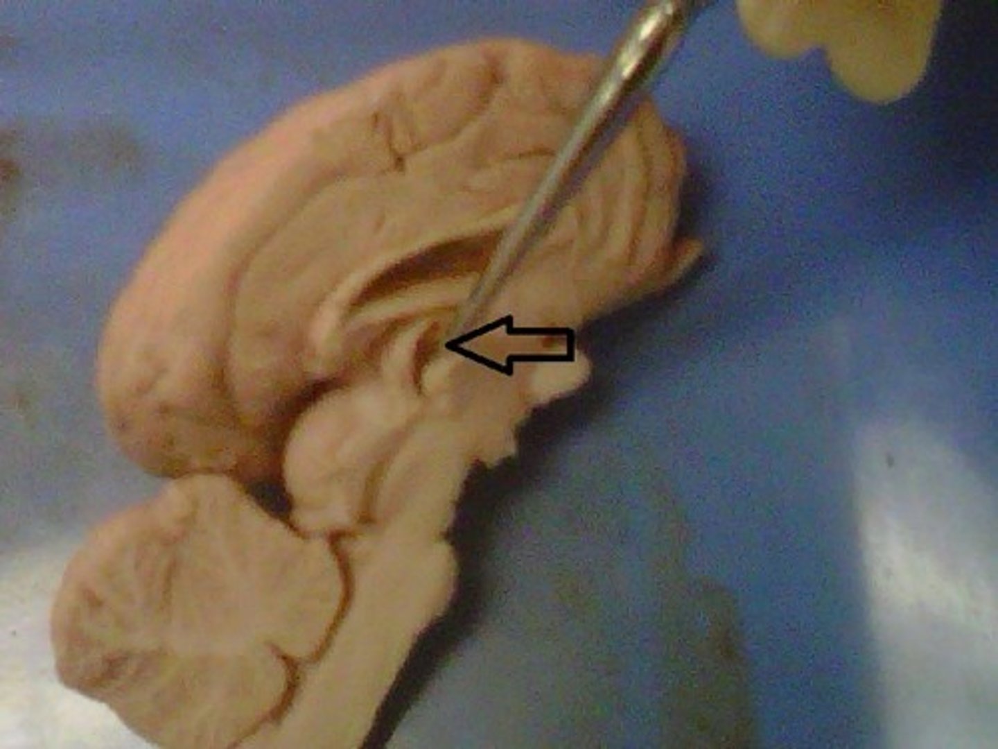
corpora quadrigemina
superior colliculi
inferior colliculi
cerebral peduncles

hypothalamus
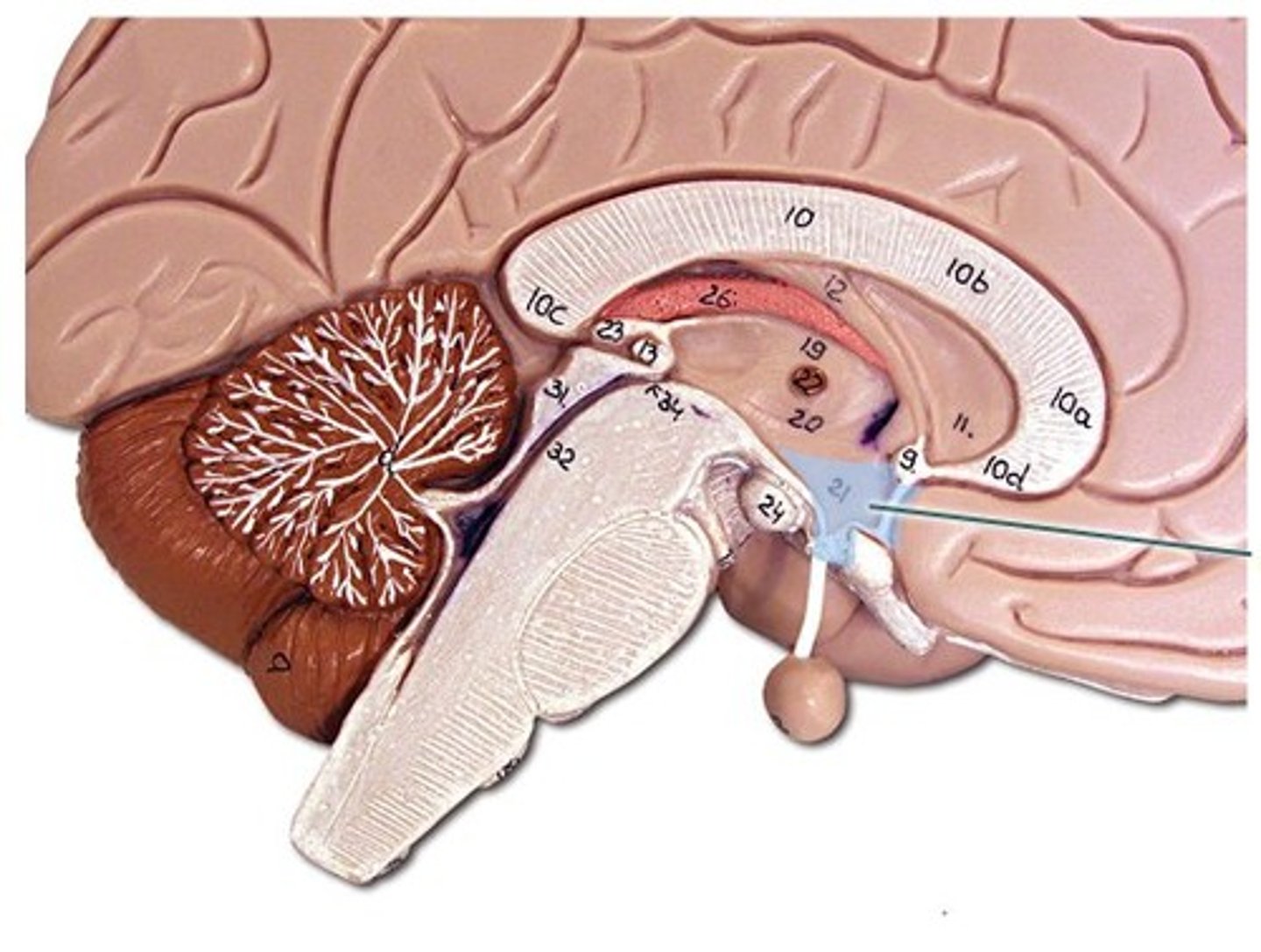
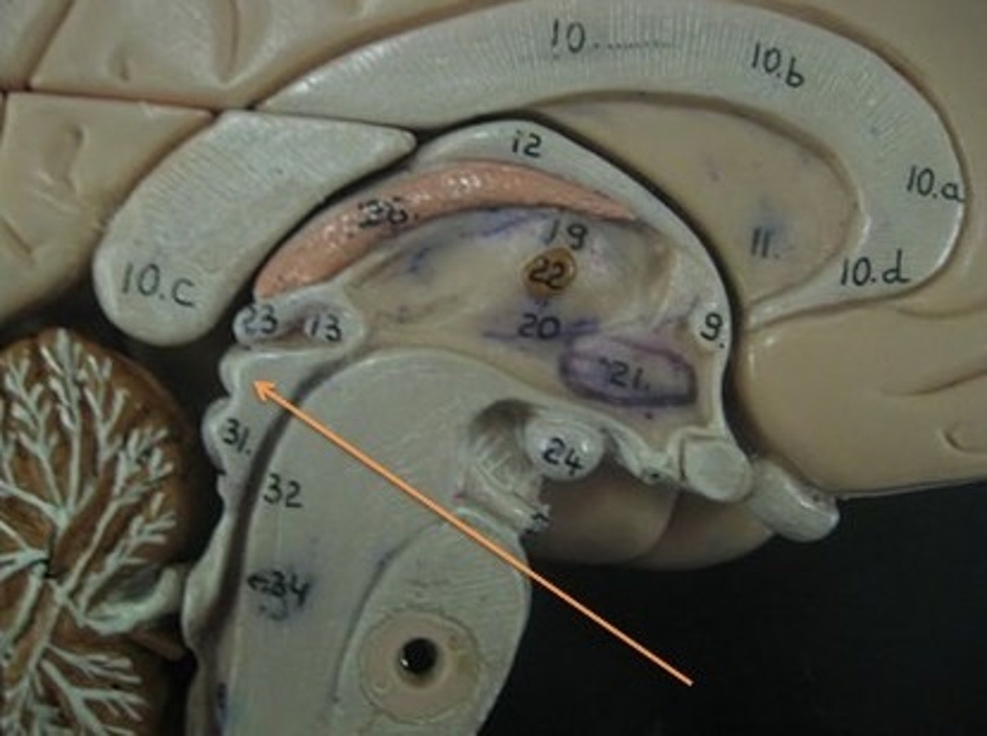
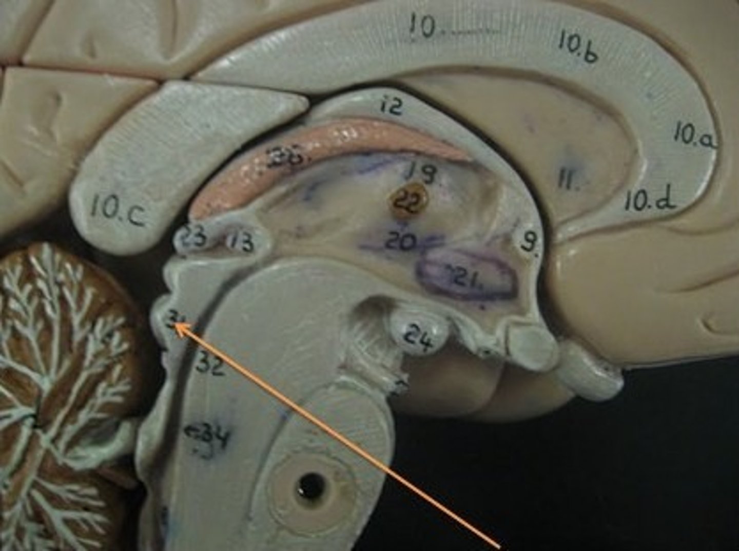
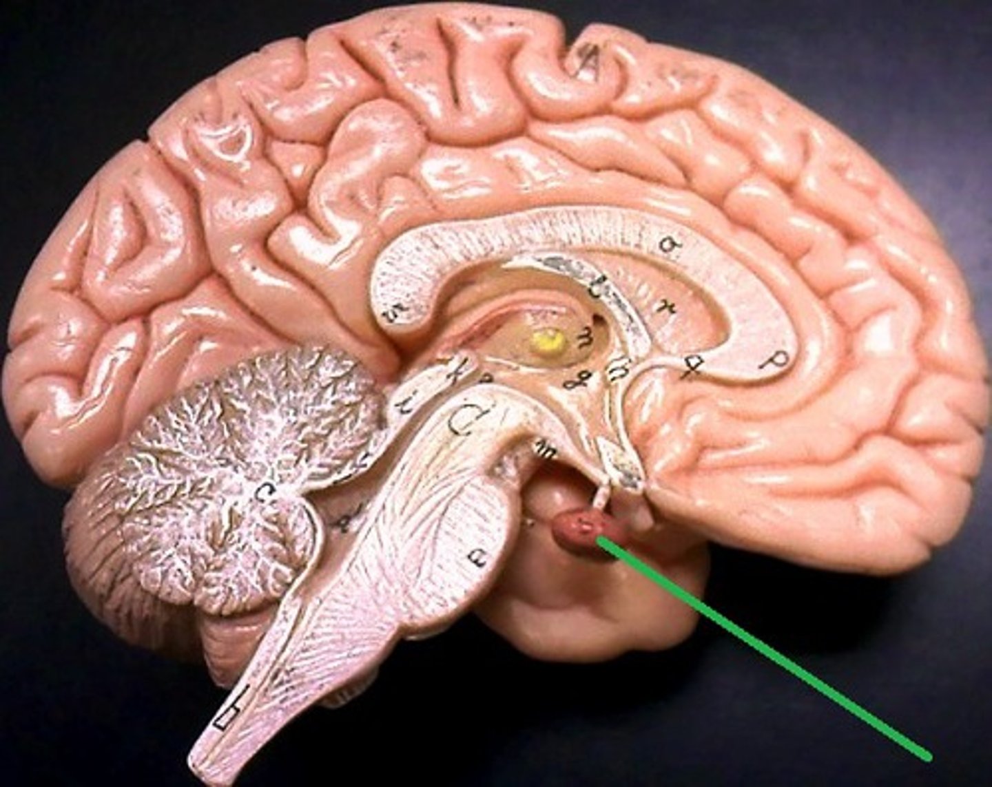
pituitary gland
thalamus
pineal body
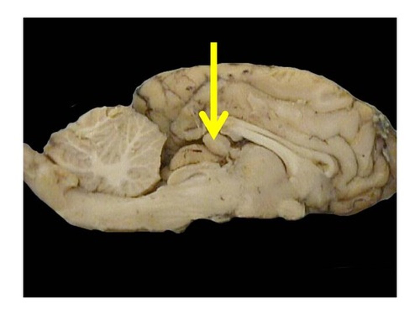
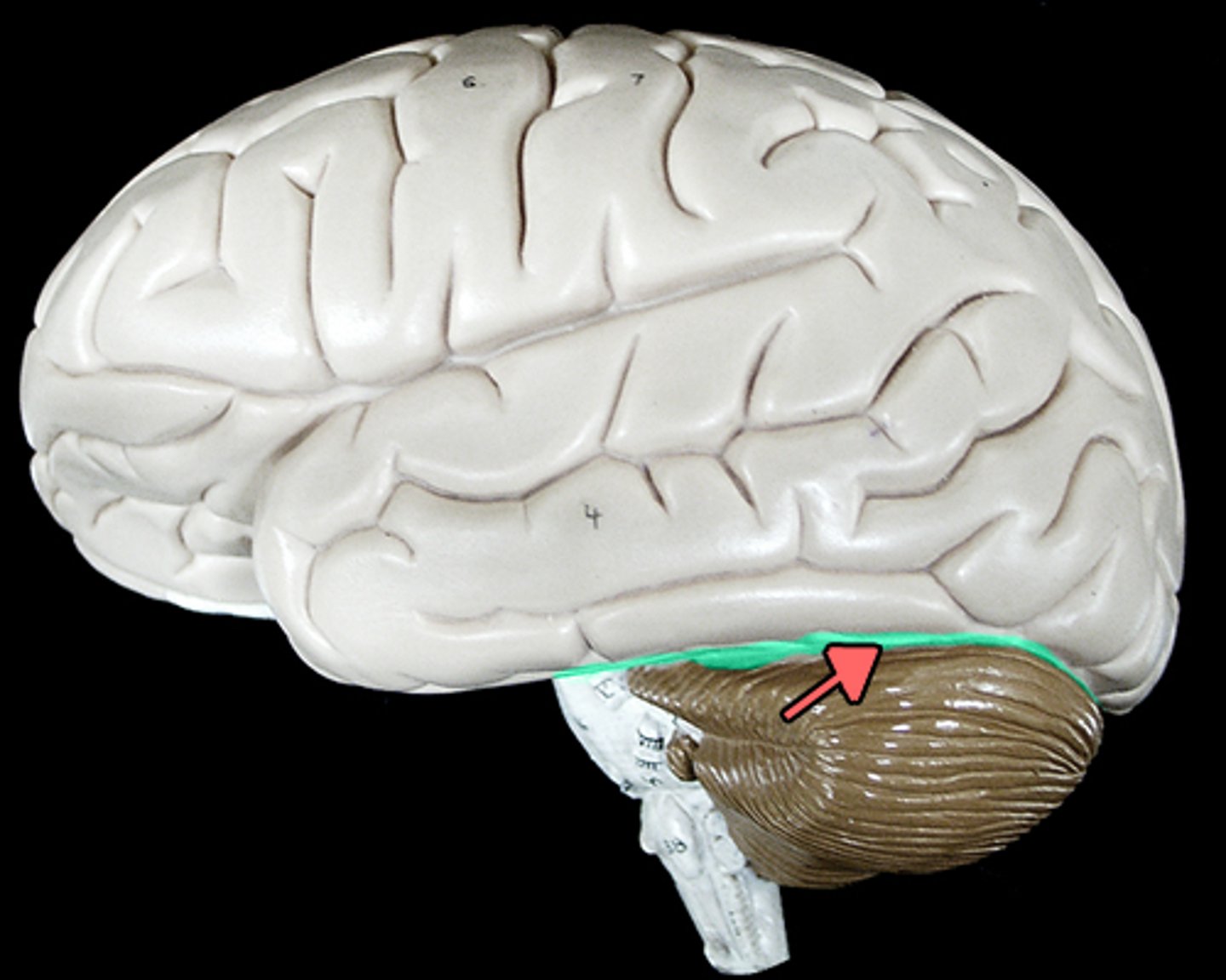
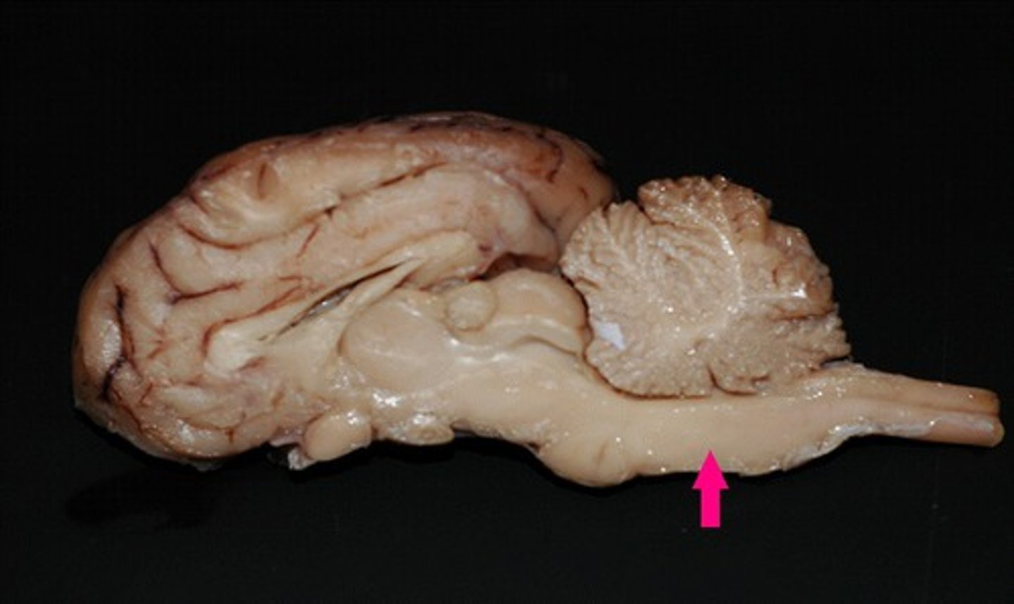
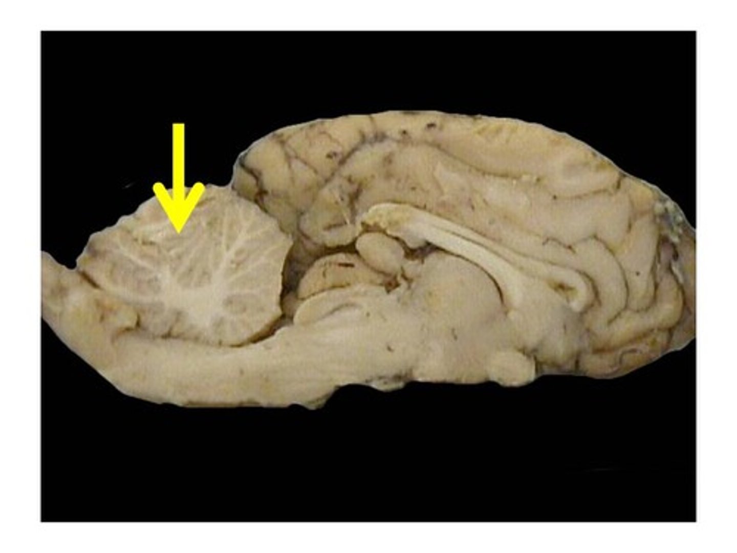
cerebellum
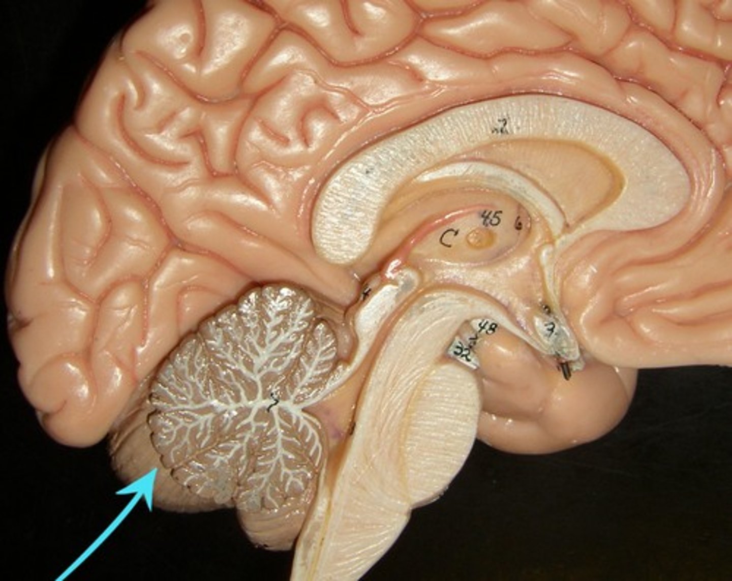
arbor vitae
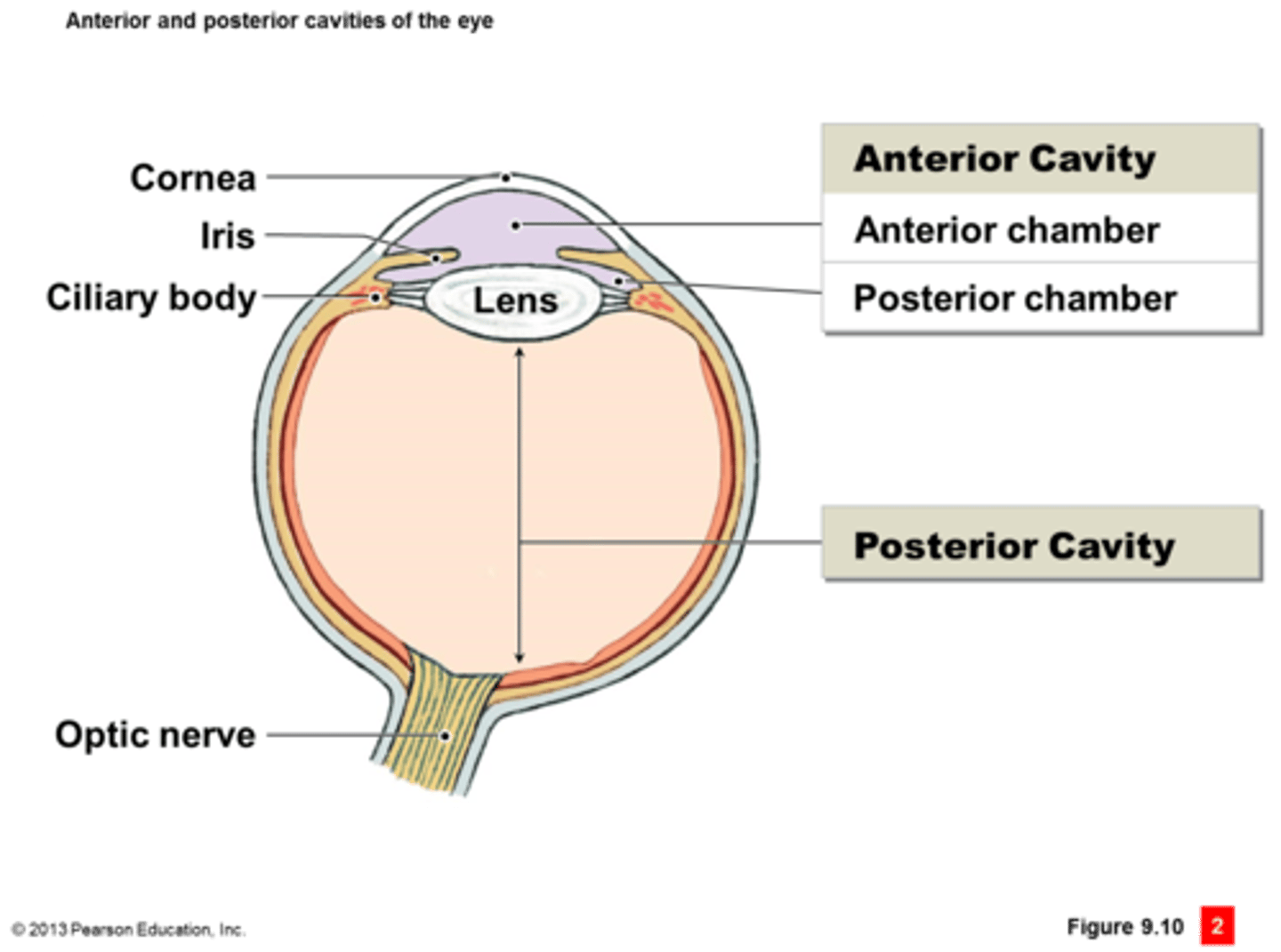
3 tunics of eye
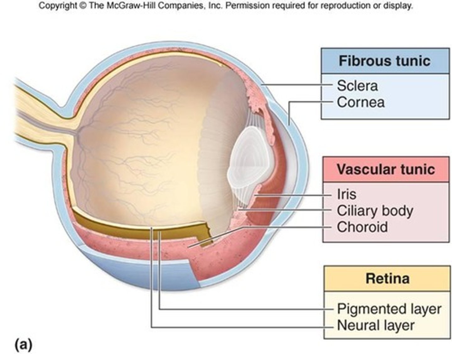
cornea
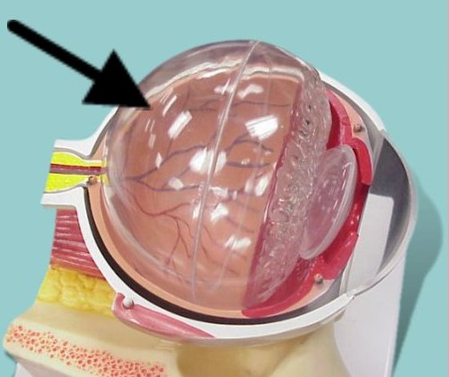
sclera
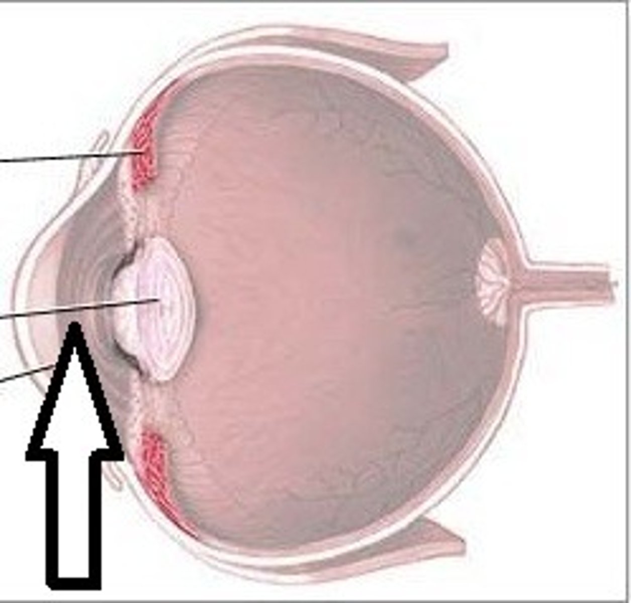
iris
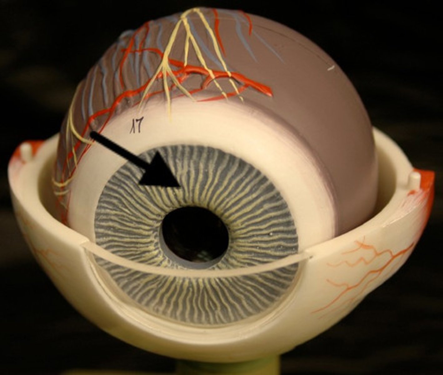
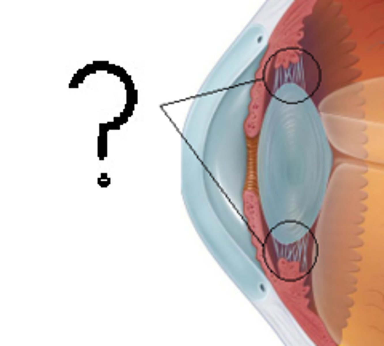
ciliary body
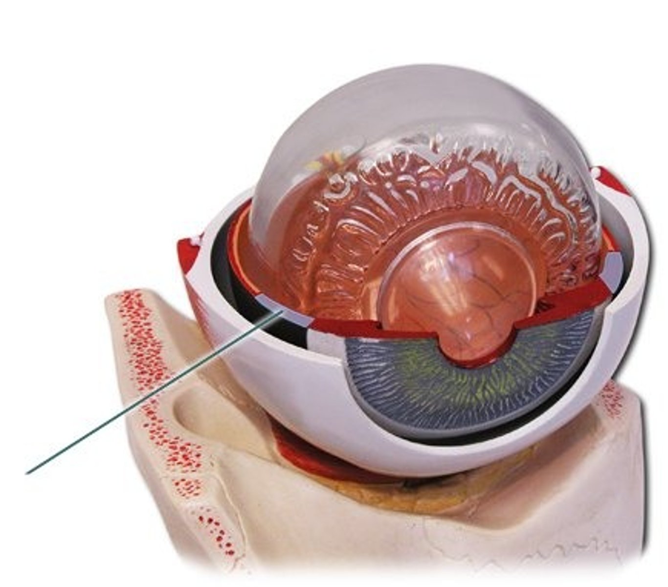
lens
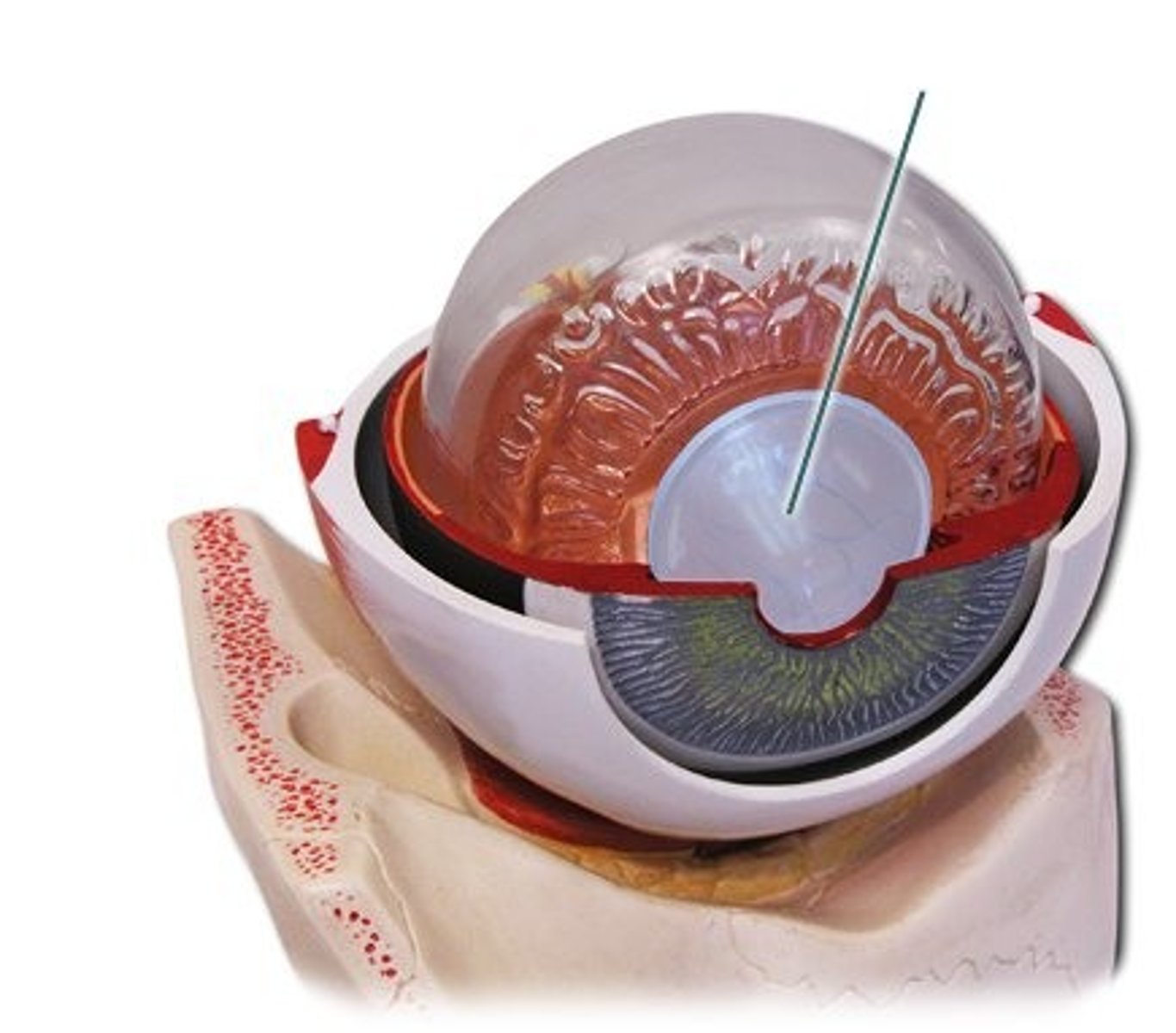
suspensory ligaments
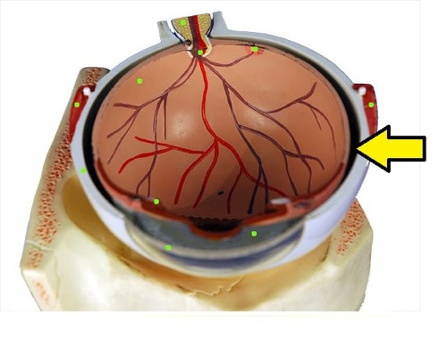
retina
optic disc
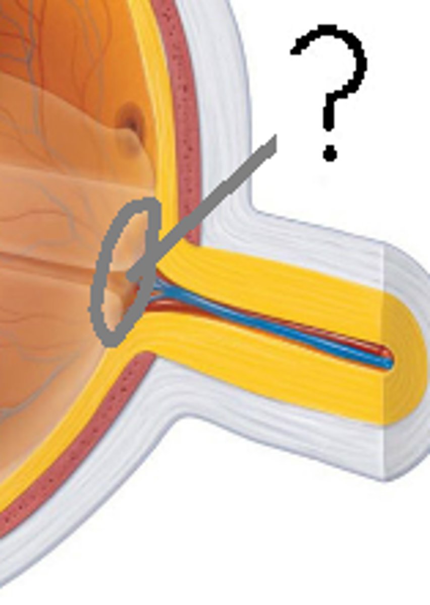
optic nerve
choroid
anterior cavity of the eye

anterior chamber
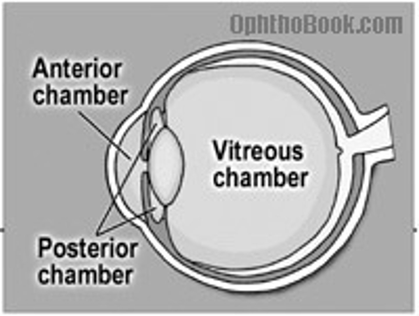
posterior chamber of the eye
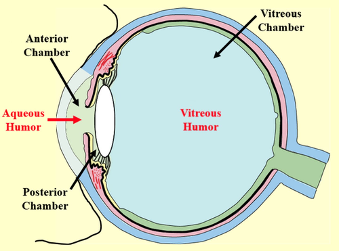
posterior cavity of the eye
aqueous humor
vitreous humor
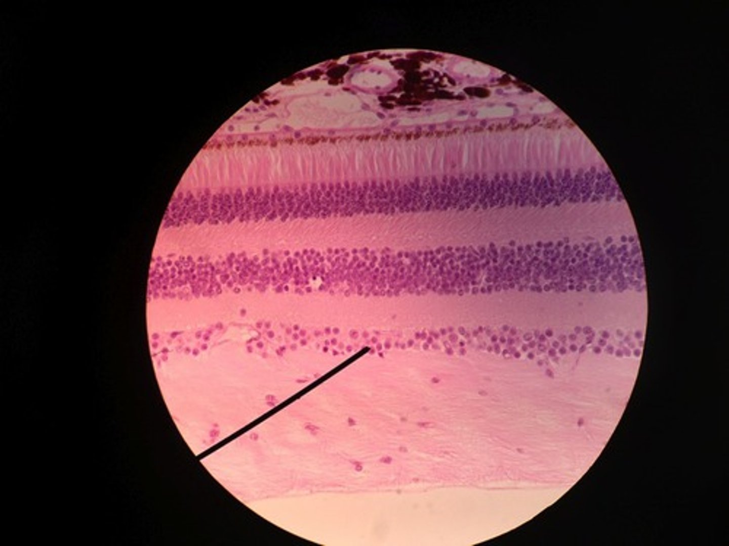
photoreceptor cell layer
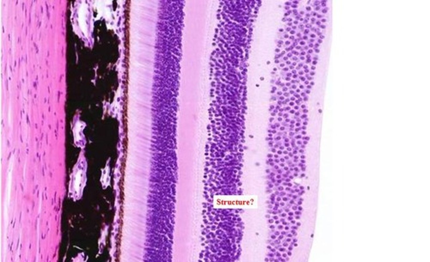
bipolar cell layer
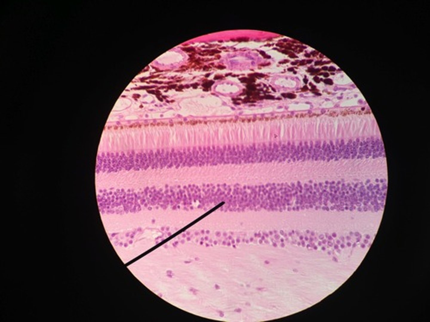
ganglion cell layer
how do you determine visual acuity
V= vision
d= distance from the chart you could read in a line correctly
D= distance from the chart that the same line of letters could be read by a person with normal vision
V=d/D
20/20 vision
if you can read correctly at 20 feet the line designated to be read at 20 feet by a person with normal vision
20/200 vision
only see large letters that a normal person can see at 200 feet
20/15 vision
can see a letter 20 feet away that a 20/20 person would need to see at 15 feet to just barely see
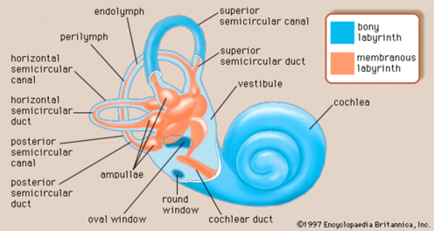
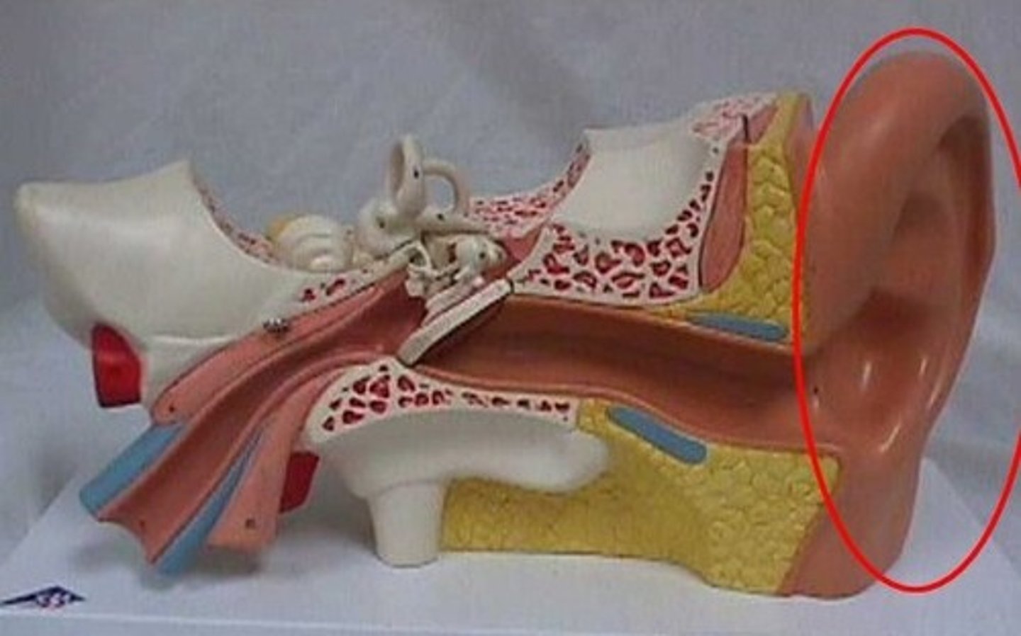
outer ear

inner ear
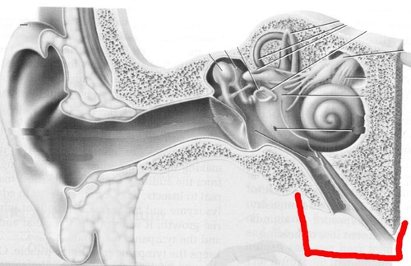
middle ear
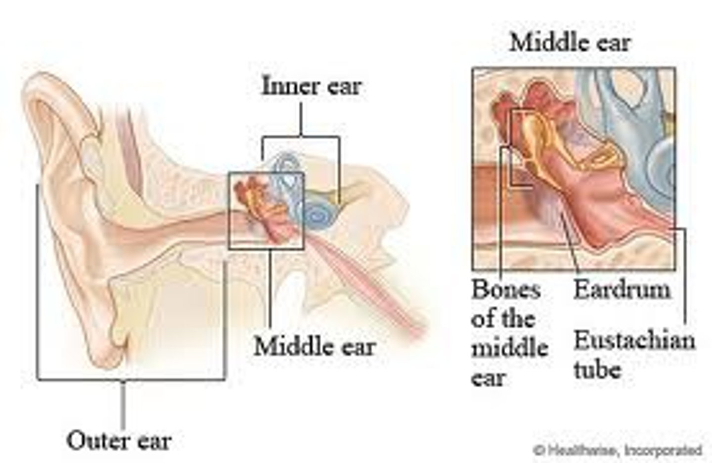
auricle or pinna
external auditory meatus
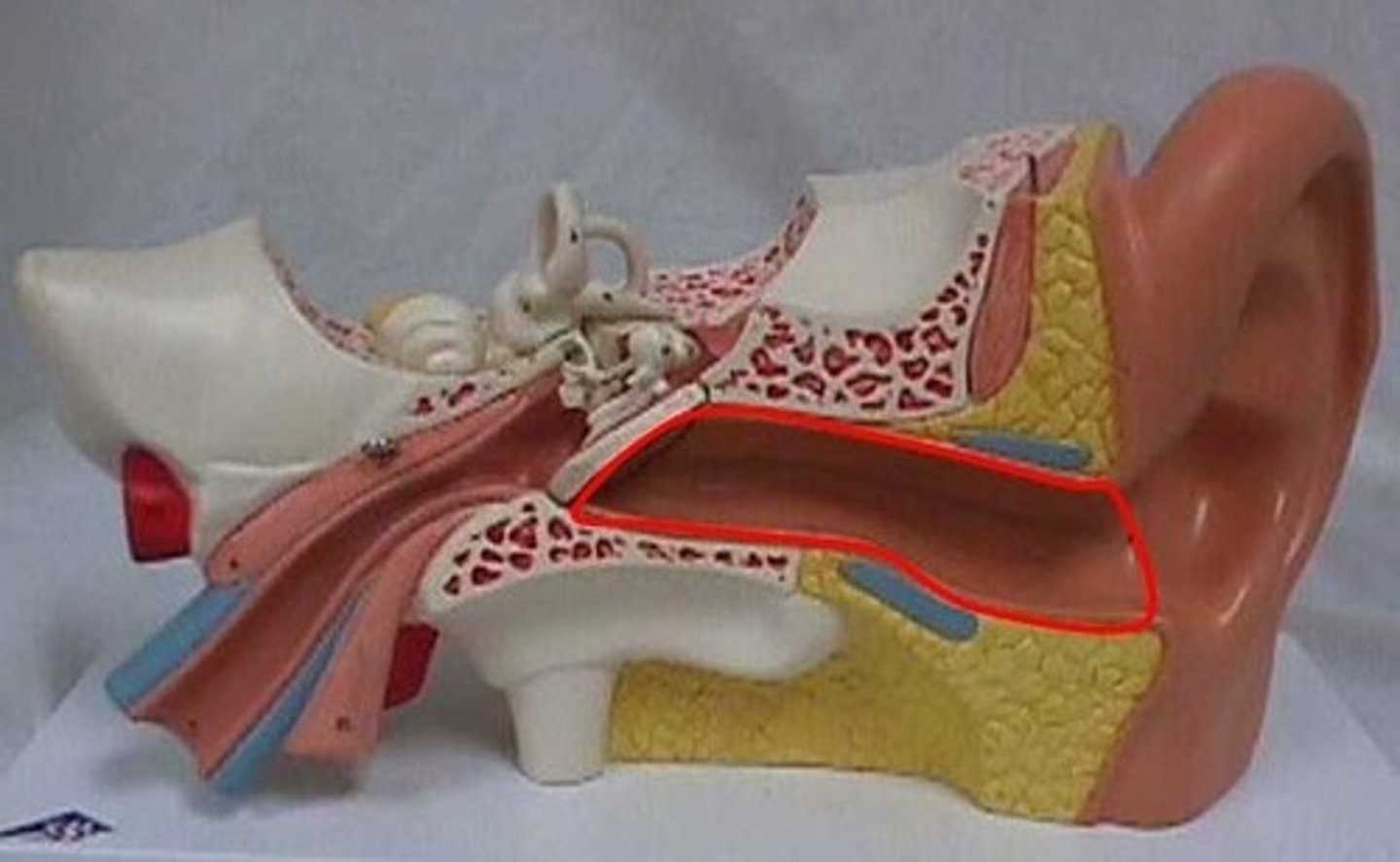
tympanic membrane
auditory or Eustachian tube
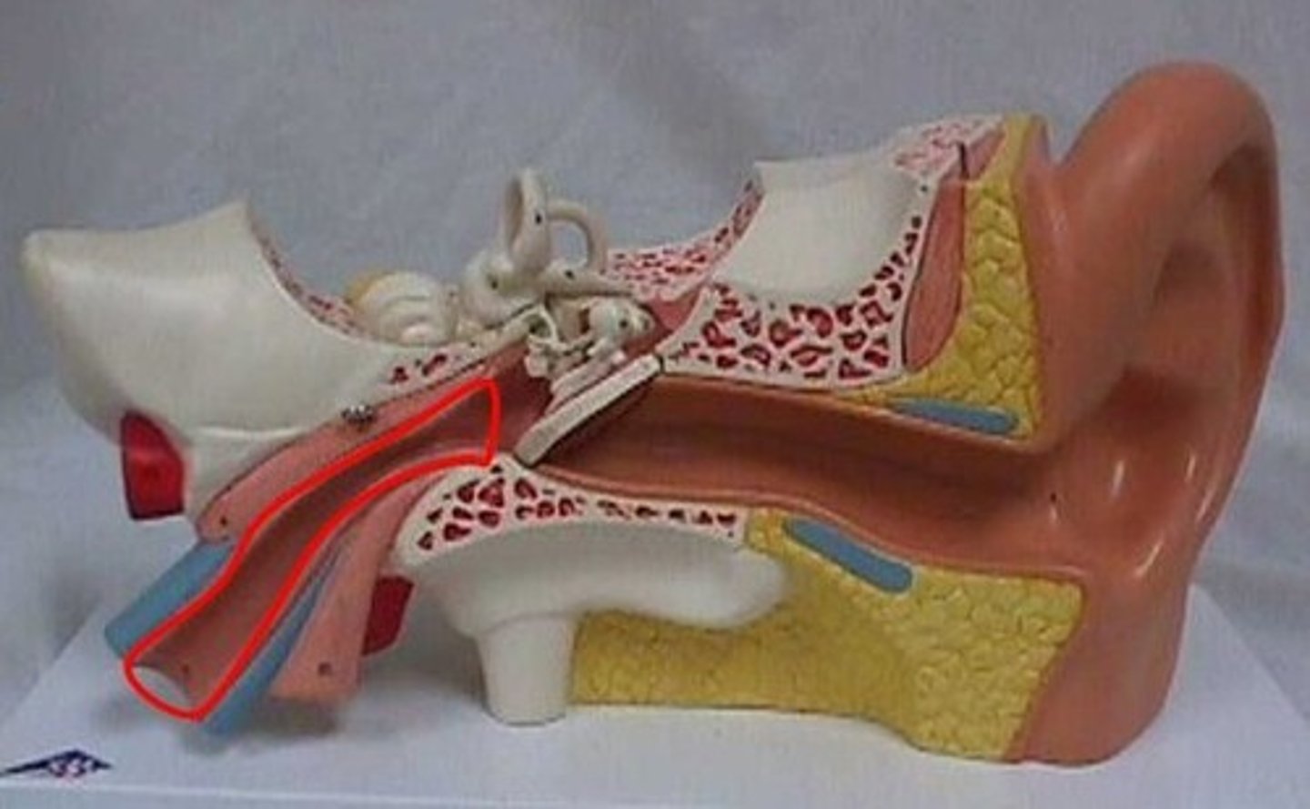
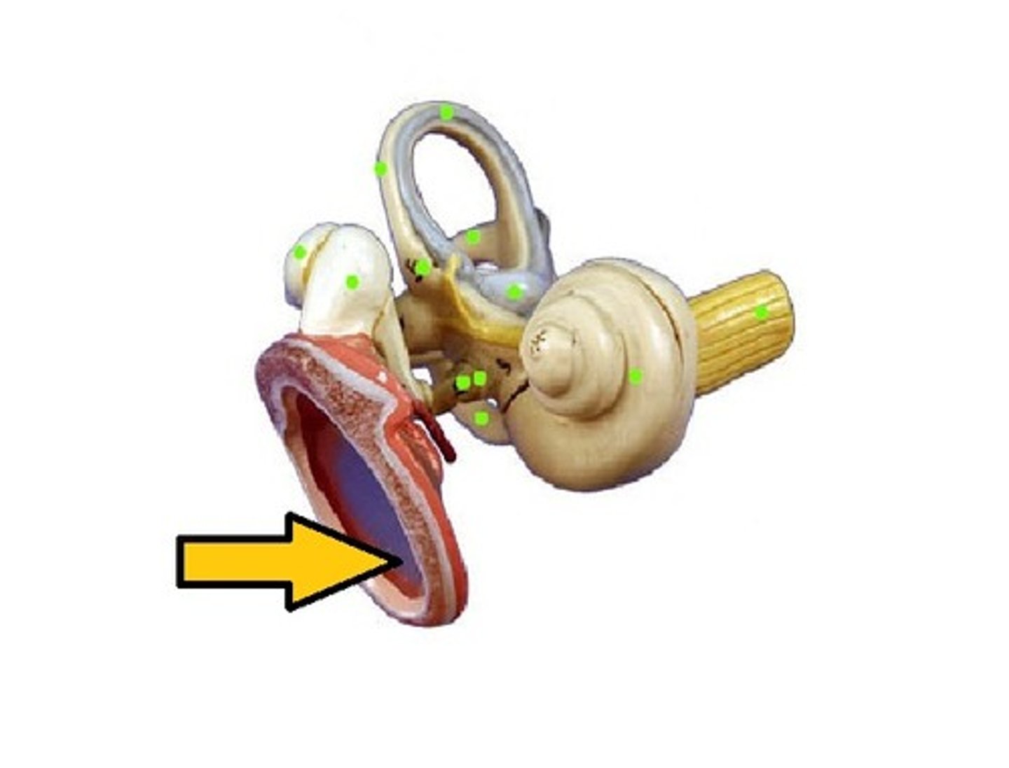
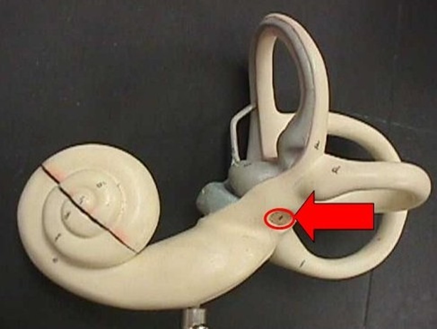
ossicles
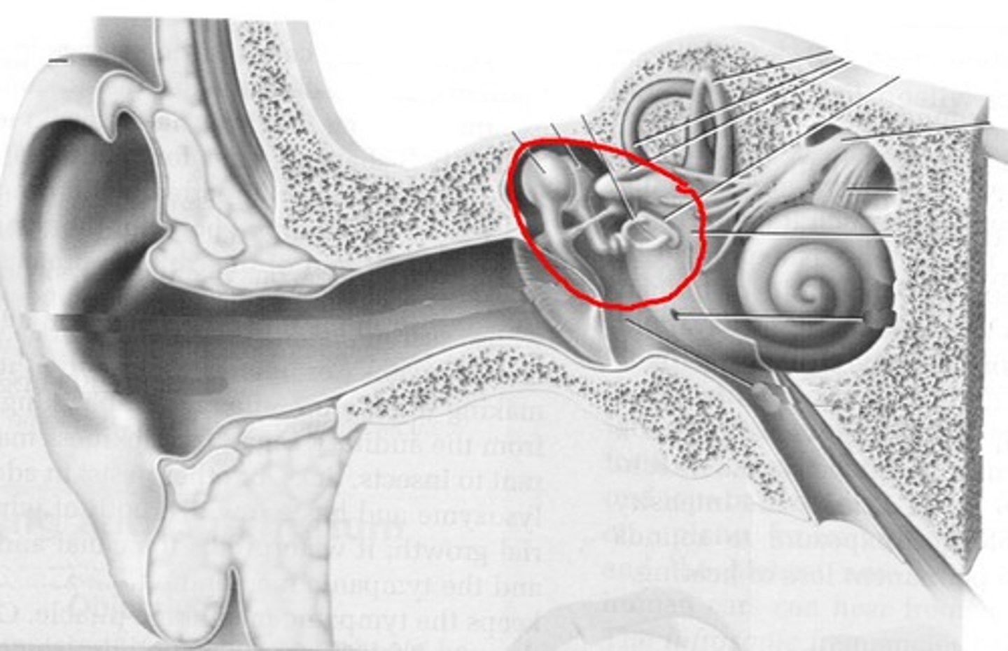
malleus
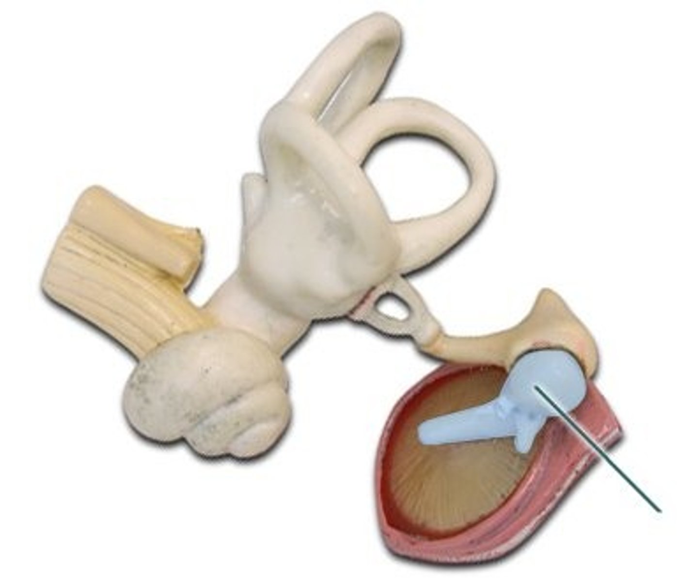
incus
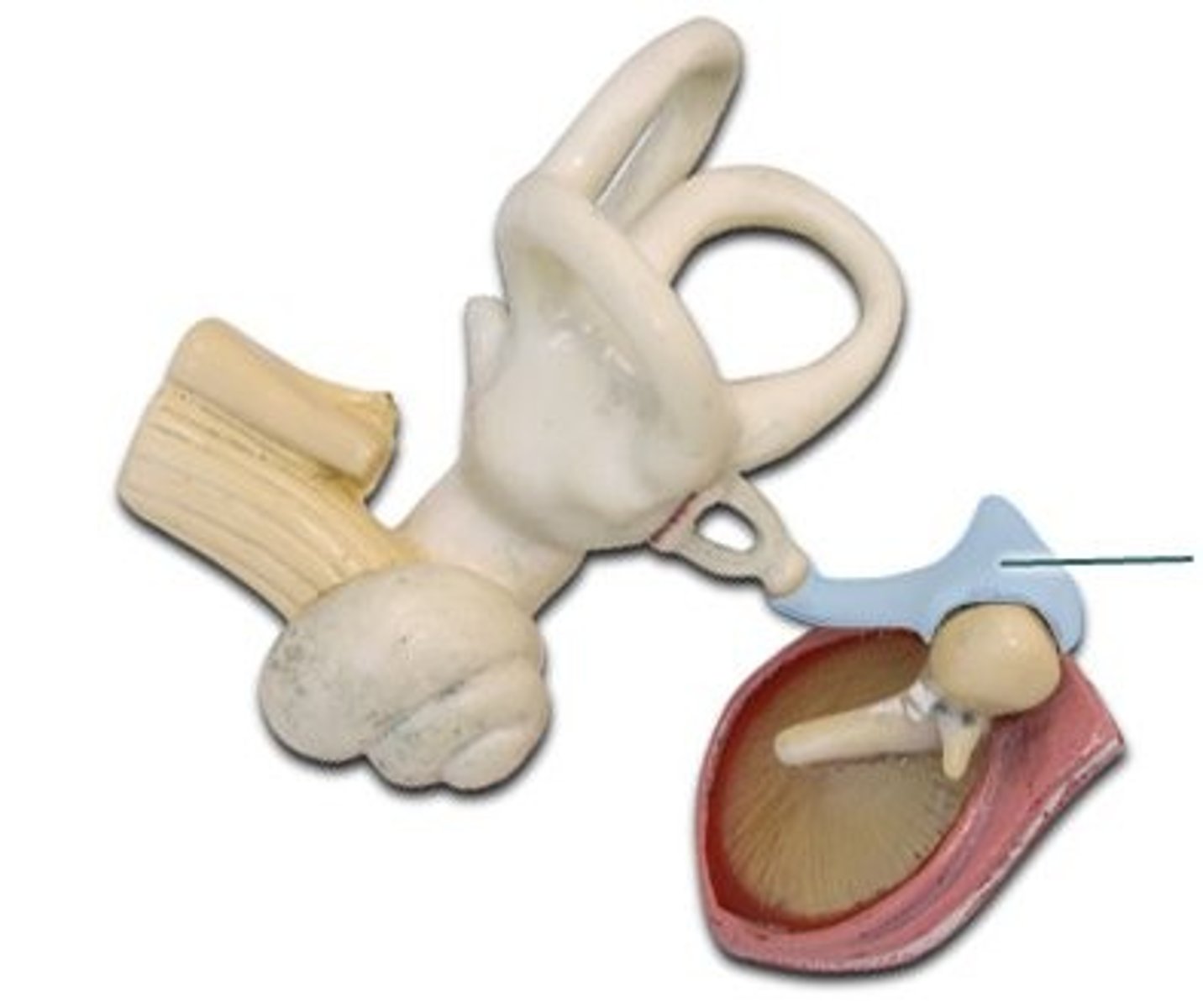
stapes
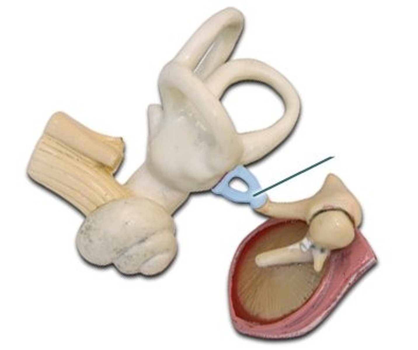
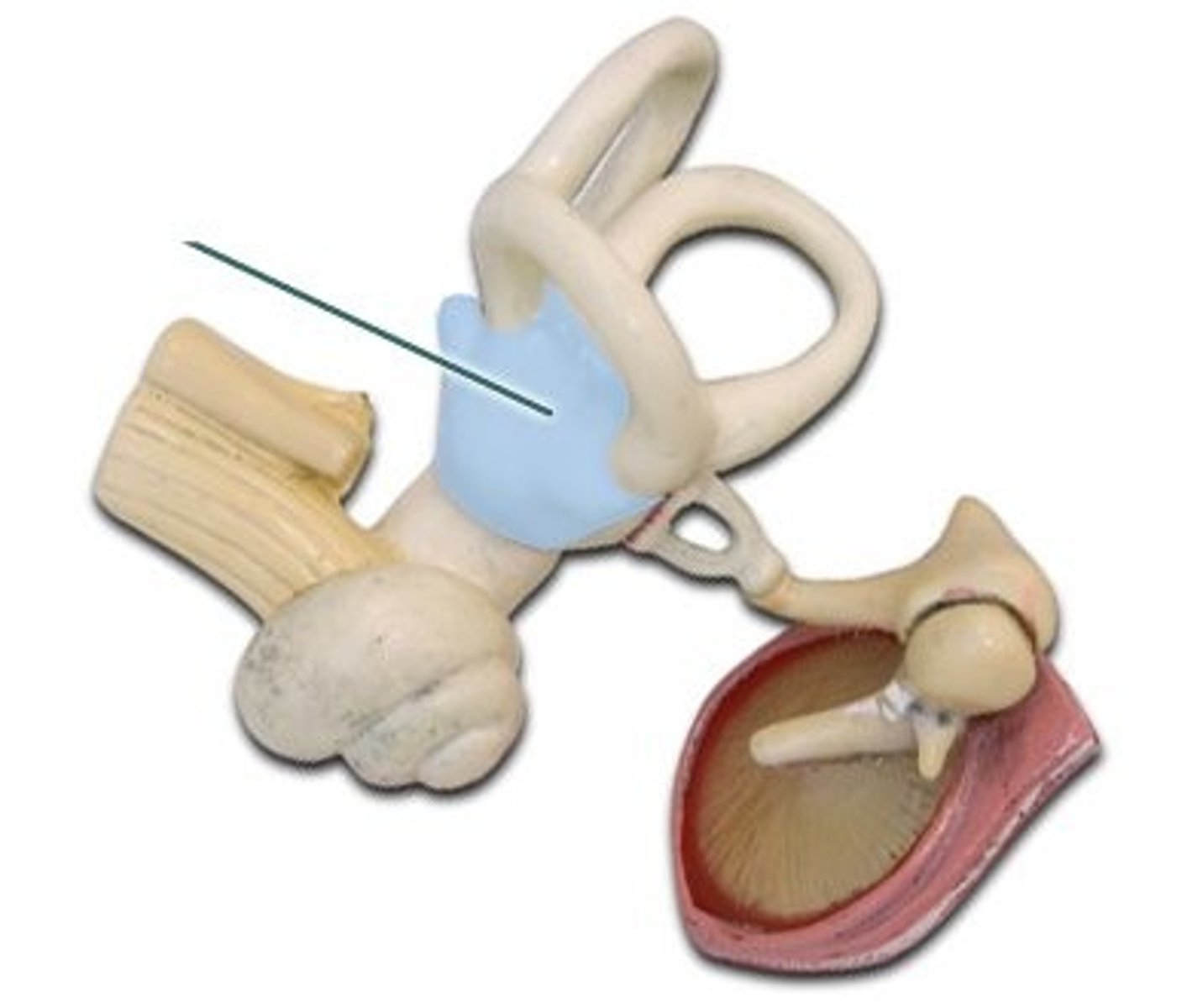
oval window
vestibule
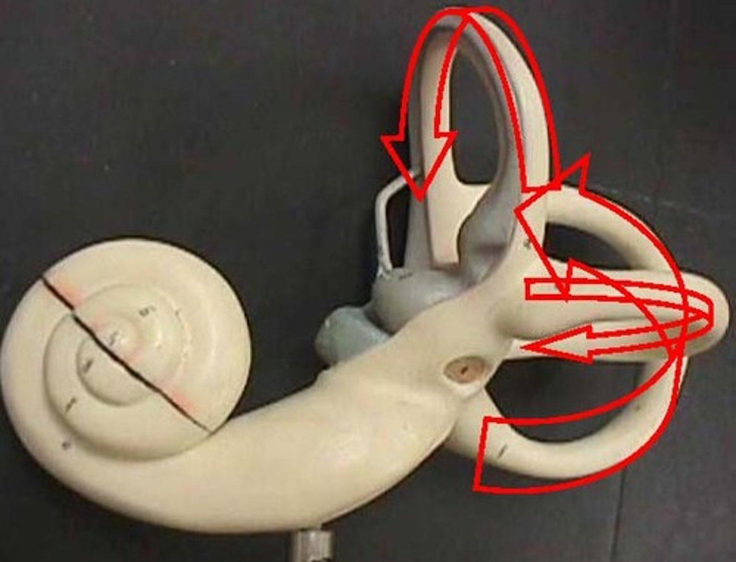
semicircular canals
cochlea
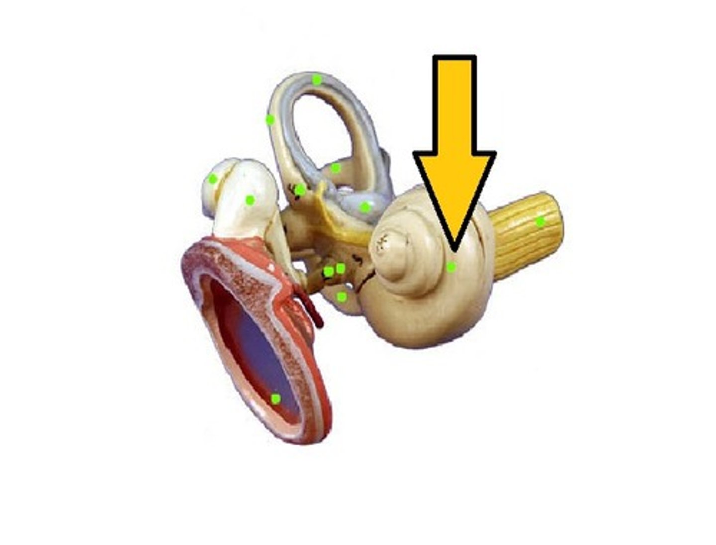
bony labyrinth vs membranous labyrinth