Hip joint, gluteal region, posterior thigh
 Call Kai
 Learn
Learn Practice Test
 Spaced Repetition
 Match
Match Flashcards
 Knowt Play
1/132

There's no tags or description
Looks like no tags are added yet.
Name | Mastery | Learn | Test | Matching | Spaced | Call with Kai |
|---|
No analytics yet
Send a link to your students to track their progress
133 Terms
Recognise the major bony features of the hip bone and upper thigh and appreciate how these bones articulate with each other.
Recognise the major muscles surrounding the hip and thigh, and explain their actions.
Identify the major features of the hip joint and understand their importance in the functioning of the lower limb.
Recognise and name the major nerves that enter the lower limb from the trunk and describe the muscle groups each one supplies.
What makes up the lower limb?
thigh, leg, foot and hip, knee ankle joints
What are the basic movements of the lower limb?
Extension / flexion
Abduction / adduction
Medial (internal) / lateral (external) rotations
Circumduction
Eversion / inversion + plantarflexion / dorsiflexion
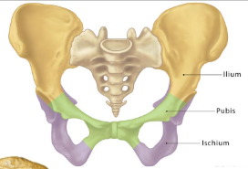
What makes up the pelvic girdle?
sacrum and coccyx, hip bones (ilium + pubis + ischium - fuse at the acetabulum)
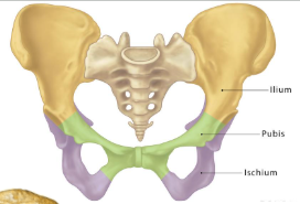
What are another names for the hip bone?
os coxae or innominate bone
What are the joints of the pelvis?
sacroiliac joint, pubic symphysis and hip joint
What does the hip joint transition between ?
trunk and lower limb
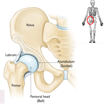
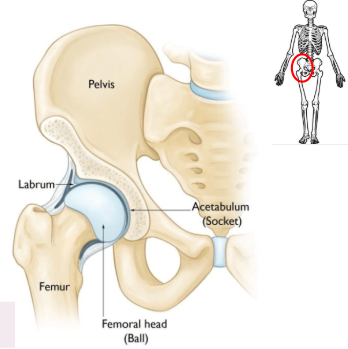
What does the hip joint join between?
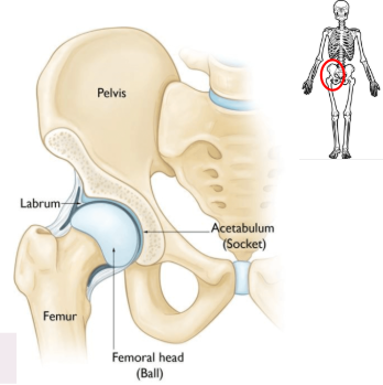
hip bone (acetabulum and labrum) and femur (femoral head)
Why is the hip joint termed multiaxial?
allows flexion, extension, abduction, adduction, external (lateral) rotation, internal (medial) rotation and circumduction
Define the labrum
ring of cartilage that surrounds the brim of the acetabulum. Stabilises and lubricates joint, and absorbs shock
What are the capsular ligaments of the hip joint?
iliofemoral, pubofemoral and ischiofemoral
What distinguishes capsular from intracapsular joints?
thickened intrinsic parts of joint that provide stability // separate structures located in the synovial capsule that guide complex movements
synovial joint meaning
most common, freely movable joint type in the human body, characterized by a fluid-filled cavity (synovial cavity) enclosed by a fibrous capsule eg shoulder, hips, knees, elbows - allow for extensive movement
synovial capsule meaning
two-layered, fibrous envelope surrounding synovial joints that separates them from other tissues, providing structural stability and enclosing the synovial cavity
What are the intracapsular ligaments of the hip joint?
transverse acetabular ligament, ligament of the head of the femur (LHF)
What are other terms for the ligament of the head of the femur?
ligamentum capitis femoris, ligamentum teres
Identify the ligaments of the hip joint
anterior capsular ligaments so iliofemoral and pubofemoral are visible
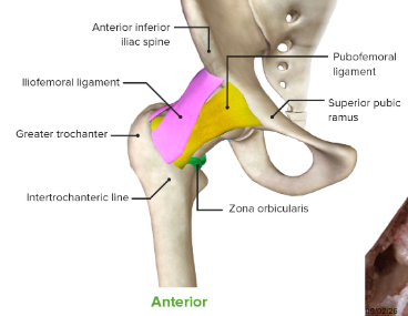
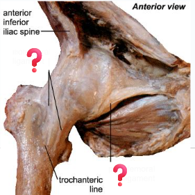
Identify the capsular ligaments of the hip joint
anterior capsular ligaments so iliofemoral and pubofemoral are visible
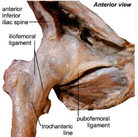
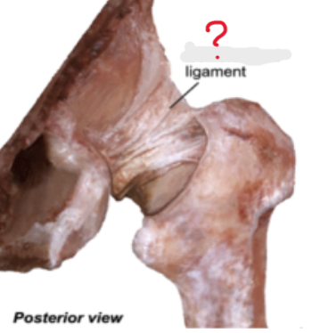
Which joint is on this photo ? What gives this away?
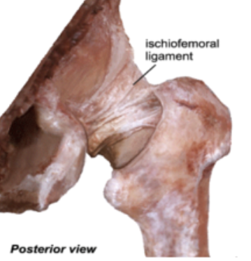
ischiofemoral ligament - posterior view
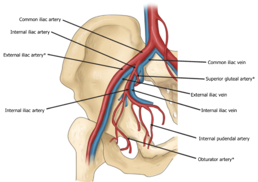
Describe the blood supply to the hip joint?
Common iliac artery → internal iliac artery → superior gluteal artery then internal iliac artery off of which comes the obturator artery + deep femoral artery from external iliac artery
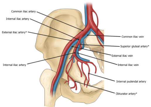
Which arteries anastomose off of the common iliac artery to provide blood to the hip joint?
deep femoral artery, obturator, superior and inferior gluteal arteries
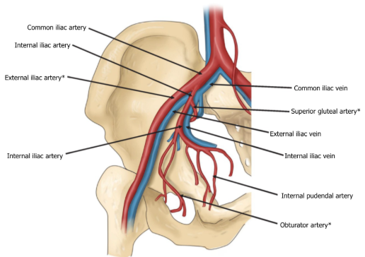
Which two arteries give rise to the arteries that supply the hip bone? Which ones?
external iliac artery - deep femoral artery
internal iliac artery - superior gluteal artery and inferior gluteal artery off of which comes the obturator artery
both from common iliac artery (L4)
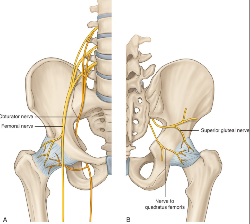
Which 4 nerves innervate the hip joint?
femoral, obturator, superior gluteal and quadratus femoris nerves
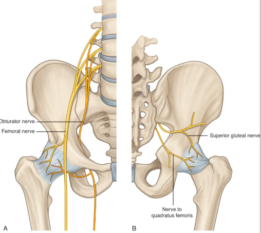
Anterior innervation of the hip joint/anterior compartment thigh
femoral nerve
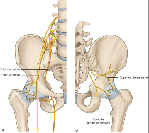
Inferior innervation of the hip joint/medial compartment thigh
obturator nerve
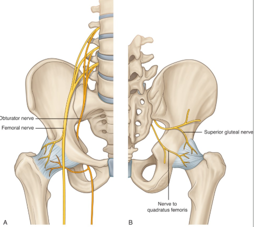
Superior innervation of the hip joint/gluteus medius and minimus mm
superior gluteal nerve
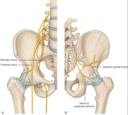
Posterior innervation of the hip joint/quadratus femoris and inferior gemellus mm
nerve to quadratus femoris nerve
Locate the gluteal region relative to the pelvic girdle, iliac crest and gluteal fold
posterior to pelvic girdle, inferior to iliac crest and superior to gluteal fold
What are the 2 muscular groups of the gluteal region?
gluteal group and lateral rotator group
Describe the size of the muscles of the gluteal group vs lateral rotator group of muscles of the gluteal region
superficial, large muscles vs deep, small muscles
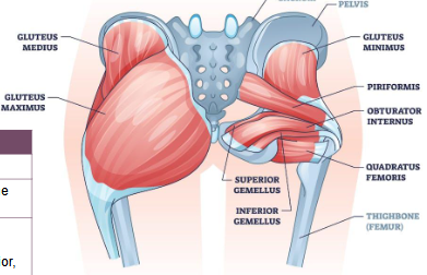
Movement gluteal group
abduct or extend the femur at the hip joint + stabilisation
Movement lateral rotator group
mainly laterally rotate the femur at the hip joint
List the muscles that make up the gluteal group of the gluteal region
Gluteus maximus, gluteus medius, gluteus minimus and tensor fascia latae
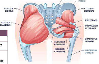
List the muscles that make up the lateral rotator group of the gluteal region
Quadratus femoris, piriformis, gemellus superior, gemellus inferior, obturator internus, and obturator externus
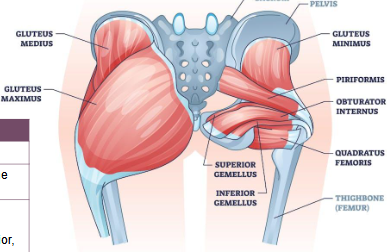
blood supply to the gluteal group of muscles of the gluteal region
superior and inferior gluteal arteries
Mvt gluteus maximus
Main extensor of the thigh, assists with lateral rotation if force is needed
Mvt gluteus medius
Abduction and medial rotation of the thigh, stabilises pelvis on locomotion (medi is medial)
Mvt gluteus minimus
Abduction and medial rotation of the thigh, stabilises pelvis on locomotion (mini is medial mvt)
Innervation gluteus maximus
inferior gluteal nerve (extends lower than the other 2 muscles)
Innervation gluteus medius
superior gluteal nerve (inserts higher than glut max)
Innervation gluteus minimus
superior gluteal nerve (inserts higher than glux max)
What is the innervation of both the tensor fascia and latae and the gluteus minimus/medius ?
superior gluteal nerve
What is the role of the tensor fascia latae?
Stabilises hip and knee joints and supports gait cycle and climbing via iliotibial band, assists gluteus medius and minimus (abduction and medial rotation of the thigh)
What is the blood supply to the gluteal group of the gluteal region and the tensor fascia latae?
superior and inferior gluteal arteries
What is the role of the iliotibial band/tract?
fibrous reinforcement of the fascia lata on the lateral thigh. Stabilises primarily the knee, but also hip joint, during locomotion and against gravity on knee flexion
What is the fascia lata ?
deep fascia of the thigh
What is the reinforcement of the fascia lata of the thigh called?
iliotibial band
Which 3 structures share a same function of stabilisation, abduction and medial rotation of the thigh ?
gluteus minimus, medius and tensor fascia latae
Nerve piriformis
nerve to piriformis
Nerve quadratus femoris
nerve to quadratus femoris
What is the movement of the piriformis ?
lateral rotation of the thigh, abduction when hip is flexed
What is the movement of the quadratus femoris ?
lateral rotation of the thigh, weak adduction of the thigh
What is the blood supply to the lateral rotator group of the gluteal region?
superior and inferior gluteal arteries
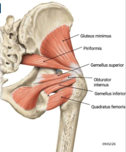
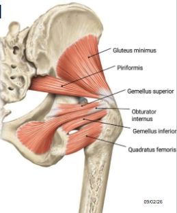
Locate the muscles of the lateral rotator group of the gluteal region on the following image
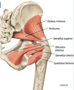
Innervation of superior gemellus
nerve to obturator internus (muscle located just above and piggy backs the innervation of obturator internus)
Innervation of obturator internus
nerve to quadratus femoris
Innervation of inferior gemellus
nerve to quadratus femoris (muscle located just above and piggy backs the innervation of quadratus femoris)
Mvt of superior gemellus
Lateral rotation of the thigh, abduction when hip is flexed
Mvt of obturator internus
Lateral rotation of the thigh, abduction when hip is flexed
Mvt of inferior gemellus
Lateral rotation of the thigh, abduction when hip is flexed
What is the longest and widest single nerve in the body?
sciatic nerve
What is the course of the sciatic nerve ?
from anterior lumbar and sacral nerve fibres from the spinal cord
nerve roots unite into single nerve anterior to piriformis muscle
exits pelvis between piriformis and superior gemellus into gluteal region
travels down into posterior thigh
Below which muscle do the inferior gluteal artery and nerve go below?
piriformis
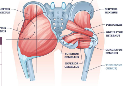
When administering a shot in the gluteal region, which site is preferred ? What are risks of doing it in a different region?
cross on the gluteus maximus - left upper corner is target region
jab can be very jerksome/can induce damage to the sciatic nerve leading to paralysis of the lower limbs
From which collection of nerves does the sciatic nerve come from? Where does it originate?
lumbosacral plexus - originates from vertebrae L4 to S3
What joints define the upper and lower thigh?
hip and knee joints
Which bone is located in the thigh?
femur
What are the 3 fascial/muscular compartments of the thigh?
anterior, medial and posterior
Which 3 muscles make up the posterior compartment of the thigh?
biceps femoris, semitendinosus and semimembranosus
What is the blood supply to the posterior muscles of the thigh?
inferior gluteal artery (proximal) and deep femoral artery
Innervation of posterior muscles of the thigh
sciatic nerve
What are the posterior muscles of the thigh called as a unit?
hamstrings
Which muscles of the thigh cross the hip vs the knee and therefore what are their movements?
long head of biceps femoris crosses over hip so extends it + semi-tendinosus/mebranosus // all three cross over the anterior knee joint so all flex it
Movements of the posterior compartment muscles
extension of hip and flexion of knee
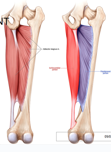
Describe the structure of the adductor magnus
posterior and anterior portions, the two portions of the muscle don’t lie laterally but at a slight angle
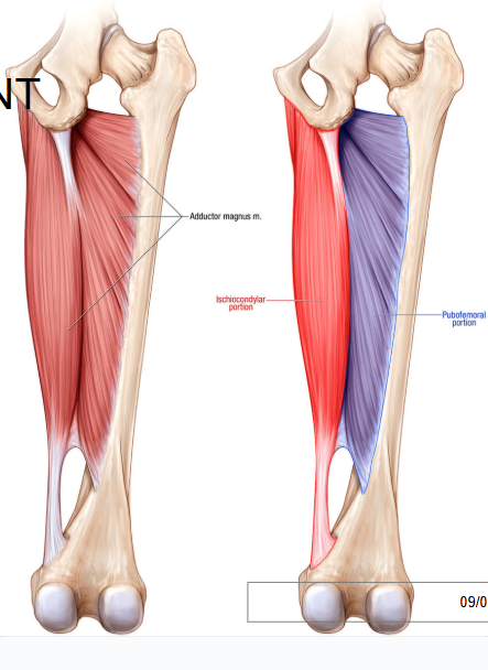
Role of pubofemoral portion of the adductor magnus of the posterior compartment of the thigh
flexion of hip
Role of ischiocondylar portion of the adductor magnus of the posterior compartment of the thigh
extension of the hip
Innervation of adductor magnus
sciatic and obturator nerves
Blood supply to adductor magnus
deep femoral artery
Which portion of the adductor magnus is innervated and has the same movement as the hamstring?
ischiocondylar portion
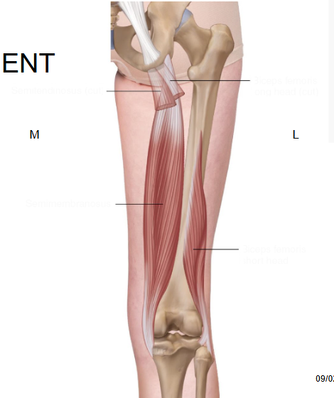
Label the hamstrings
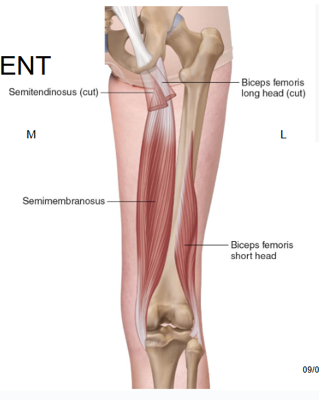
What is an easy way to distinguish semitendinosus and semimembranosus muscles of the posterior compartment of the thigh?
tendinosus looks like a tendon, membranosus is flat and looks like a membrane
What is the course of the sciatic nerve on the posterior thigh?
from gluteal region → between adductor magnus and biceps femoris, lateral to semimembranosus/semitendinosus
divides at apex of popliteal fossa (behind knee)
What are the 2 divisions of the sciatic nerve on the posterior thigh?
tibial and common fibular nerves
What role do gluteus medius and minimus have in walking?
stabilise the pelvis while one leg is up - more pronounced in women bc of the larger pelvis, allow counter balance when walking
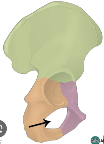
Describe the obturator foramen
large paired opening below acetabulum, covered by thin fibrous membrane leaving an obturator canal through which the obturator nerve, artery and vein pass
Which artery gives the deep femoral artery (similar to the arm blood supply)
femoral artery comes off external iliac artery and deep brachial artery comes off and wraps around to posterior compartment of the thigh (like deep brachial artery in arm)
What is an easy way to identify the ischiofemoral ligament of the hip joint?
only posterior one, other 2 are anterior + has horizontal fibres
What is the role of LHF? What differs between this ligament in children vs in adulthood ?
anchors head of femur, making up for the horseshoe shape of the acetabulum and maintaining stability at the joint
in children has its own artery // in adulthood regresses to a ligament
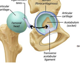
What is the aspect of pubofemoral and iliofemoral ligaments of the hip joint?
both melded in cadavers, only distinction is relative positions, overlap in the middle where they join
congruent meaning
degree to which opposing articular surfaces of a joint fit together
How congruent are the acetabulum and head of femur?
very ! removing makes a popping sound, fit very well together, head of femur is very round and goes deep into socket
At which point could the hip joints open? Under action of what?
during childbirth only - fibres or joint relax and cartilage enlarge - under action of relaxin hormone
At what stage of life do the bones of the pubis fuse forming the socket of the hip bone (acetabulum)?
adolescence
What is the most anterior part of the pelvis?
pubic symphysis cartilage
What is the leg?
between knee and ankle joints
What are the 2 surfaces of the ala ?
pelvic (anterior) and dorsal (posterior)