chapter 50 sense reception
1/26
There's no tags or description
Looks like no tags are added yet.
Name | Mastery | Learn | Test | Matching | Spaced | Call with Kai |
|---|
No analytics yet
Send a link to your students to track their progress
27 Terms
sense receptors
mechanoreceptors, thermoreceptors, proprioceptors, nociceptors, chemopreceptors, photoreceptrs, electroreceptors, and magnetoreceptors
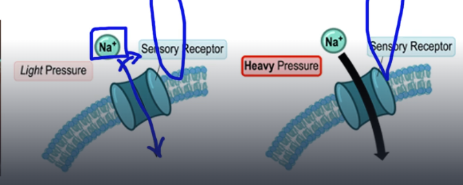
Hair cell and stereocilia
used by auditory and vestibular system, respond to physical stimulation to open ion channels. Stereocilia: projections on hair cells, that can be physically manipulated to change membrane potentials. bending opens ion channels, creating a graded potential, to transduce a signal.
statocyst, statolith
statocyst: balance sensory receptor, uses gravity to stimulate hair cells in dac like structure.statolih: mineral ctstals that touch and stimulate hair cells.
hearing:
ear transducing pressure waves in the air
sound, frequency, and pitch
sound: vibration that sends pressure wave through air. Frequency: cycles per second of the wave. Pitch: perception of frequency
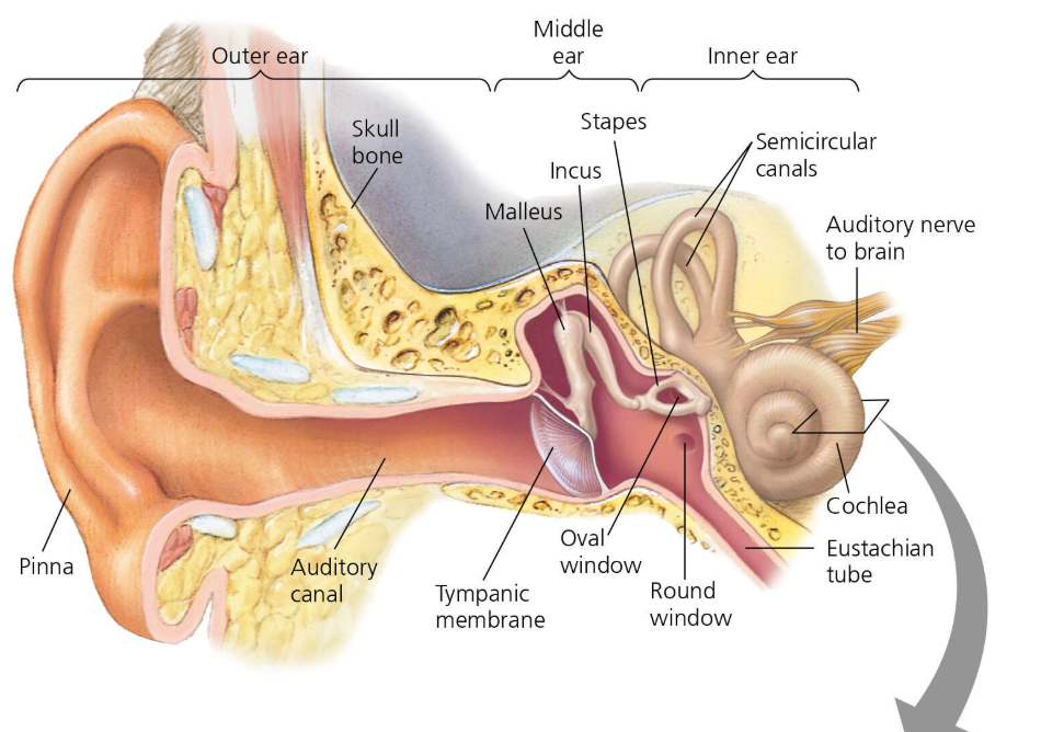
Tympanic membrane:
ear drum: thin membrane that seperates outer and middle ear.

middle ear: bones that amplify sound
Eustachian tube, ossicles, oval window
Eustachian tube, ossicles, oval window
Eustachian tube: connects middle ear to nasopharynx. Ossicles: bones that amplify sound in middle ear: Malleus, incus, and stapes. Oval window : membrane covered opening connects middle to inner ear.
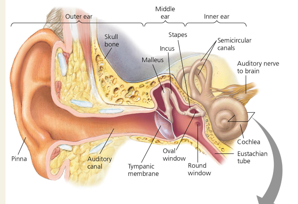
Inner ear
contains hair cells receptors responsible for sound detection and balance.
Vestibular system
Balance system: uses fluid to stimulate hair cells. Semicircular canals: fluid filled canals that detect motion.
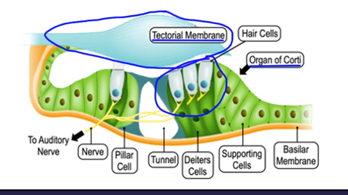
Cochlea
spiral shaped cavity containing hair cells that detects sounds
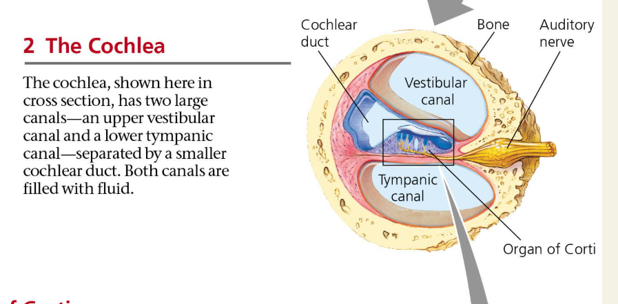
Cochlear membrane:
three ducts: Basilar membrane: membrane in the middle that sits under the hair cilia. organ of corti: sits on basilar membrane and contains many hair cells that detect sound!!. Tectorial membrane: located, like an awning, above the organ of corti.
Round window
dampens waves. located in inner ear
Main parts of the eye
Sclera, cornea, iris, pupil, lens, Retina,
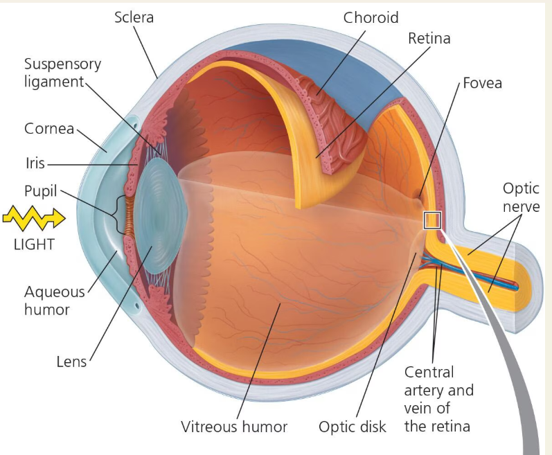
sclera
white of the eye: collagen and elastic fiber
Cornea
fluid filled transparent cover, over iris and pupil,
Iris
controls puil diameter and lens shape
pupil
hole lets light into lens
lens
changes shape to focus light from cornea
Retina
back of the eye containing photorecepots
photoreceptors in retina
Rods and cones
RODS
detect low levels of light and they are found around edges of the retina
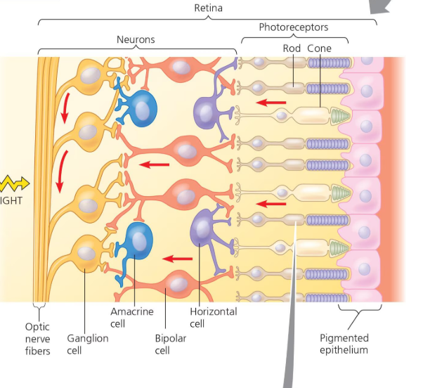
rhodopsin
light sensitive receptor protein in rods
Retinal/opsin
Retinal: light absorbing molecule: Opsin: light sensitive protein: acts as g receptor protein that leads to the opening of NA+ ion channels.
cones
best for color vision
Retina composition
made of photoreceptors, bipolar cells, and ganglion cells. REceptor potential is generated by hyperpolarization of from opening of NA+ channels
Photo receptors and bipolarcells have graded potentials, ganglion cells have action poentials.
Ganglion cells, receive input from multiple rods and cones