2 - Methods & Microbe Host interactions
1/117
There's no tags or description
Looks like no tags are added yet.
Name | Mastery | Learn | Test | Matching | Spaced | Call with Kai |
|---|
No analytics yet
Send a link to your students to track their progress
118 Terms
b. Simple microscope
[Methods - Microscopy]
Microscope with a single lens.
a. Compound microscope
b. Simple microscope
c. Electron microscope
d. Fluorescence microscope
b. Compound microscope
[Methods - Microscopy]
Microscope with multiple lenses for higher magnification.
a. Simple microscope
b. Compound microscope
c. Light microscope
d. Stereomicroscope
b. Scanning electron microscopy
[Methods - Microscopy]
Type of electron microscopy that produces three-dimensional images.
a. Transmission electron microscopy
b. Scanning electron microscopy
c. Confocal microscopy
d. Dark-field microscopy
b. Transmission electron microscopy
[Methods - Microscopy]
Type of electron microscopy that produces two-dimensional images.
a. Scanning electron microscopy
b. Transmission electron microscopy
c. Bright-field microscopy
d. Phase-contrast microscopy
c. Bright-field microscopy
[Methods - Microscopy]
Microscopy technique with a bright background and darker organism.
a. Dark-field microscopy
b. Fluorescence microscopy
c. Bright-field microscopy
d. Phase-contrast microscopy
b. Dark-field microscopy
[Methods - Microscopy]
Microscopy principle in which organisms appear light against a dark background.
a. Bright-field microscopy
b. Dark-field microscopy
c. Fluorescence microscopy
d. Transmission electron microscopy
c. Dark-field microscopy
[Methods - Microscopy]
Microscopy technique applicable for visualizing spirochetes.
a. Bright-field microscopy
b. Phase-contrast microscopy
c. Dark-field microscopy
d. Fluorescence microscopy
b. Treponema pallidum
[Methods - Microscopy]
Spirochete causative agent of syphilis.
a. Borrelia burgdorferi
b. Treponema pallidum
c. Leptospira interrogans
d. Spirillum minus
c. Genital chancre
[Methods - Microscopy]
BEQ: Primary lesion of syphilis caused by Treponema pallidum.
a. Papule
b. Pustule
c. Genital chancre
d. Vesicle
d. Scraping
[Methods - Microscopy]
BEQ: Specimen collection method for early diagnosis of syphilis from genital chancre.
a. Swabbing
b. Biopsy
c. Culture
d. Scraping
b. Dark-field microscopy
[Methods - Microscopy]
Microscopy technique used for early diagnosis of syphilis from scraped genital chancre material.
a. Bright-field microscopy
b. Dark-field microscopy
c. Fluorescence microscopy
d. Electron microscopy
b. Simple stain
[Methods - Stain / Dye]
Staining technique using a single dye.
a. Differential stain
b. Simple stain
c. Composite stain
d. Negative stain
c. Chromophore
[Methods - Stain / Dye]
The component of a dye responsible for giving color in staining.
a. Chromophobe
b. Auxochrome
c. Chromophore
d. Mordant

b. Basic dye
[Methods - Stain / Dye]
Type of dye with cationic (+) charge used in simple staining of bacteria.
a. Acidic dye
b. Basic dye
c. Neutral dye
d. Amphoteric dye
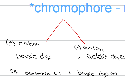
c. Acidic dye
[Methods - Stain / Dye]
Type of dye with anionic (-) charge in simple staining.
a. Basic dye
b. Cationic dye
c. Acidic dye
d. Zwitterionic dye
b. Bacteria CS + basic dye
[Methods - Stain / Dye]
Example of simple staining where bacteria interact with cation and basic dye.
a. Bacteria + acidic dye
b. Bacteria CS + basic dye
c. Bacteria + mordant
d. Bacteria + decolorizer
c. Differential stain
[Methods - Stain / Dye]
Staining technique that can be used to classify and differentiate organisms.
a. Simple stain
b. Negative stain
c. Differential stain
d. Shadow stain
Gram stain
Acid fast stain
[Methods - Stain / Dye]
Types of differential stain [2]
c. Gram stain
[Methods - Stain / Dye]
Differential stain used to differentiate Gram-positive and Gram-negative bacteria.
a. Acid fast stain
b. Methylene blue stain
c. Gram stain
d. Malachite green stain
c. Acid fast stain
[Methods - Stain / Dye]
Differential stain used to identify acid-fast organisms such as Mycobacterium.
a. Gram stain
b. Simple stain
c. Acid fast stain
d. Capsule stain
Ziehl - Neelsen
Kinyoun method
Acid fast staining technique [2]
b. Ziehl-Neelsen method
[Methods - Stain / Dye]
Acid fast staining technique that uses heat during the staining process.
a. Kinyoun method
b. Ziehl-Neelsen method
c. Loeffler method
d. Albert method
c. Kinyoun method
[Methods - Stain / Dye]
Acid fast staining technique that does not require heat application.
a. Ziehl-Neelsen method
b. Loeffler method
c. Kinyoun method
d. Modified acid fast method
b. Ziehl-Neelsen uses heat; Kinyoun uses cold
[Methods - Stain / Dye]
Key difference between Ziehl-Neelsen and Kinyoun acid fast staining techniques.
a. Different mordants used
b. Ziehl-Neelsen uses heat; Kinyoun uses cold
c. Different decolorizers used
d. Ziehl-Neelsen requires extended incubation
Carbolfuchsin
Heat
Alcohol
Methylene blue
= Acid fast
📌Mnemonic: “CHAMBA”
[Methods - Stain / Dye]
Steps in acid fast staining [4]
Mycobacterium spp.
Nocardia spp.
[Methods - Stain / Dye]
Acid-fast organisms include ______ [2]
a. Staphylococcus spp.
b. Nocardia spp.
c. Streptococcus spp.
d. E. coli
b. Feulgen stain
[Methods - Stain / Dye]
Special staining technique used to visualize bacterial nuclei.
a. Methylene blue stain
b. Feulgen stain
c. Malachite green stain
d. Carbol-fuchsin stain
a. India ink stain / Nigrosin
[Methods - Stain / Dye]
BEQ: Staining technique used to visualize bacterial capsules.
a. India ink stain
b. Malachite green stain
c. Feulgen stain
d. Gram stain
b. Cryptococcus Neoformans
[Methods - Stain / Dye]
Encapsulated organism commonly stained using India ink or Nigrosin for capsule visualization.
a. Staphylococcus aureus
b. Cryptococcus Neoformans
c. Streptococcus pyogenes
d. Bacillus anthracis
c. Welch method
[Methods - Stain / Dye]
Capsule staining method involving precipitation of glacial acetic acid.
a. Gram method
b. India ink method
c. Welch method
d. Loeffler method
Schaeffer-Fulton method
Malachite green stain
📌Mnemonic: “SM”
[Methods - Stain / Dye]
Spore staining technique [2]
a. Malachite green stain
[Methods - Stain / Dye]
Special stain used to visualize bacterial spores.
a. Malachite green stain
b. Nigrosin stain
c. Feulgen stain
d. Methylene blue stain
b. Schaeffer-Fulton method
[Methods - Stain / Dye]
Spore staining technique that uses malachite green and heat.
a. Kinyoun method
b. Schaeffer-Fulton method
c. Welch method
d. Loeffler method
Bacillus spp.
Clostridium spp.
📌Mnemonic: “BC”
[Methods - Stain / Dye]
Two spore-forming bacteria genera commonly identified using Schaeffer-Fulton staining:
a. Bacillus and Streptococcus
b. Bacillus and Clostridium
c. Clostridium and Pseudomonas
d. Bacillus and Salmonella
b. Carbolfuschsin stain
[Methods - Stain / Dye]
Staining technique used to visualize bacterial flagella.
a. Malachite green stain
b. Carbolfuschsin stain
c. India ink stain
d. Nigrosin stain
c. Culture studies
[Methods - Culture Studies]
Gold standard for bacterial infections.
a. Gram stain
b. Sensitivity testing
c. Culture studies
d. PCR
c. Takes long time (matagal ang result)
[Methods - Culture Studies]
Primary disadvantage of culture studies for bacterial infection diagnosis.
a. Low sensitivity
b. Requires expensive equipment
c. Takes long time
d. Cannot identify organism type

b. Inoculation into culture medium
[Methods - Culture Studies]
Initial step in bacterial culture studies from a clinical sample.
a. Identification of organism
b. Inoculation into culture medium
c. Antibiotic susceptibility testing
d. Gram staining

b. Allow organism growth
[Methods - Culture Studies]
Purpose of culture medium inoculation in bacterial culture studies.
a. Kill contaminating organisms
b. Allow organism growth
c. Perform antibiotic sensitivity
d. Identify organism morphology

b. Identification of organism
[Methods - Culture Studies]
After growth is observed in culture, the next step is:
a. Antibiotic susceptibility testing
b. Identification of organism
c. Gram staining only
d. Report results

c. Identification antibiotic resistance and susceptibility
[Methods - Culture Studies]
Final step in culture studies to determine appropriate antibiotic therapy.
a. Organism identification
b. Colony morphology assessment
c. Identification antibiotic resistance and susceptibility
d. Repeat culture
Blood
CSF
Pleural fluid
Pericardial fluid
Synovial fluid
[Methods - Culture Studies]
Example of sterile body fluids [5]
c. Basal/Simple medium
[Methods - Culture Studies]
Culture medium type with minimal nutrients
a. Enriched medium
b. Selective medium
c. Basal/Simple medium
d. Differential medium
c. Basal/Simple medium
[Methods - Culture Studies]
Culture medium type that supports non-fastidious organisms.
a. Enriched medium
b. Selective medium
c. Basal/Simple medium
d. Differential medium
c. Nutrient agar
[Methods - Culture Studies]
Example of a basal/simple culture medium.
a. Chocolate agar plate
b. Lowenstein-Jensen medium
c. Nutrient agar
d. Blood agar plate
b. Enriched medium
[Methods - Culture Studies]
Culture medium type with added growth factors for fastidious organisms.
a. Basal medium
b. Enriched medium
c. Selective medium
d. Indicator medium
Lowenstein-Jensen
Chocolate Agar Plate
[Methods - Culture Studies]
Example of Enriched culture medium [2]
c. Lowenstein-Jensen medium
[Methods - Culture Studies]
Enriched culture medium used for Mycobacterium tuberculosis.
a. Nutrient agar
b. Chocolate agar plate
c. Lowenstein-Jensen medium
d. Blood agar plate
c. Chocolate agar plate
[Methods - Culture Studies]
Enriched culture medium used for Haemophilus influenzae.
a. Nutrient agar
b. Lowenstein-Jensen medium
c. Chocolate agar plate
d. MacConkey agar
c. Selective medium
[Methods - Culture Studies]
Culture medium type that inhibits some organisms while allowing others to grow.
a. Basal medium
b. Enriched medium
c. Selective medium
d. Differential medium
c. Colistin-Nalidixic acid
[Methods - Culture Studies]
Example of Selective Culture medium
a. Chocolate agar plate
b. Anaerobic media
c. Colistin-Nalidixic acid
d. MacConkey agar
c. Colistin-Nalidixic acid
[Methods - Culture Studies]
Selective medium that allows only Gram-positive organisms to grow.
a. Chocolate agar plate
b. Anaerobic media
c. Colistin-Nalidixic acid
d. MacConkey agar
d. Indicator/Differential medium
[Methods - Culture Studies]
Culture medium type that distinguishes organisms based on their characteristics.
a. Basal medium
b. Selective medium
c. Enriched medium
d. Indicator/Differential medium
Anaerobic media
MCA,EMB
BAP
[Methods - Culture Studies]
Example of an indicator/differential culture medium [3]
b. Anaerobic medium
[Methods - Culture Studies]
Type of differential medium that is reducing in nature.
a. Lactose fermenter medium
b. Anaerobic medium
c. Blood agar plate
d. MacConkey agar
c. Candle jar method
[Methods - Culture Studies]
Example of an anaerobic medium technique used in microbiology.
a. Streak plate method
b. Pour plate method
c. Candle jar method
d. Spread plate method
b. MacConkey Agar
[Methods - Culture Studies]
Full name of MCA used to identify lactose fermenters.
a. Modified Culture Agar
b. MacConkey Agar
c. Mannitol Crystal Agar
d. Modified Chrome Agar
a. Eosin-Methylene Blue
[Methods - Culture Studies]
Full name of EMB used to identify lactose fermenters.
a. Eosin-Methylene Blue
b. Erythrosine-Methylene Blue
c. Eosin-Malachite Blue
d. Eosin-Methyl Blue
c. Pink
[Methods - Culture Studies]
Color produced by lactose fermenters on MacConkey Agar and EMB.
a. Colorless
b. Blue
c. Pink
d. Yellow
d. Colorless
[Methods - Culture Studies]
Color produced by non-lactose fermenters on MacConkey Agar and EMB.
a. Pink
b. Green metallic
c. Red
d. Colorless
c. E. coli
[Methods - Culture Studies]
Lactose fermenter that produces green metallic color on EMB.
a. Klebsiella
b. Serratia
c. E. coli
d. Enterobacter
E.coli
Klebsiella
Enterobacter
Citrobacter
Serration
📌Mnemonic: “EKECS
[Methods - Culture Studies]
Examples of Lactose Fermenter [4]
Shigella
Salmonella
Yersinia
Proteus
Pseudomonas
📌Mnemonic: “SS” Y “PP”
[Methods - Culture Studies]
Examples of Non - Lactose Fermenter [5]
c. Blood agar plate
[Methods - Culture Studies]
Differential medium used to observe hemolytic pattern of Streptococcus spp.
a. MacConkey agar
b. EMB agar1
c. Blood agar plate
d. Nutrient agar
d. Alpha hemolysis
[Methods - Culture Studies]
Hemolysis pattern characterized by partial hemolysis and green discoloration.
a. Beta hemolysis
b. Gamma hemolysis
c. Delta hemolysis
d. Alpha hemolysis
b. Beta hemolysis
[Methods - Culture Studies]
Hemolysis pattern characterized by complete hemolysis and clear zone.
a. Alpha hemolysis
b. Beta hemolysis
c. Gamma hemolysis
d. Delta hemolysis
c. Gamma hemolysis
[Methods - Culture Studies]
Hemolysis pattern characterized by no hemolysis and pink discoloration.
a. Alpha hemolysis
b. Beta hemolysis
c. Gamma hemolysis
d. Delta hemolysis
d. Green
[Methods - Culture Studies]
Color associated with alpha hemolysis on blood agar plate.
a. Clear
b. Pink
c. Yellow
d. Green
c. Clear
[Methods - Culture Studies]
Color associated with beta hemolysis on blood agar plate.
a. Green
b. Pink
c. Clear
d. Yellow
d. Pink
[Methods - Culture Studies]
Color associated with gamma hemolysis on blood agar plate.
a. Clear
b. Green
c. Yellow
d. Pink
c. Symbiosis
[Microbe Host Interactions - Symbiosis]
A type of relationship between microbe and host.
a. Infection
b. Competition
c. Symbiosis
d. Colonization
d. Mutualism
[Microbe Host Interactions - Symbiosis]
Type of symbiosis where both organisms benefit from each other.
a. Commensalism
b. Parasitism
c. Predation
d. Mutualism
b. Normal colonic flora
[Microbe Host Interactions - Symbiosis]
Example of mutualism between microbe and host.
a. Staphylococcus epidermidis
b. Normal colonic flora
c. Balatindium coli
d. Bdellovibrio bacteriovorus

c. Vitamin K
[Microbe Host Interactions - Symbiosis]
Vitamin produced by normal colonic flora benefiting the host.
a. Vitamin A
b. Vitamin C
c. Vitamin K
d. Vitamin D
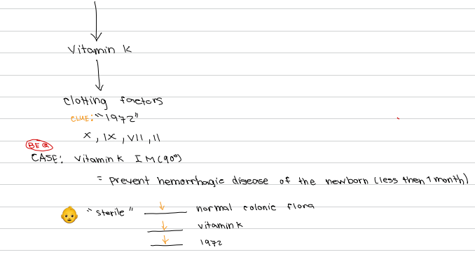
d. X, IX, VII, II
📌Clue: “1972”
[Microbe Host Interactions - Symbiosis]
Clotting factors produced through Vitamin K from normal colonic flora.
a. I, II, III, IV
b. V, VI, VIII, IX
c. XI, XII, XIII, XIV
d. X, IX, VII, II

d. Hemorrhagic disease of the newborn
[Microbe Host Interactions - Symbiosis]
BEQ: Condition prevented by Vitamin K IM (90°) given to newborns less than 1 month old.
a. Neonatal jaundice
b. Neonatal sepsis
c. Hemolytic disease of the newborn
d. Hemorrhagic disease of the newborn

Decrease normal colonic flora
Decrease Vitamin K
Decrease Clotting factors X, IX, VII, II (1972)
[Microbe Host Interactions - Symbiosis]
In a sterile newborn → ______ [3]
b. Commensalism
[Microbe Host Interactions - Symbiosis]
Type of symbiosis where one organism benefits while the other does not benefit.
a. Mutualism
b. Commensalism
c. Parasitism
d. Predation
c. Staphylococcus epidermidis
[Microbe Host Interactions - Symbiosis]
Normal flora of the skin that exemplifies commensalism.
a. Normal colonic flora
b. Balatindium coli
c. Staphylococcus epidermidis
d. Bdellovibrio bacteriovorus
d. Parasitism
[Microbe Host Interactions - Symbiosis]
Type of symbiosis where one organism benefits while the other gets hurt.
a. Mutualism
b. Commensalism
c. Predation
d. Parasitism
d. Balatindium coli
[Microbe Host Interactions - Symbiosis]
Protozoan parasite that causes dysentery and exemplifies parasitism.
a. Giardia lamblia
b. Entamoeba histolytica
c. Trichomonas vaginalis
d. Balatindium coli
c. Protozoa
[Microbe Host Interactions - Symbiosis]
Classification of Balatindium coli as a microorganism.
a. Bacteria
b. Fungi
c. Protozoa
d. Helminth
c. Dysentery
[Microbe Host Interactions - Symbiosis]
Disease caused by Balatindium coli in parasitism.
a. Malaria
b. Meningitis
c. Dysentery
d. Pneumonia
d. Predation
[Microbe Host Interactions - Symbiosis]
Type of symbiosis where one organism benefits while the other dies.
a. Commensalism
b. Parasitism
c. Mutualism
d. Predation
d. Bdellovibrio bacteriovorus
[Microbe Host Interactions - Symbiosis]
Organism that acts as predator against gram negative bacteria.
a. Staphylococcus epidermidis
b. Normal colonic flora
c. Balatindium coli
d. Bdellovibrio bacteriovorus
c. Pathogenicity
[Microbe Host Interactions - Features of Infectious Diseases]
Ability to cause disease.
a. Virulence
b. Toxigenicity
c. Pathogenicity
d. Invasiveness
d. Virulence
[Microbe Host Interactions - Features of Infectious Diseases]
Quantitative ability to cause disease.
a. Pathogenicity
b. Invasiveness
c. Toxigenicity
d. Virulence
b. Opportunistic pathogen
[Microbe Host Interactions - Features of Infectious Diseases]
Type of pathogen that causes disease in immunocompromised or malnourished individuals.
a. Primary pathogen
b. Opportunistic pathogen
c. Obligate pathogen
d. Commensal pathogen
a. Malnutrition = immunocompromise
[Microbe Host Interactions - Features of Infectious Diseases]
Condition that predisposes a host to opportunistic pathogens.
a. Malnutrition = immunocompromise
b. Obesity
c. Hypertension
d. Diabetes mellitus
b. Pneumocystis carinii
[Microbe Host Interactions - Features of Infectious Diseases]
Old name of Pneumocystis jiroveci.
a. Pneumocystis pneumoniae
b. Pneumocystis carinii
c. Pneumocystis aeruginosa
d. Pneumocystis fumigatus
c. Pneumocystis jiroveci
[Microbe Host Interactions - Features of Infectious Diseases]
New name of Pneumocystis carinii.
a. Pneumocystis pneumoniae
b. Pneumocystis fumigatus
c. Pneumocystis jiroveci
d. Pneumocystis aeruginosa
d. PCP
[Microbe Host Interactions - Features of Infectious Diseases]
Pneumonia caused by Pneumocystis jiroveci in HIV patients.
a. CAP
b. HAP
c. VAP
d. PCP
b. Cotrimoxazole
[Microbe Host Interactions - Features of Infectious Diseases]
Drug of choice for PCP pneumonia.
a. Amoxicillin
b. Cotrimoxazole
c. Azithromycin
d. Doxycycline
Invasiveness
Toxigenicity
[Microbe Host Interactions - Features of Infectious Diseases]
Determinants for Virulence [2]
c. Invasiveness
[Microbe Host Interactions - Features of Infectious Diseases]
Determinant of virulence referring to ability to spread from one part of the body to another.
a. Toxigenicity
b. Pathogenicity
c. Invasiveness
d. Virulence
d. Toxigenicity
[Microbe Host Interactions - Features of Infectious Diseases]
Determinant of virulence referring to ability to cause toxin production.
a. Invasiveness
b. Pathogenicity
c. Virulence
d. Toxigenicity
c. Reservoir
Continual source of an organism in infectious disease transmission.
a. Host
b. Vector
c. Reservoir
d. Fomite
Humans
Animals
Example of Living reservoir of infectious organisms include _____ [2]
c. Zoonotic infections
Infections transmitted from animals to humans.
a. Nosocomial infections
b. Opportunistic infections
c. Zoonotic infections
d. Iatrogenic infections
d. Cows
Animal reservoir of Brucella spp.
a. Pigs
b. Rats
c. Wild birds
d. Cows