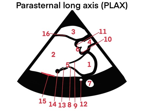
Echo Views and Measurements
1/94
There's no tags or description
Looks like no tags are added yet.
Name | Mastery | Learn | Test | Matching | Spaced | Call with Kai |
|---|
No analytics yet
Send a link to your students to track their progress
95 Terms
7
DAo

8
posterior MV leaflet

9
anterior MV leaflet

10
aortic valve, non coronary cusp

11
aortic valve, right coronary cusp

12
coronary sinus

15
posterior wall
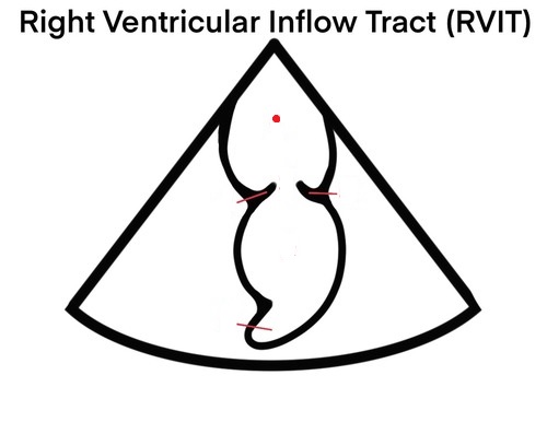
RV
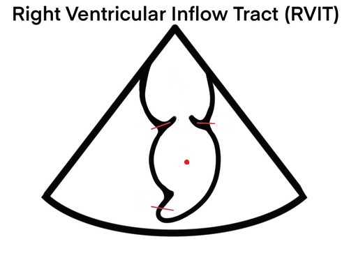
RA

TV
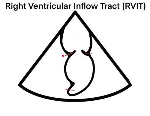
posterior TV leaflet
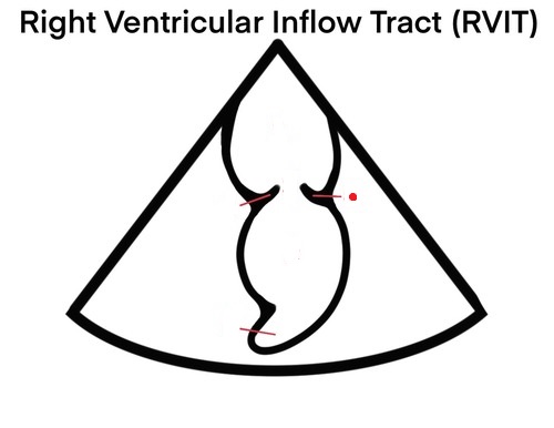
Anterior TV leaflet
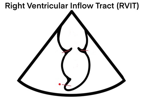
IVC
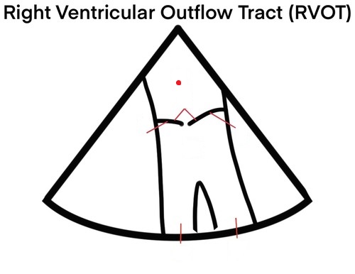
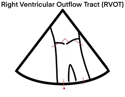
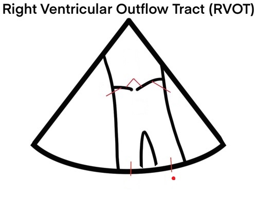
RV

Main pulmonary artery
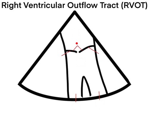
PV
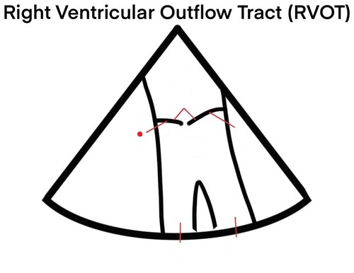
Right PV cusp

Anterior PV cusp
Right pulmonary artery
left pulmonary artery
LV
LV
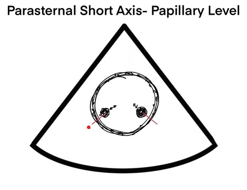
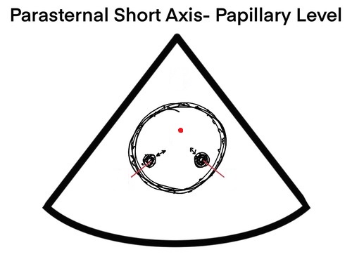
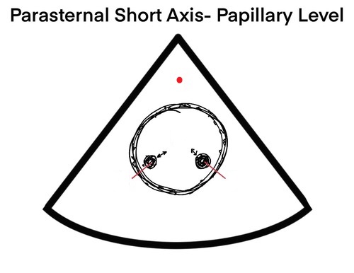
posteriomedial papillary muscle
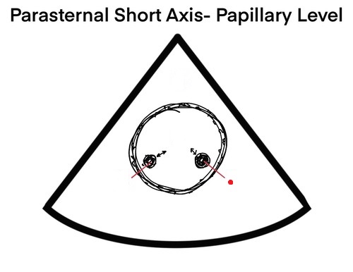
anteriolateral papillary muscle
RV

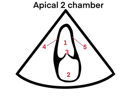
1
LV

2
Anterior MV leaflet

3
posterior MV leaflet

4
IVS
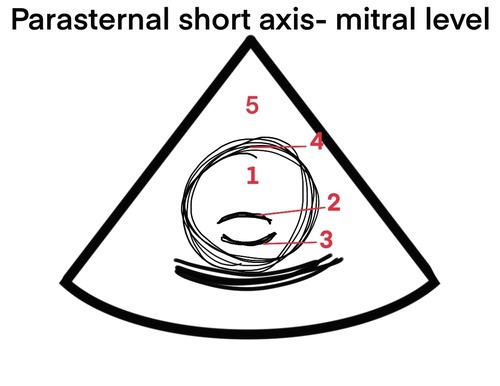
5
RV

1
LA

2
RA

3
RV

4
Pulmonary artery

5
TV

6
PV

7
Aortic valve, non coronary cusp

8
Aortic valve, right coronary cusp

9
Aortic valve, left coronary cusp

10
anterior TV leaflet

11
septal/medial TV leaflet
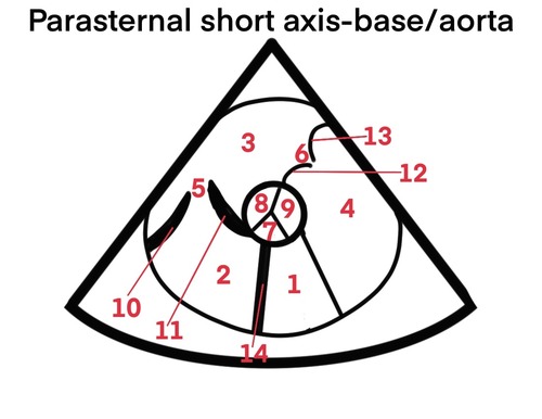
12
Right PV cusp

13
anterior PV cusp

14
IAS

7
anterior TV leaflet

8
septal/medial TV leaflet

9
anterior MV leaflet

10
posterior MV leaflet

14
RV free wall

15
inferior septal wall (LV)
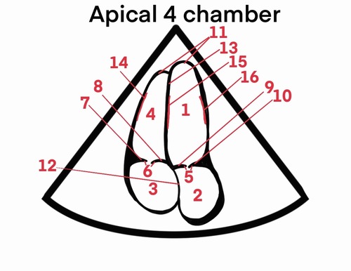
16
anterior lateral wall (LV)

5
aorta
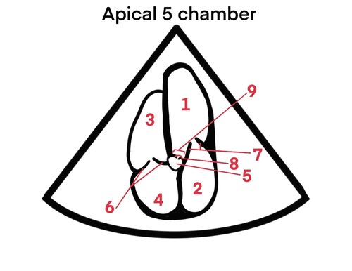
8
aortic valve

9
LVOT
4
inferior wall

5
anterior wall

1
RA

2
IVC
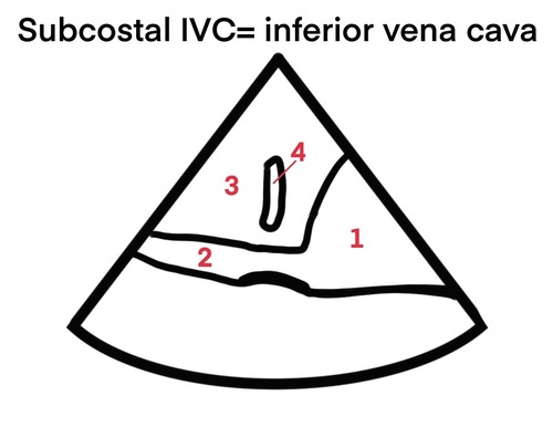
3
liver

4
hepatic vein
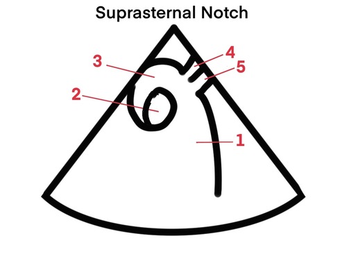
1
DAo

2
IVC

3
Ascending aorta

4
left common carotid artery

5
left subclavian artery
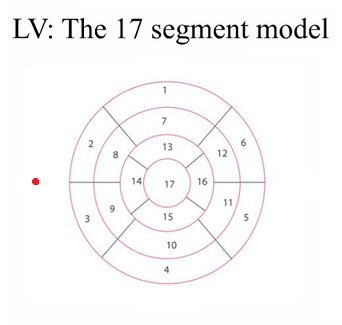
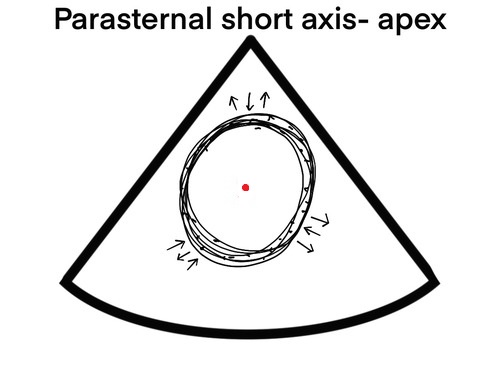
septal
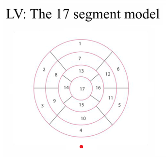
inferior
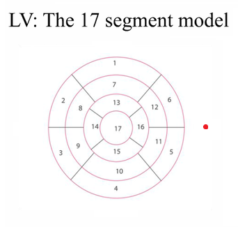
lateral
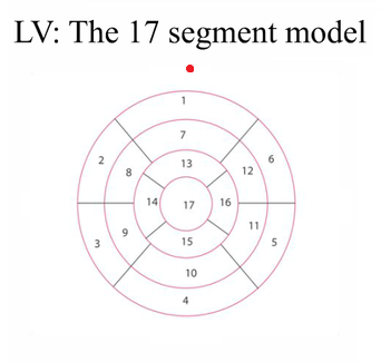
anterior
parabolic flow
Has a bullet shaped profile, Velocity is highest in the center of the lumen and gradually decreases to its minimum at the vessel wall.
plug flow
Occurs when all of the layers and blood cells travel at the same velocity.
More common with intracardiac flow due to tapering of flow streams through the valves.
laminar flow
Movement of fluid along well defined parallel stream line (layers) with uniform flow velocities
Reynold’s number
Predicts the likelihood that turbulence will occur.
demodulation
The process of extracting the low Doppler frequency from the higher transducer frequency
Negative Doppler shift
Color flow is away from the tx and the spectrum is below the baseline (retrograde flow).
Positive Doppler shift
Color flow is toward the tx and the spectrum is above the baseline (antegrade flow).
antegrade flow
Another word for Positive Doppler shift
Spectral Doppler
allows us to measure the velocity (speed and direction) of blood flow (traveling toward or away from the tx); displayed in the form of a time/velocity graph.
On a Spectral Doppler graph, what does the horizontal plane represent?
Time
On a Spectral Doppler graph, what does the vertical plane represent?
Velocity
Peak velocity (PSV)
Maximum velocity
Velocity time integral (VTI)
mean velocity or mean pressure gradient
Planimetry
cross section of a specific intracardiac location and a specific time point in the cardiac cycle
Power Doppler
Measures the presence of moving blood cells by detecting a Doppler shift. Non-directional; all vessels are the same color. The strength (amplitude) of the reflected signal is processed - the amount of moving blood cells. Also called Color Angio.
Advantages of Power Doppler
1. no aliasing
2. unaffected by Doppler angle
3. Very sensitive to low flow.
Disadvantages of Power Doppler
1. susceptible to motion
2. lower frame rate than conventional color flow Doppler
3. You don't get direction
Bi-directional Doppler
distinguishes the direction of blood flow (phase quadrature processing)
Advantage of CW Doppler
No aliasing; able to accurately measure very high velocities.
Disadvantage of CW Doppler
Range ambiguity - inability to determine the exact location of the moving RBCs being Dopplered.
What is CW Doppler used for?
Stenotic valve gradients, and calculations of pressures - 4(v²)
Pedoff Probe
A blind, standalone Doppler probe (no 2D image, not steerable). Easily fits into rib spaces and suprasternal notch.
Aliasing
Blood cells moving at high velocities are displayed as moving in the wrong direction. Occurs when velocity of blood exceeds the Nyquinst limit equals 1/2 the pulse repetition frequency. Only occurs with PW, never with CW.
Nyquist limit formula
PRF/2
How to reduce aliasing
increase the Doppler scale
use a lower freq tx
use a CW tx
change to a shallower imaging depth (high PRF)
drop the baseline
Color flow Doppler
Converts recorded flow frequency into different colors, allowing us to examine the direction and velocity of the blood flow through the chambers and valves. Allows for real time demonstration of flow patterns (velocities) superimposed on a gray scale image.