Hon Anatomy - Skin + The Digestive System
1/94
There's no tags or description
Looks like no tags are added yet.
Name | Mastery | Learn | Test | Matching | Spaced | Call with Kai |
|---|
No analytics yet
Send a link to your students to track their progress
95 Terms
Integumentary System
The enveloping organ of the body that includes the epidermis, dermis, sudoriferous and sebaceous glands, nails, and hair.
What are the functions of skin?
1) Water Retention
2) Produce Vitamin D
3) Thermal Regulation
4) UV Protection
5) Waste Removal (urea, uric acid, salts)
6) Sensory Aspects (Touch, Texture, Temp, Pressure, Vibration, Pain)
Keratin
A tough protein found in hair and nails that adds structural strength and protect the skin against damage from harmful chemicals. (Keratin + oils also act as a water barrier)
Melanocytes / Melanin
Melanocytes are specialized cells in the skin that produce melanin, a pigment that helps protect the body against the harmful effects of ultraviolet ray damage from sunlight
Epidermis
The outer layer of the skin. (Not Vascularized)
Dermis
The layer of skin that underlies the epidermis (between the epidermis and hypodermis). It includes nerve endings, glands, and hair follicles. (Vascularized)
Hypodermis / Subcutaneous Fascia
The innermost layer beneath the dermis. It is not part of the skin, but it connects the skin to the underlying tissues and provides cushioning and insulation, as well as serving as a storage repository for fat. (Vascularized)
What are the five layers of the epidermis?
1) Stratum Corneum
2) Stratum Lucidum
3) Stratum Granulosum
4) Stratum Spinosum
5) Stratum Basale
Function of the Stratum Corneum
The first and most superficial (outer) layer of the Epidermis. It is the layer of dead, flattened skin cells that are constantly being removed.
Function of the Stratum Lucidum
The second layer of the epidermis. It is a specialized clear layer of skin found only on the palms and soles, and it is what gives us our fingerprints.
Function of the Stratum Granulosum
The third layer of the epidermis. It prepares cells to become the new layer of the stratum corneum by killing, flattening, and dehydrating keratinocytes.
Function of the Stratum Spinosum
The fourth layer of the epidermis. It is the thickest layer and produces keratin.
Function of the Stratum Basale
The fifth and deepest layer of the epidermis. It rests upon the papillary layer of the dermis and has melanocytes that produce melanin. As the innermost layer, it absorbs nutrients from the underlying dermis and constantly produces new skin cells which are pushed upwards.
Keratinocytes
Cells within the epidermis that produce keratin.
Epidermal Dendritic Cells
Specialized cells within the epidermis are associated with the immune and nervous systems. They respond to the presence of foreign bacteria or viruses by initiating an immune system response, which brings in other specialized cells to attack the foreign invaders.
Merkel Cells (Merkel-Ranvier Cells)
Located in the stratum basale, these cells function as touch receptors. They form junctions with sensory nerve endings that relay information about touch to the brain.
Papillary Layer
The first and outer layer of the dermis, named after the dermal papillae that protrude from its surface up into the epidermis, which create fingerprints. Responsible for the sense of touch.
Reticular Layer
The second layer of the dermis underneath the papillary layer; Contains sudoriferous, sebaceous, and eccrine/apocrine glands.
Sudoriferous Glands
Sweat Glands; Distributed in the dermis over the entire body, with larger concentrations in the axilla, on the palms of the hands and soles of the feet, and on the forehead. Two types: eccrine and apocrine.
Eccrine Glands
The major sweat glands of the body; they cover most of the body and open directly onto the skin, producing a clear acidic fluid that is mainly water, but also contain waste products such as urea, uric acid, salts, and vitamin C.
Apocrine Glands
Sweat glands located in the genital and axillary areas that begin to function during puberty. They are larger than eccrine glands and secrete a milky fluid directly into the hair follicles, consisting of sweat, fatty acids, and proteins.
Sebaceous Glands
Located all over the body except for the palms of the hands and the soles of the feet; secrete sebum.
Sebum
Oily substance secreted by sebaceous glands that helps keep the skin and hair soft.
Hair follicles
Bulb-shaped structures in the dermis that produce hair.
Arrector Pili
Tiny muscles that connect both sides of the hair follicle to the epidermis, which then contract, pulling the hair upright and causing goosebumps.
Nail bed
A specialized region of the stratum basale. Nail growth occurs within the proximal end of the nail bed in a thickened region called the nail matrix or growth zone.
Decibitus Ulcers
Pressure ulcers; skin injuries caused by an area of localized pressure that restricts blood flow to one or more areas of the body which causes the skin cells to die.
First-Degree Burns
Affects only the epidermal layer of skin; Causes skin reddening and mild pain. (ex. minor sunburn).
What are common causes of burns?
Exposure to excessive heat, corrosive chemicals, electricity, or ultraviolet radiation. (Which cause tissue damage and cell death).
Second-Degree Burns
Involves damage to both the epidermis and the upper portion of the underlying dermis; Characterized by blisters, fluid filled pockets that form between the epidermal and dermal layers.
Third-Degree Burns
Destroy the entire thickness of the skin (AKA full-thickness burns); The affected area appears grayish-white or blackened.
Fourth-Degree Burns
Destroy all layers of skin and also some of the underlying tissues (including: nerve endings, muscle, tendon, ligament, and bone)
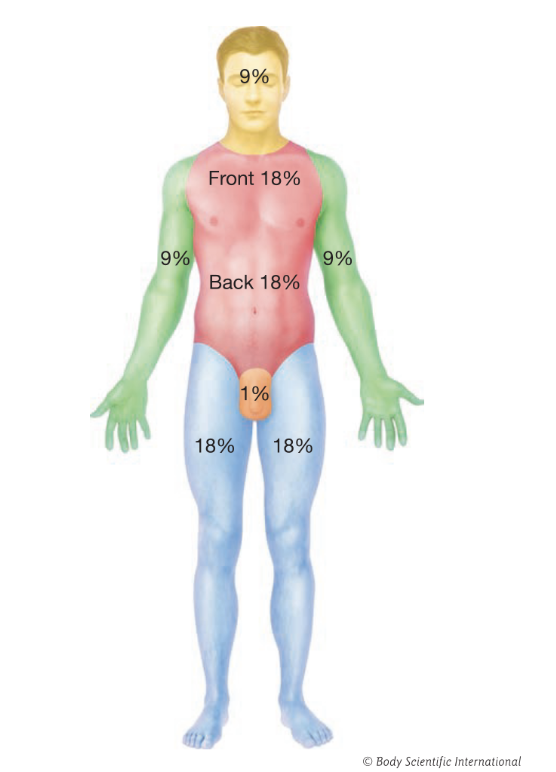
Rule of Nines
Used to estimate the extent of burned tissue when a large region of skin has been burned:
9% for both the anterior and posterior of the back and neck
18% for the anterior and 18% for the posterior of the torso
9% for both the anterior and posterior of each arm
18% for both the anterior and posterior of each leg
1% for the genital region
Herpes
A viral infection that produces small, painful, blister-like sores.
Herpes Varicella (Chickenpox) - A common childhood disease that is highly contagious and causes itchy fluid-filled blisters
Herpes Zoster (Shingles) - Causes an extremely painful, blistering rash accompanied by headache, fever, and a general feeling of unwellness
Herpes Simplex Virus Type 1 (HSV-1) - Associated with the common cold, generates cold sores / fever blisters around the mouth
Herpes Simplex Virus Type 2 (HSV-2) - The genital form of herpes
Human Papillomavirus (HPV)
A group of more than 150 related viruses that cause warts
Plantar warts - Develop on the soles of the foot and grow inward
Fungal Infections / Tinea
Occur in areas of the body that are moist
Athletes Foot (Tinea Pedis) - Characterized by cracked, flaky skin between the toes or on the side of the foot
Jock Itch (Tinea Cruris) - An itchy red rash on the genita, inner thighs, or buttocks that primarily affect males
Ringworm (Tinea Corporis) - Characterized by a red, ring-shaped rash with a pale center that resembles the shape of a worm
Toenail Fungus (Tinea Unguium) - A fungal infection under the nails of the fingers or toes that causes discoloration and thickening of the infected nail
Bacterial Infections
Impetigo - A highly contagious staphylococcus infection that causes pink, blister-like bumps around the mouth and nose
Cellulitis - A staphylococcus infection that causes an inflamed area of skin that is red, swollen, and painful
Inflammatory Conditions
Pleurisy - An inflammation of the pleura, the membrane that lines the thoracic cavity and lungs
Peritonitis - An inflammation of the peritoneum, the membrane that lines the inner wall of the abdomen and covers the abdominal organs
Psoriasis - A common skin disorder that speeds up the life cycle of skin cells and causes cells to build up rapidly on the surface of the skin
Basal Cell Carcinoma
The most common and least malignant form of skin cancer. Caused by the overproduction of cells in the stratum basale that push upward forming dome-shaped bumps.
Squamous Cell Carcinoma
Caused by the overproduction of cells in the stratum spinosum layer of the epidermis. Appears as a scaly, reddened patch that progresses to an ulcer-like mass with a raised border.
Melanoma
The most dangerous form of skin cancer, a cancer of the melanocytes. Typically dark colored and irregular in shape, but can appear pink, red, or ‘fleshy’.
ABCD Rule
The parameters the American Cancer Society advocated for determining the presence of melanoma:
A - Asymmetry: The shape of the mole is irregular
B - Border Irregularity: The outside borders are not smooth
C - Color: More than one color is present
D - Diameter: The mole size is larger than about one-quarter of an inch in diameter, or larger than the diameter of a pencil
Gastrointestinal Tract (GI Tract)
(AKA the Alimentary Canal) The tube (stomach, small intestine, large intestine) that runs through the body, beginning with the mouth and ending with the anus.
Ingestion
Intake of food and liquids via the mouth; Involved the mouth, teeth, lips, and tongue.
Propulsion
The movement of food through the gastrointestinal tract is stimulated by swallowing at the pharynx and peristalsis.
Peristalsis
The symmetrical contraction of muscles that move food along the remainder of the GI Tract.
Mechanical Breakdown
Reduces food into smaller pieces and increased the surface area of the food. (Chewing, churning in the stomach, and further churning by muscular contraction in the small intestine)
Chemical Breakdown
(Digestion) Enzymes in the lumen — the central opening of the GI tract into the stomach — and on the walls of the GI tract break large food molecules into smaller molecules
Absorption
Involves the movement of small food molecules from the lumen of the small intestine into the blood. Once this has occurred, the blood carries food to other parts of the body.
Defecation
The expulsion of food that was not absorbed. Waste matter/feces exits the body via the anus.
What are the four layers of the GI Tract? (Inside-Out)
1) Mucosa
2) Submucosa
3) Muscularis Externa
4) Serosa
Mucosa
(Mucous Membrane) The innermost layer of the GI tract is made of epithelial tissue, and its surface is covered by mucus secreted by cells or glands.
Submucosa
Below the muscosa; A layer of irregular dense connective tissue containing blood vessels, lymphatic vessels, and nerves. Can secrete substances that aid in digestion and absorption
Muscularis Externa
Surrounds the submucosal layer; In most of the GI tract it has two layers of smooth muscle. It propels food through the GI tract via peristalsis and also churns food mechanically
Serosa
The outermost layer of the GI tract; a thin serous membrane that minimizes friction between organs and between organs and the body cavity wall (known as the peritoneum is the abdominopelvic cavity)
Peritoneum
Divided into two layers: The Parietal and Visceral layers
Parietal lines the body wall
Visceral wraps around the organs and forms the outer layer of those organs
(Connected to each other via the Mesentary, a double layer of peritoneum, which helps hold the organs in place)
What are the four activities of digestion that the mouth is involved in?
1) Ingestion
2) Mechanical Breakdown
3) Chemical Breakdown
4) Propulsion
Deglutition
The tongue manipulating food in the mouth and moving chewed food to the back of the mouth; Swallowing
What is the function of the uvula?
Prevent swallowed food from entering the nasal cavity.
Gums / Gingiva
The soft tissue that covers the necks of the teeth and the maxilla and mandible.
Mastication
Teeth beginning the mechanical breakdown of food by grinding or crushing the food after it enters the mouth; Chewing.
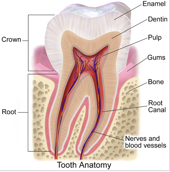
Tooth Anatomy
(Know basic structure)
Salivary Glands
The first accessory organs of digestion that contribute to the chemical breakdown of food; Saliva is mainly composed of water but it also contains mucus, antibodies, and several enzymes including salivary amylase.
What structure does the pharynx connect the mouth and nasal cavity to?
The trachea and esophagus.
What is the function of the epiglottis?
It acts as a switch between the larynx and esophagus, folding down during swallowing to prevent food and liquid from entering the airway and lungs.
Esophagus
A muscular tube that connects the pharynx to the stomach. When food enters the top of the esophagus during the act of swallowing, a wave of peristalsis begins, which pushes the food downward and into the stomach
Stomach
A reservoir in which food is broken down both mechanically and chemically before it enters the small intestine
What are the three major regions of the stomach?
1) Cardia
2) Fundus
3) Pyloric Region
What is the internal volume of an empty stomach?
50 mL; The folds (rugae) inside of the stomach flatten as the stomach stretches to increase the stomachs internal volume. An average stomach can hold up to two liters or more.
What layer of muscle helps the stomach churn food?
The oblique muscle sits below the circular and longitudinal muscle layers and helps the stomach churn food.
Goblet Cells
Secrete mucus onto the intestinal lining.
Gastric Pits
Tiny openings in the stomach linings; secrete gastric juice.
What is the pH of the stomach?
1.5-2.5
What are the mucus secreting cells that line the gastric pits and what do they secrete?
Parietal Cells - Secrete Hydrochloric Acid and Intrinsic Factor (helps with vitamin B absorption)
Chief Cells - Secrete pepsinogen (a protein-digesting enzyme)
Enteroendocrine Cells - Produce gastrin, which stimulates the secretion of more gastric juice
How does the body prevent its own tissues from being digested?
Mucus is constantly produced to line the stomach to prevent it from being dissolved by HCL or digested by enzymes.
Maceration
Food that enters the stomach mixes with gastric juice to form chyme.
What is the function of the pyloric sphincter
(located at the top of the duodenum/pyloric region of the stomach) It prevents stomach acid from entering the small intestine.
What is the primary function of the small intestine?
All food absorption and most water absorption.
What are the three segments of the small intestine?
1) Duodenum
2) Jejunum
3) Ileum
(Chemical digestion, absorption, and propulsion occur in all three segments)
Villi
Finger-like projections on the circular folds that cover the inner surface of the small intestine that maximize nutrient absorption by increasing surface area.
Emulsification
When chyme enters the duodenum from the stomach, it mixes with bile and pancreatic juice. (Bile (produced by the liver) helps with this process by breaking down large fat particles into smaller, more evenly distributed particles;
The breakdown of large fat particles into much smaller ones, aided by bile
Why are bile and pancreatic juices important?
They help with emulsification and neutralize chyme.
Gallbladder
The digestive organ that stores bile and delivers it to the duodenum when needed.
Liver + Gallbladder Function
Make bile (liver), store it (gallbladder), and deliver it the duodenum where it is used for the chemical breakdown of lipids.
Liver Functions
Maintenance of normal blood concentrations of glucose, lipids, and amino acids
Conversion of one nutrient type to another
Synthesis and storage of glycogen and secretion of cholesterol, plasma proteins, and clotting factors
Storage of iron, lipids, and fat-soluble vitamins
Absorption and inactivation of toxins, hormones, immunoglobulins, and drugs
Bile
A watery solution containing bile salts, which aids in emulsification.
What is the pancreas’s digestive and metabolic function?
Digestive - Make and secrete pancreatic juice into the duodenum
Metabolic - Make the hormones insulin and glucagon and secrete them into the blood stream
Why are pancreatic proteases produced and secreted in an inactive form?
To prevent them from self-digesting the pancreas.
How does the pancreas regulate glucose levels in the blood?
When glucose levels are high the pancreas secretes insulin into the blood, which causes hepatocytes to extract and convert glucose into glycogen. When blood glucose levels are low, glucagon (a hormone secreted by the pancreas) binds to receptors throughout the body to promote the conversion of glycogen to glucose in liver cells.
What are the four major segments of the large intestine?
1) Cecum
2) Colon
3) Rectum
4) Anal Canal
What are the main functions of the large intestine?
Propulsion and Elimination of Waste.
What allows defecation to occur?
Once waste reaches the rectum it empties into the anal canal, where anal sphincters (under voluntary control) initiate contractions that push the waste toward the anus, allowing the waste to exit.
Chyme
The mixture of food and digestive juice in the stomach and duodenum
Bolus
A mass of chewed food mixed with saliva that is ready to be swallowed
What are the functions of the Kidneys?
Remove toxins, urea, and excess salts from the bloodstream to produce urine
Regulate the body’s water balance and maintain levels of minerals
Release enzymes to manage blood pressure