Anatomy, Everything you need to know about Week 4
1/116
Earn XP
Description and Tags
I promise these aren't too difficult (that's why there's so many)
Name | Mastery | Learn | Test | Matching | Spaced | Call with Kai |
|---|
No analytics yet
Send a link to your students to track their progress
117 Terms
what are the 2 bones in the leg?
tibia and fibula
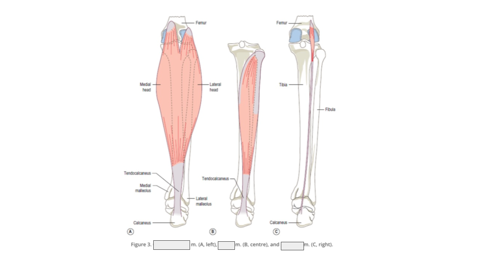
What are the 3 muscles in the posterior superficial compartment of the leg?
gastrocnemius, plantaris and soleus
what action does the gastrocnemius perform?
flexion of the knee and plantarflexion of the foot
the calcaneal tendon is also known as the?
achilles tendon
what is the function of the calcaneal tendon?
attaches the gastrocnemius and soleus muscles (sometimes plantaris too) to the calcaneus bone
what is the thickest tendon in the body?
calcaneal tendon
what supplies blood to the gastrocnemius, plantaris and popliteus?
popliteal artery
what supplies innervation to the superficial and deep posterior muscles of the leg?
tibial nerve
what are the heads of the gastrocnemius called?
lateral and medial head
the foot movement that ballerinas often do is called?
plantarflexion
what supplies blood to the gastrocnemius, soleus, tibialis posterior, flexor hallucis longus and flexor digitorum longus?
posterior tibial artery
the gastrocnemius is superficial to which muscle?
soleus
what action does plantaris perform?
flexion of the knee and plantarflexion of the foot
what movement does the soleus perform?
plantarflexion of the foot
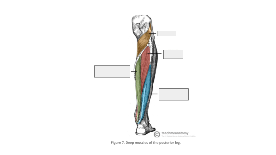
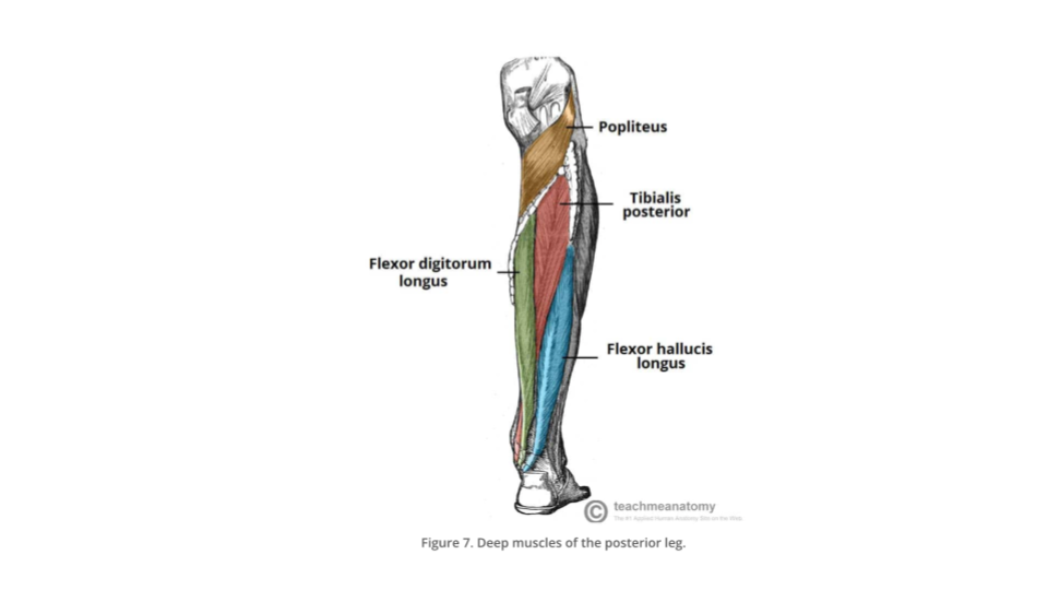
what are the 4 muscles of the posterior deep compartment of the leg?
popliteus, tibialis posterior, flexor hallucis longus and flexor digitorum longus
what action does the popliteus perform?
Initiates the flexion of the fully extended (“locked”) knee (“key to unlock the knee”)
(think of a ‘pop’ sound before flexion occurs, don’t know if that makes sense)
what action does the tibialis posterior perform?
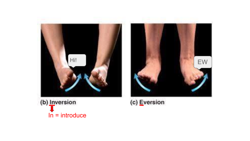
Plantarflexion of ankle and inversion (where the soles of your feet INtroduce eachother) of the foot
describe what eversion is:
where the soles of your feet face the sides (imagine the soles of your feet are saying ‘ew’ and facing the other way, idk bro)
appreciate the image. it took me a while to do.
what action does the flexor hallucis longus perform?
flexion of the hallux and plantarflexion of the ankle. assists with inversion of foot
what action does the flexor digitorum longus perform?
flexion of toes 2-5 and plantarflexion of ankle. assist with inversion of foot (in = introduce = “hi other foot!”)
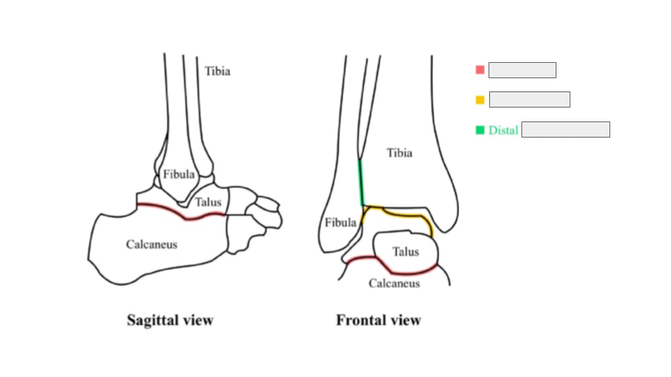
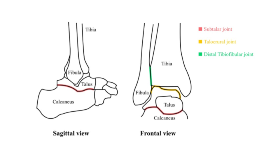
what are the 3 articulating surfaces in the ankle joint?
talocrural joint, subtalar joint and distal tibiofibular joint
you know the drill, fill in the blanks:
the ankle joint performs 4 movements making it multiaxial: d__________, p_________, e________, i__________
dorsiflexion, plantarflexion, eversion, inversion
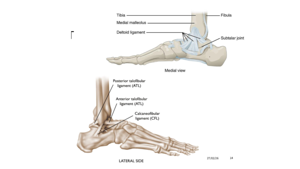
there is only one ligament on the medial side of the ankle joint! what is it called?
deltoid ligament
there are 3 ligaments on the lateral side of the ankle joint ☹ what are these called?
anterior talofibular ligament, posterior talofibular ligament and calcaneofibular ligament (this kind of makes sense since the fibula is on the lateral side of the leg)

fill in the blanks:
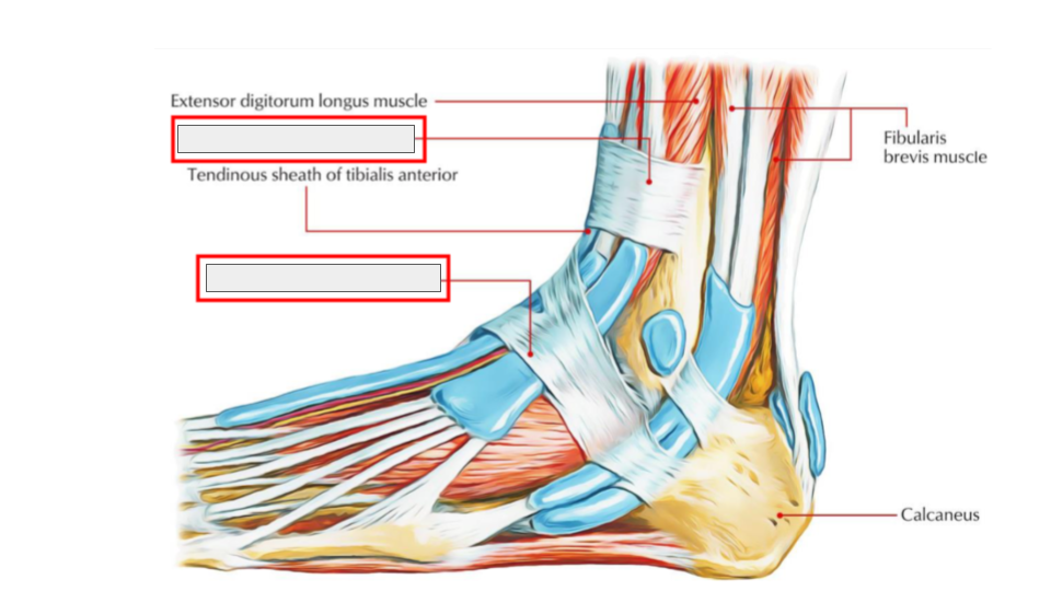
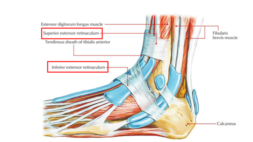
define the retinacula:
A broad band of ligaments that stabilise tendons on the foot
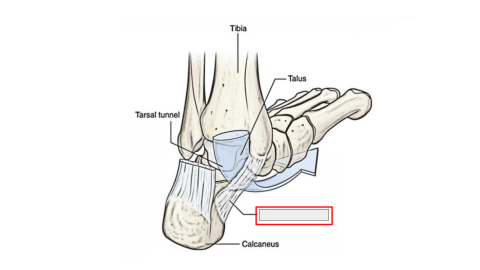
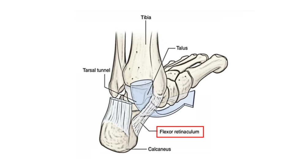
name the 3 types retinaculum:
superior extensor retinaculum, inferior extensor retinaculum and flexor retinaculum
which retinaculum is Y-shaped in front of the ankle joint, attaching the lateral calcaneus to the medial malleolus of the tibia and plantar aponeurosis?
inferior extensor retinaculum
Which retinaculum is on the medial side, attaching the medial calcaneus to the medial malleolus of tibia?
flexor retinaculum
which retinaculum is above the ankle joint, attaching fibula and tibia
superior extensor retinaculum
fill in the blanks:
fill in the blank:
easy peasy lemon squeezy
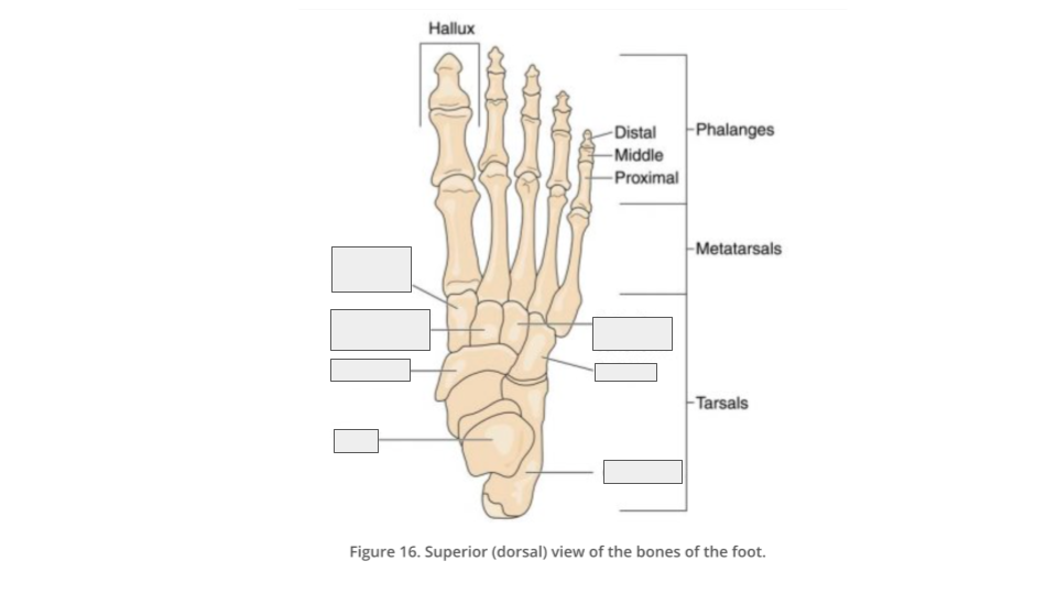
how many bones are in the foot?
26 (or just remember the date of my birthday ;))
what are the 3 types of bones in the foot?
tarsals, metatarsals and phalanges
how many tarsals are in the foot?
7
how many metatarsals are in the foot/
5
how many phalanges are in the foot
14
how many intrinsic muscles does the foot have?
10
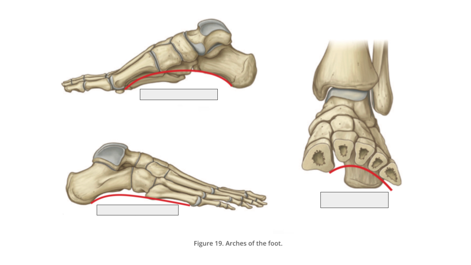
how many arches does the foot have?
3
what are the 2 types of arches?
longitudinal and transverse
how many longitudinal arches does our foot have?
2
how many transverse arches does our foot have?
1
what is the function of the arches of the foot?
weight-bearing, movement, shock-absorption and propulsion
(you’re gonna love this) how many muscles does the dorsal foot have?
2
what is the function of the extensor hallucis brevis?
extension of hallux
what is the action of the extensor digitorum brevis?
extension of toes 2-4 (not 2-5 because….I said so)
what nerve supplies innervation to the dorsal foot muscles?
deep fibular nerve
(you’re gonna hate this) how many layers of muscles does the plantar foot have?
4! (there’s a code for how many muscles in each layer)
how many muscles are there in the first layer (most superficial) of the plantar foot?
3
how many muscles are there in the second layer of the plantar foot?
2
how many muscles are there in the third layer of the plantar foot?
3
how many muscles are there in the fourth layer (the deepest) of the plantar foot?
2 (so from the first to fourth layer (superficial to deep), the number of muscles in each layer go: 3,2,3,2)
what is the names of the muscles in the 1st layer?
abductor hallucis, flexor digitorum brevis and abductor digiti minimi (all the abductors in the foot are in this layer)
describe the action of the abductor hallucis:
abduction and flexion of hallux
describe the action of the flexor digitorum brevis:
flexion of toes 2-5
describe the action of the abductor digiti minimi:
abduction and flexion of toe 5 (little toe)
what is the plantar aponeurosis/fascia?
A thick band of fascia that runs from the heel to the toes on the plantar surface. It supports the arches of the foot and protects the structures underneath.
describe the action of quadratus plantae:
assists flexor digitorum longus in flexion of toes 2-5
describe the location and action of lumbricals:
located on toes 2-5, flexion at the metatarsophalangeal joints and extension of the interphalangeal joints
describe the action of the flexor hallucis brevis:
flexion of hallux at the metatarsophalangeal joints
describe the action of adductor hallucis:
adduction of hallux
Describe the action of the flexor digiti minimi brevis:
flexion of toe 5 at metatarsophalangeal joint
describe the action of the dorsal interossei:
on toes 2-5, they abduct and flex at the metatarsophalangeal joints
describe the action of plantar interossei:
on toes 3-5 (not 2-5 because then life would be too easy), they adduct and flex at the metatarsophalangeal joints
all the muscles in the plantar foot are innervated by which nerve?
tibial nerve
the muscles in the posterior compartment of the leg are commonly known as?
plantarflexors
the tibial nerve is a _______ branch of the sciatic nerve;
terminal
The posterior tibial artery is a branch of the?
tibiofibular trunk
which muscle am I describing:
The biggest and most superficial muscle in this compartment.
Its two heads arise from the distal end of the femur, medial and lateral sides, and combine into a very big muscle belly that inserts on the calcaneus via the calcaneal tendon (also known as the Achilles tendon).
Because of its size and proximity to the popliteal fossa, it receives blood supply from branches of the popliteal artery.
Actions: flexion of the knee and plantarflexion of the foot.
gastrocnemius
as soleus and gastrocnemius muscles occupy roughly the same space and inserts together in same tendon sometimes these muscles are called the?
triceps surae (tri-ceps, 3 heads + surae, means calf = 3 heads of the calf)
which muscle am I describing:
has a thin muscle belly and a long thin tendon (the longest tendon in the human body!).
It comes from the inferolateral part of the femur, just slightly superior to the origin of the lateral head of gastrocnemius. Its tendon inserts either into the calcaneal tendon or just medial to it, on the calcaneus.
Because of its location so near the knee joint, it receives blood supply from branches of the popliteal artery.
It flexes the knee and plantarflexes the foot. However, its actions are weak when compared to the other superficial posterior muscles of the leg.
plantaris 🌱🌱
fill in the blanks:

which muscle am I describing:
It is the most central of the deep muscles of the posterior leg, situated between the flexor hallucis longus and the flexor digitorum longus muscles.
Comes from the posterior surface of the fibula and the interosseous membrane and inserts on sole of the foot.
It plantarflex the ankle and inverts the foot.
tibialis posterior
which muscle am I describing:
A small, triangular band-like muscle on the lower aspect of the back of the knee, hence, it is the only muscle in the posterior compartment of the lower leg that acts just on the knee and not on the ankle.
Because of its location, it is the main stabiliser of the posterior knee region, receiving blood supply from branches of the popliteal artery.
It plays an important role in the gait cycle by initiating the flexion of the fully extended (“locked”) knee. In other words, it acts unlocking the knees when walking by laterally rotating the femur on the tibia. This is why it is referred to as the “key to unlock the knee”.
popliteus
Which muscle am I describing:
Situated lateral to the tibialis posterior.
Comes from the posterior surface of the fibula and inserts into the plantar surface of the hallux (big toe).
It flexes all the joints in the hallux and plantarflexes the ankle. Because of the location of its tendon, it also assists with inversion of the foot.
flexor hallucis longus
which muscle am I describing:
The most medial of the deep posterior leg muscles.
It arises from the posterior tibia and inserts to the plantar surface of toes 2-5.
It flexes toes 2-5 and plantarflexes the ankle. Because of the location of its tendon, it also assists with inversion of the foot.
flexor digitorum longus
fill in the blanks:
what am I describing:
Also known as ankle proper joint, this joint is the only mortise and tenon joint in the human body.
It is formed between the inferior aspect of the distal end of the tibia and fibula and the superior aspect of the talus.
This is one of the weight-bearing components of the ankle joint.
talocrural joint
what am I describing:
This joint, also known as the talocalcaneal joint, occurs at the meeting point of the inferior aspect of the talus and the superior aspect of the calcaneus.
This is one of the weight-bearing components of the ankle joint.
subtalar joint
what am I describing:
This joint, also known as the inferior tibiofibular joint and tibiofibular syndesmosis, is formed between the medial side of the distal end of the fibula, and the lateral side of the distal tibia.
This is not a weight-bearing joint.
distal tibiofibular joint
what am I describing:
It is attached at the medial malleolus of the tibia and connects via 4 lesser ligaments to the talus, calcaneus and navicular bones.
It supports the ankle joint and also resists excessive eversion of the foot.
deltoid ligament
what ligaments am I describing:
It supports the lateral side of the joint from the lateral malleolus of the fibula to the anterior and posterior aspects of the talus.
They prevent the foot from sliding forward or in relation to the tibia.
anterior and posterior talofibular ligaments
what am I describing:
A narrow, rounded cord.
It runs from the lateral malleolus of the fibula downward and slightly backward to the calcaneus.
It opposes the hyperinversion of the subtalar joint, which is a common type of ankle sprain.
calcaneofibular ligament
which toe digit doesn’t have 3 phalanges?
the hallux
fill in the blanks:

there are 3 groups for the 7 tarsal bones: the proximal group, the intermediate group and the distal group, what are the bones in each?
proximal: calcaneus and talus
intermediate: navicular
distal: cuboid, lateral cuneiform, intermediate cuneiform
what am I describing:
The largest tarsal bone, lying inferior to the talus. It protrudes posteriorly and forms the heel. The posterior aspect is marked by the calcaneal tuberosity, to which the calcaneal (Achilles) tendon attaches.
calcaneus
what am I describing:
It articulates with the talus posteriorly, all three cuneiform bones anteriorly, and the cuboid bone laterally.
navicular bone
what am I describing:
The most superior of these bones. It transmits the weight of the entire body from the tibia to the calcaneus.
talus
Metatarsal 1 is related to the ______ and 2-5 are related to the toes ____, respectively.
hallux, 2-5
what is the metatarsophalangeal (MTP) joint?
The joint between each metatarsal and the proximal phalanx
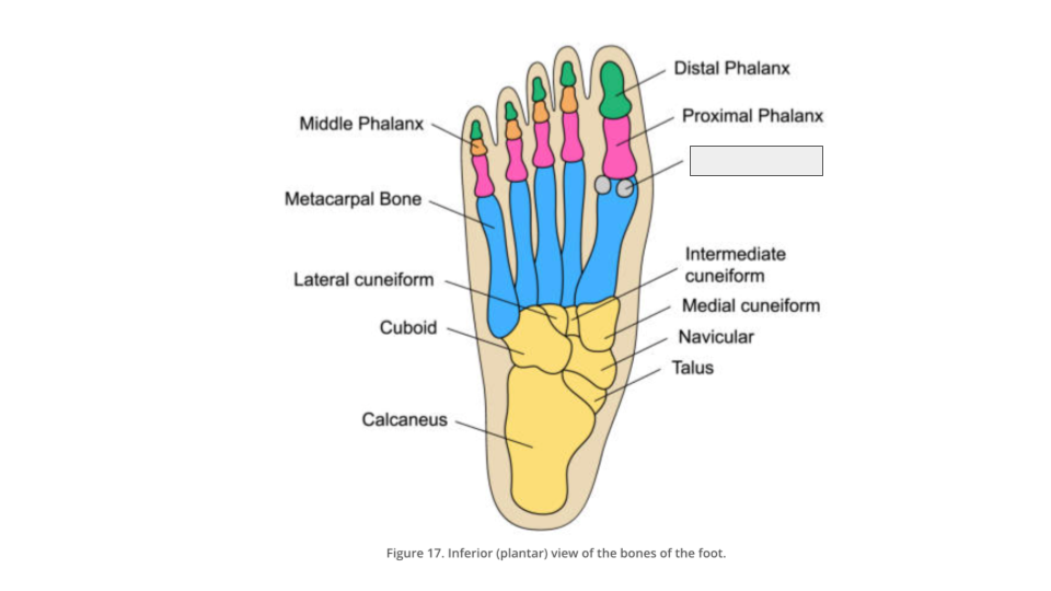
The first metatarsal bone usually has two _________ bones on the inferior (plantar) side, near the MTP joint. Both these bones can be found within the tendon of flexor hallucis brevis muscle.
sesamoid

what am I describing:
This arch is the flatter of the two longitudinal arches (it touches the ground when you're standing).
It is formed by the calcaneus, cuboid and 4th and 5th metatarsal bones.
lateral longitudinal arch
what am I describing:
This arch is the higher of the two longitudinal arches.
It is formed by the calcaneus, talus, navicular, three cuneiforms and first three metatarsal bones.
medial longitudinal arch
what am I describing:
This arch is located in the coronal plane of the foot (it is not a line, but a plane).
It is formed by the metatarsal bases, the cuboid and the three cuneiform bones.
transverse arch
fill in the blanks:

the foot is supplied with what 2 tibial arteries which form arches around the dorsum and sole of the foot?
anterior and posterior
at the dorsum foot there are 4 arteries to consider what are they?
lateral and medial tarsal arteries, dorsalis pedis artery and arcuate artery
the lateral and medial tarsal arteries supply the dorsal aspects of the tarsal bones, they are branches of which artery?
anterior tibial artery